. 1997 Oct 15;17(20):7754-62.
doi: 10.1523/JNEUROSCI.17-20-07754.1997.

Neurons promote the translocation of peripheral myelin protein 22 into myelin

Affiliations

Neurons promote the translocation of peripheral myelin protein 22 into myelin

S Pareek et al. J Neurosci. .

Abstract

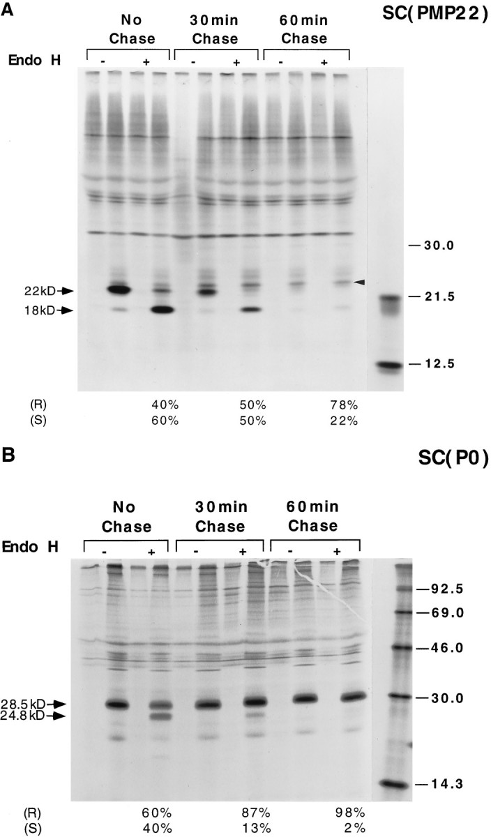
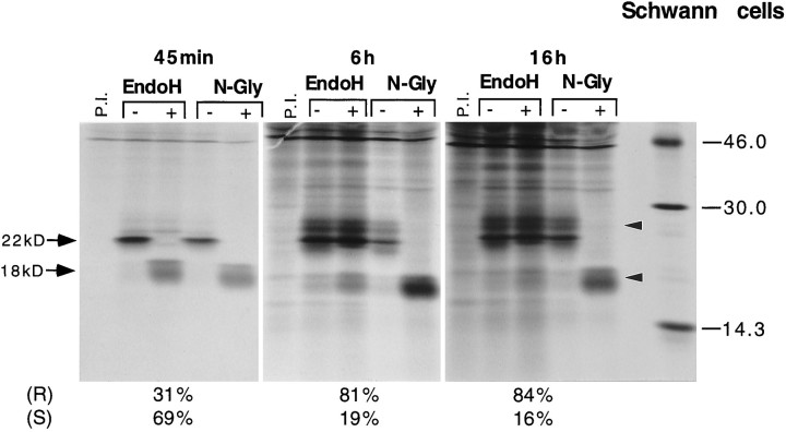
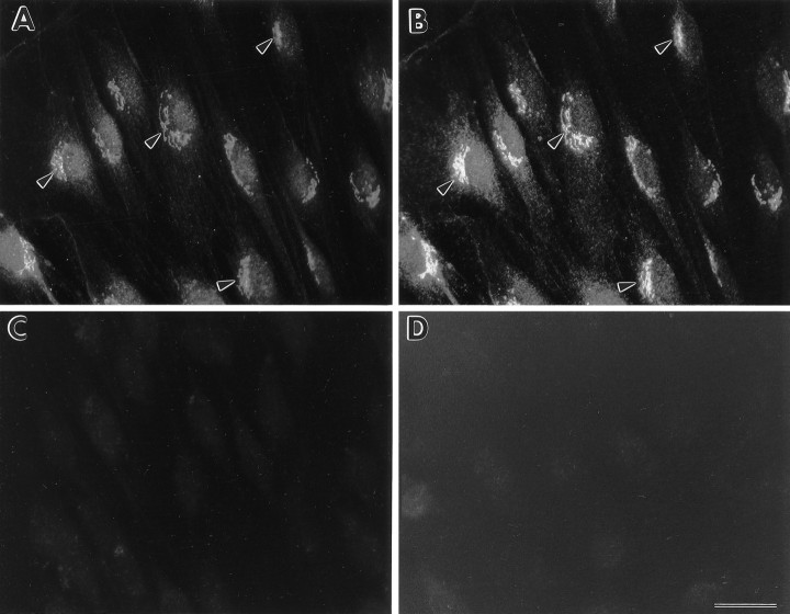
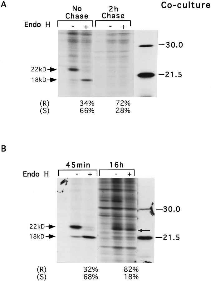
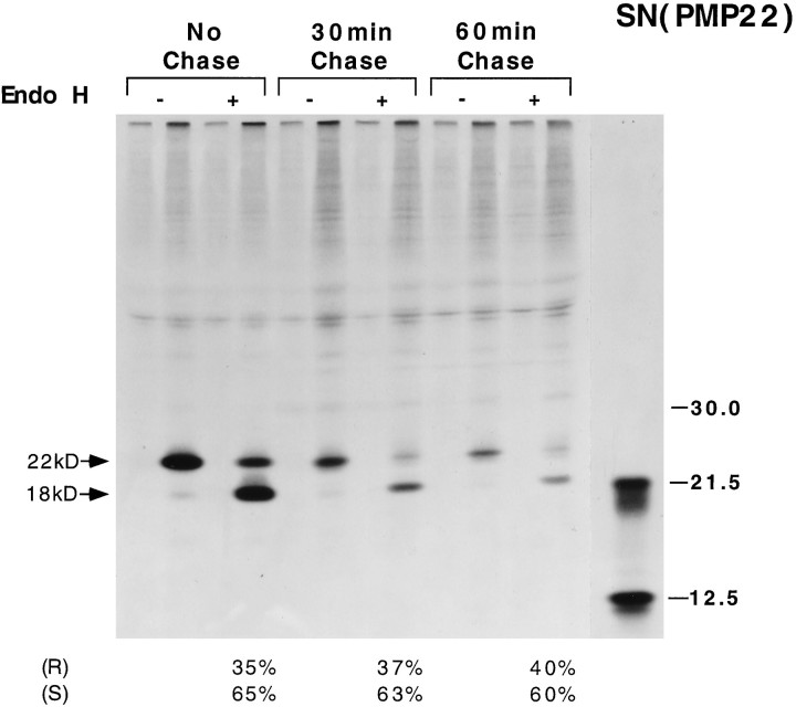
Schwann cells express low levels of myelin proteins in the absence of neurons. When Schwann cells and neurons are cultured together the production of myelin proteins is elevated, and myelin is formed. For peripheral myelin protein 22 (PMP22), the exact amount of protein produced is critical, because peripheral neuropathies result from its underexpression or overexpression. In this study we examined the effect of neurons on Schwann cell PMP22 production in culture and in peripheral nerve using metabolic labeling and pulse-chase studies as well as immunocytochemistry. Most of the newly synthesized PMP22 in Schwann cells is rapidly degraded in the endoplasmic reticulum. Only a small proportion of the total PMP22 acquires complex glycosylation and accumulates in the Golgi compartment. This material is translocated to the Schwann cell membrane in detectable amounts only when axonal contact and myelination occur. Myelination does not, however, alter the rapid turnover of PMP22 in Schwann cells. PMP22 may therefore be a unique myelin protein in that axonal contact promotes its insertion into the Schwann cell membrane and myelin without altering its rapid turnover rate within the cell.


Figures

Fig. 1.
Fig. 1.
Pulse-chase analysis and Endo H digestion of PMP22 (A) and P0 (B) in SC. SC were metabolically labeled with trans-35S label for 30 min and chased for 30 and 60 min with cold methionine and cysteine. SC lysates were immunoprecipitated with either PMP22 (A) or P0 (B) antibodies, and the immunoprecipitates were incubated with (+) or without (−) Endo H at 4°C for 16–18 hr. After immunoprecipitation, cell lysates were analyzed by SDS-PAGE and fluorography. Duplicate samples were precipitated with preimmune serum (first lane in each group). The migration positions of PMP22 and P0 are indicated byarrows. Top arrows show the glycosylated proteins, and bottom arrows show the unglycosylated proteins. The right arrow in A shows the Endo H resistant PMP22. Molecular weight markers are shown at the extreme right in both A andB. Endo H resistant (R) and sensitive (S) PMP22 (A) and P0 (B) are shown as the percentage of total protein at different time intervals.
Fig. 2.
Fig. 2.
Comparison of short- and long-term labeling of cultured SC. SC were metabolically labeled withtrans-35S label for 45 min, 6 hr, or 16 hr. Cell lysates were immunoprecipitated with PMP22 antibodies, and the immunoprecipitates were incubated with (+) or without (−) Endo H or PNGase F (N-Gly) at 37°C for 16–18 hr. After immunoprecipitation, cell lysates were analyzed by SDS-PAGE and fluorography. Duplicate samples were treated with preimmune serum (first lane in each group). The migration positions of glycosylated 22 kDa PMP22 (top arrow) and the 18 kDa core peptide (bottom arrow) are indicated. The top right arrow shows the Endo H resistant accumulated PMP22 protein, and the bottom right arrowshows the 18 kDa core protein that appears after PNGase F digestion. Molecular weight markers are shown at the extreme right. Endo H resistant (R) and sensitive (S) PMP22 are shown as the percentage of total protein at different time intervals.
Fig. 3.
Fig. 3.
Immunocytochemical localization of PMP22 in cultured SC. SC cultures were fixed in 4.0% paraformaldehyde and permeabilized by treatment with methanol. Samples were double-stained with PMP22 antisera visualized by Texas Red-conjugated anti-rabbit IgG and monoclonal Golgi marker (FITC). The distribution of PMP22 (B) and the 58 kDa Golgi protein (A) are shown. Most of the PMP22-like immunoreactivity co-localized with the Golgi marker (arrows in A, B). Controls are shown in which the samples were incubated with PMP22 peptide-adsorbed antiserum (C) or preimmune serum (D). Scale bar (shown in D), 22 μm.
Fig. 4.
Fig. 4.
Pulse-chase analysis (A) and short- and long-term labeling (B) of PMP22 in myelinating co-cultures of SC and neurons. DRG-SC co-cultures were grown in the presence of ascorbic acid and progesterone. Cells were metabolically labeled with trans-35S label for 45 min and chased for 2 hr with cold methionine and cysteine (A). For comparison of short- and long-term labeling studies (B), myelinating co-cultures were metabolically labeled with trans-35S label for either 45 min or 16 hr. Cell lysates were immunoprecipitated with PMP22 antibodies, and the immunoprecipitates were incubated with (+) or without (−) Endo H at 4°C for 16–18 hr. After immunoprecipitation, the lysates were analyzed by SDS-PAGE and fluorography. Duplicate samples were precipitated with preimmune serum (first lane in each group). The migration positions of glycosylated PMP22 (top arrows) and the 18 kDa core peptide (bottom arrows) are indicated. Theright arrow in B indicates the accumulated Endo H resistant PMP22 protein. Molecular weight markers are shown at the extreme right. Endo H resistant (R) and sensitive (S) PMP22 are shown as the percentage of total protein at different time intervals.
Fig. 5.
Fig. 5.
Immunolocalization of PMP22 in short- and long-term myelinating co-cultures. One-week-old SC and neuron co-cultures were double-stained with monoclonal anti-NF (A) and polyclonal PMP22 antiserum (B). Arrows point to SC (B) that are in contact with neuronal processes (A). After 4 weeks in medium that promotes myelination, PMP22 (D) co-localizes with P0 reactive myelin segments (C).Arrows point to the cell bodies of elongated SC (D) with uniform PMP22 staining over the cell membrane. Scale bars: A, B (shown in B), 22 μm; C, D (shown in D), 25 μm.
Fig. 6.
Fig. 6.
Pulse-chase analysis of ex vivo-labeled sciatic nerve explants. Ten-day-old sciatic nerve segments were metabolically labeled withtrans-35S label for 30 min and chased for 30 and 60 min with cold methionine and cysteine. Nerve homogenates were immunoprecipitated with PMP22 antibodies, and the immunoprecipitates were incubated with (+) or without (−) Endo H at 4°C for 16–18 hr. After immunoprecipitation, nerve samples were analyzed by SDS-PAGE and fluorography. Duplicate samples were precipitated using preimmune serum (first lane in each group). The migration positions of glycosylated 22 kDa PMP22 (top arrow) and the 18 kDa core peptide (bottom arrow) are indicated. Molecular weight markers are shown at the extreme right. Endo H resistant (R) and sensitive (S) PMP22 are shown as the percentage of total protein at different time intervals.
Fig. 7.
Fig. 7.
Western blot analysis of PMP22 in in vitro and in vivo samples. PMP22 in nonmyelinating SC (50 μg/lane total protein), 8-week-old myelinating co-cultures (50 μg/lane), 10-d-old sciatic nerve (10 μg/lane), and purified myelin (10 μg/lane) was tested for the presence of high-mannose and complex carbohydrates by PNGase F (N) and Endo H (H) treatments as described in Materials and Methods. Control samples incubated without the addition of enzymes are analyzed in lanes C. Molecular masses are indicated in kilodaltons.

Similar articles

Cited by

References

    1. Adlkofer K, Martini R, Aguzzi A, Zielasek J, Toyka KV, Suter U. Hypermyelination and demyelinating peripheral neuropathy in Pmp22-deficient mice. Nat Genet. 1995;11:274–280. - PubMed
    1. Bonifacino JS, Cosson P, Shah N, Klausner RD. Role of potentially charged transmembrane residues in targeting proteins for retention and degradation within the endoplasmic reticulum. EMBO J. 1991;10:2783–2793. - PMC - PubMed
    1. Bosse F, Zoidl G, Wilms S, Gillen CP, Kuhn HG, Müller HW. Differential expression of two mRNA species indicates a dual function of peripheral myelin protein PMP22 in cell growth and myelination. J Neurosci Res. 1994;37:529–537. - PubMed
    1. Brockes JP, Fields KL, Raff MC. Studies on cultured rat Schwann cells. I. Establishment of purified populations from cultures of peripheral nerve. Brain Res. 1979;165:105–118. - PubMed
    1. Brunden KR. Age-dependent changes in the oligosaccharide structure of the major myelin glycoprotein, P0. J Neurochem. 1992;58:1659–1666. - PubMed

Publication types

LinkOut - more resources