. 1997 Oct 15;17(20):7796-804.
doi: 10.1523/JNEUROSCI.17-20-07796.1997.

Pathfinding by identified zebrafish motoneurons in the absence of muscle pioneers

Affiliations

Pathfinding by identified zebrafish motoneurons in the absence of muscle pioneers

E Melançon et al. J Neurosci. .

Abstract

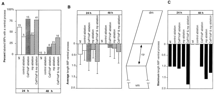
To identify the cellular cues that guide zebrafish neuronal growth cones to their targets, we examined interactions between identified motor growth cones and identified muscle fibers and tested whether these fibers were required for growth cone navigation. Caudal primary motoneurons (CaPs) and middle primary motoneurons (MiPs) are identified motoneurons that innervate cell-specific regions of the myotome. Growth cones of both cells initially extend along a common pathway and then pause at a set of identified muscle fibers, called muscle pioneers, before diverging along cell-specific pathways. Muscle pioneers are intermediate targets of both CaP and MiP (Westerfield et al., 1986; Liu and Westerfield, 1990); both motoneurons extend their growth cones directly to the muscle pioneers on which the first functional neuromuscular contacts form, suggesting that muscle pioneers may provide guidance information to these growth cones. We tested this idea by ablating muscle pioneers and observing the resulting motor axonal trajectories. Both CaP and MiP ultimately formed normal axonal arbors after muscle pioneer ablation, showing that muscle pioneers are unnecessary for formation of correct axonal trajectories; however, although final cellular morphology was correct in the absence of muscle pioneers, MiP growth cones branched abnormally or extended ventrally beyond the common pathway. Ablation of CaP and the muscle pioneers together increased the aberrant behavior of the MiP growth cone. Our results provide evidence that an intermediate target, the muscle pioneers, affects motor axonal extension without altering target choice, suggesting that other cues also contribute to proper pathway navigation.


Figures

Fig. 1.
Fig. 1.
Schematic diagrams of neuromuscular organization in developing zebrafish embryos. A, Side view of four myotomes at different stages of development; rostral to the left and dorsal to the top in this and all subsequent side views. The myotome on the left (1) shows the extent of axonal outgrowth by CaP (blue) and MiP (orange) at ∼20 h. The CaP axon has reached the distal end (broken line) of the common pathway (a), whereas the MiP growth cone has not exited the spinal cord (sc). The second myotome (2) shows CaP and MiP at ∼21 h. The CaP axon has extended ventrally beyond the common pathway onto its cell-specific pathway (b), whereas MiP has a ventral process on the common pathway and a dorsal collateral axon on the MiP pathway (c). Myotome 3 shows CaP and MiP at ∼26 h. CaP innervates ventral myotome and MiP innervates dorsal myotome; MiP has retracted its ventral process from the common pathway. By 16 h (myotome 4), three to six identifiable muscle pioneers (mps) are recognizable at the distal end of the common pathway. B, Transverse section of developing zebrafish embryo showing the positions of CaP axons relative to the muscle pioneers (mps) at ∼20 h.nc, Notochord. Scale bar, 25 μm.
Fig. 3.
Fig. 3.
Early muscle contractions are produced by cholinergic activation. The muscle pioneers are the first fibers to twitch. Video micrographs were recorded from a 19.5 h embryo before (A) and 300 msec after (B) the onset of a spontaneous twitch. The only fibers actively contracting in this myotome were the muscle pioneers, which are located at thearrows. This result was observed in 18 of 20 myotomes in 15 embryos. C, Intracellular recordings were obtained from a muscle pioneer at 19 h, a time at which the CaP growth cone had reached the muscle pioneers and contractions were observed. Spontaneous muscle activity was recorded in normal saline (top trace) but was blocked (bottom trace) by adding the cholinergic antagonist curare (10−4m). Resting potential, −68 mV. Calibration: 2 mV, 10 msec. Scale bar (for A and B): 20 μm.
Fig. 4.
Fig. 4.
The CaP axon (ma) contacts the muscle pioneers (mp). Electron micrograph of somite 12 in a 19 h embryo. Electron density in CaP axon is attributable to znp-1 labeling. Organized contractile elements (small arrows) are present in muscle pioneers but not in other surrounding muscle cells at this time. Large arrows show regions of close apposition between the CaP growth cone and muscle pioneers. The top left inset shows one of these regions (asterisk) at higher magnification. The cell (mnc) between the notochord (nc) and motor axon (ma) seems to be migrating. Two cell types have been described to migrate through this region: sclerotome (Morin-Kensicki and Eisen, 1997) and neural crest (Raible et al., 1992). On the basis of the timing of these cellular migrations and the stage of the embryo shown here, this cell is probably a neural crest cell. Bottom right inset shows the thick section from which this thin section was cut, and the arrow points to the region shown in the electron micrograph. Scale bars: bottom right inset, 20 μm; electron micrograph, 2 μm; top left inset, 1 μm.
Fig. 7.
Fig. 7.
MiP retains its ventral process in the absence of muscle pioneers. A, An intracellularly labeled control MiP at 24 h in a living embryo; this cell had retracted its ventral process and extended an axon dorsally. B, An intracellularly labeled experimental MiP in a living embryo at 48 h in the absence of muscle pioneers. The ventral process extended beyond the distal limit of the common pathway (broken line) onto the CaP pathway and had not retracted. Ectopic branching (arrows) was observed in 6 of 27 embryos at 48 h. Scale bar, 10 μm.

Similar articles

Cited by

References

    1. Altman J, Bayer SA. The development of the rat spinal cord. Adv Anat Embryol Cell Biol. 1984;85:1–164. - PubMed
    1. Ball EE, Ho RK, Goodman CS. Development of neuromuscular specificity in the grasshopper embryo: guidance of motoneuron growth cones by muscle pioneers. J Neurosci. 1985;5:1808–1819. - PMC - PubMed
    1. Beattie CE, Eisen JS. Notochord alters the permissiveness of myotome for pathfinding by an identified motoneuron in embryonic zebrafish. Development. 1997;124:713–720. - PubMed
    1. Bentley D, Caudy M. Pioneer axons lose directed growth after selective killing of guidepost cells. Nature. 1983;304:62–65. - PubMed
    1. Bentley D, Keshishian H. Pathfinding by peripheral pioneer neurons in grasshoppers. Science. 1982;218:1082–1088. - PubMed

Publication types

LinkOut - more resources