

Serotonin promotes the differentiation of glutamate neurons in organotypic slice cultures of the developing cerebral cortex
- PMID: 9315907
- PMCID: PMC6793899
- DOI: 10.1523/JNEUROSCI.17-20-07872.1997
Serotonin promotes the differentiation of glutamate neurons in organotypic slice cultures of the developing cerebral cortex
Abstract
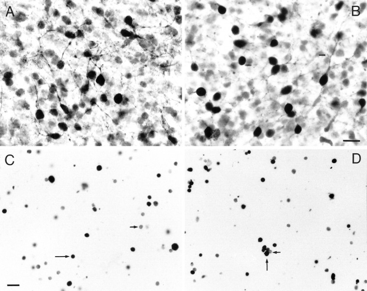
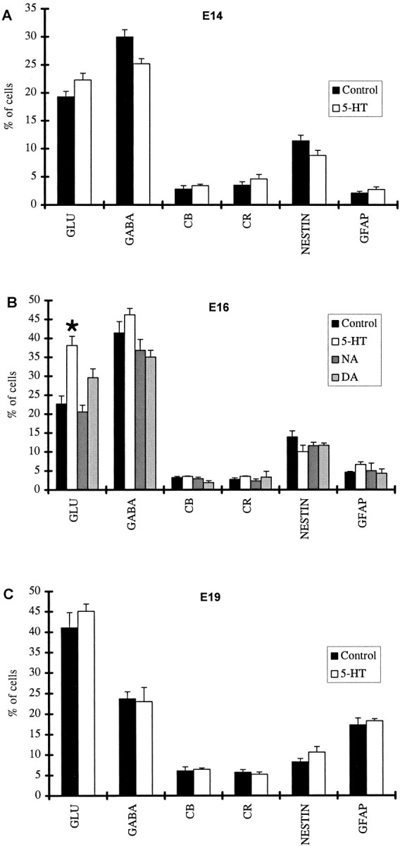
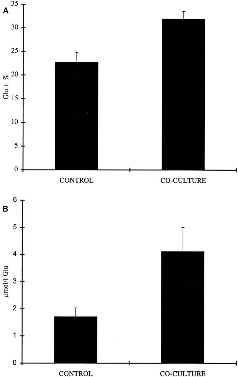
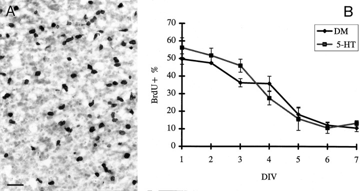
The monoamines serotonin (5-HT), noradrenaline (NA), and dopamine (DA), which are present in the developing brain apparently before they assume their neurotransmitter functions, are regarded as strong candidates for a role in the maturation of the cerebral cortex. Here we sought to investigate their effects on the generation and differentiation of cortical cell types. Slice cultures, prepared from the cortices of embryonic day (E) 14, E16, and E19 rat fetuses, were kept in defined medium or in defined medium plus 5-HT for 7 d. E16 cortices were also exposed to NA or DA for the same period. At the end of this period, the proportions of the neuronal [glutamate (Glu)-, GABA-, calbindin-, calretinin-labeled], glial (GFAP), and neuroepithelial (nestin) cell types were estimated for all conditions. We found that in E16 cultures, application of 5-HT, but not of NA or DA, significantly increased the proportion of Glu-containing neurons without affecting the overall neuronal population or the proportions of any other cell types. A similar effect was observed in co-cultures of E16 cortex with slices through the midbrain raphe nuclei of E19 rats. The total amount of cortical Glu, as measured with HPLC, was also increased in these co-cultures. To investigate whether the effect of 5-HT was the result of changes in cell proliferation, we exposed slices to bromodeoxyuridine (BrdU) and found that the proportion of BrdU-labeled cells was similar in the 5-HT-treated and control slices. These results indicate that 5-HT promotes the differentiation of cortical Glu-containing neurons without affecting neuroepithelial cell proliferation.
Figures
Similar articles
-
Serotonin promotes the survival of cortical glutamatergic neurons in vitro.Exp Neurol. 1997 Nov;148(1):205-14. doi: 10.1006/exnr.1997.6633. Exp Neurol. 1997. PMID: 9398462
-
GABA receptor antagonists modulate postmitotic cell migration in slice cultures of embryonic rat cortex.Cereb Cortex. 2000 Sep;10(9):899-909. doi: 10.1093/cercor/10.9.899. Cereb Cortex. 2000. PMID: 10982750
-
Formation and preservation of cortical layers in slice cultures.J Neurobiol. 1992 Sep;23(7):783-802. doi: 10.1002/neu.480230702. J Neurobiol. 1992. PMID: 1431845
-
Serotonin promotes region-specific glial influences on cultured serotonin and dopamine neurons.Glia. 1992;5(4):306-17. doi: 10.1002/glia.440050408. Glia. 1992. PMID: 1350272
-
The role of serotonin in early cortical development.Dev Neurosci. 2003 Mar-Aug;25(2-4):245-56. doi: 10.1159/000072272. Dev Neurosci. 2003. PMID: 12966221 Review.
Cited by
-
Cell-cycle kinetics of neocortical precursors are influenced by embryonic thalamic axons.J Neurosci. 2001 Jan 1;21(1):201-14. doi: 10.1523/JNEUROSCI.21-01-00201.2001. J Neurosci. 2001. PMID: 11150337 Free PMC article.
-
Plasma membrane transporters of serotonin, dopamine, and norepinephrine mediate serotonin accumulation in atypical locations in the developing brain of monoamine oxidase A knock-outs.J Neurosci. 1998 Sep 1;18(17):6914-27. doi: 10.1523/JNEUROSCI.18-17-06914.1998. J Neurosci. 1998. PMID: 9712661 Free PMC article.
-
Modulatory Effects of Gut Microbiota on the Central Nervous System: How Gut Could Play a Role in Neuropsychiatric Health and Diseases.J Neurogastroenterol Motil. 2016 Apr 30;22(2):201-12. doi: 10.5056/jnm15146. J Neurogastroenterol Motil. 2016. PMID: 27032544 Free PMC article. Review.
-
Expressing Constitutively Active Rheb in Adult Dorsal Root Ganglion Neurons Enhances the Integration of Sensory Axons that Regenerate Across a Chondroitinase-Treated Dorsal Root Entry Zone Following Dorsal Root Crush.Front Mol Neurosci. 2016 Jul 5;9:49. doi: 10.3389/fnmol.2016.00049. eCollection 2016. Front Mol Neurosci. 2016. PMID: 27458339 Free PMC article.
-
Serotonergic regulation of the dopaminergic system: Implications for reward-related functions.Neurosci Biobehav Rev. 2021 Sep;128:282-293. doi: 10.1016/j.neubiorev.2021.06.022. Epub 2021 Jun 15. Neurosci Biobehav Rev. 2021. PMID: 34139249 Free PMC article. Review.
References
-
- Aitken AR, Törk I. Early development of serotonin-containing neurons and pathways as seen in wholemount preparations of the fetal rat brain. J Comp Neurol. 1988;274:32–47. - PubMed
-
- Altman J, Bayer SA. Atlas of the prenatal rat brain development. CRC; Boca Raton, FL: 1995.
-
- Baimbridge KG, Celio MR, Rogers JH. Calcium-binding proteins in the nervous system. Trends Neurosci. 1992;15:303–308. - PubMed
-
- Berger B, Verney C. Development of the catecholamine innervation in rat neocortex: morphological features. In: Descarries L, Reader TR, Jasper HH, editors. Monoamine innervation of cerebral cortex. Alan R. Liss; New York: 1984. pp. 95–121.
Publication types
MeSH terms
Substances
Grants and funding
LinkOut - more resources
Full Text Sources
Other Literature Sources
Miscellaneous