. 1997 Oct 15;17(20):8049-60.
doi: 10.1523/JNEUROSCI.17-20-08049.1997.

Inflammation increases the distribution of dorsal horn neurons that internalize the neurokinin-1 receptor in response to noxious and non-noxious stimulation

Affiliations

Inflammation increases the distribution of dorsal horn neurons that internalize the neurokinin-1 receptor in response to noxious and non-noxious stimulation

C Abbadie et al. J Neurosci. .

Abstract

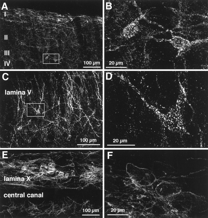
Although the neurokinin-1 (NK-1)/substance P (SP) receptor is expressed by neurons throughout the spinal dorsal horn, noxious chemical stimulation in the normal rat only induces internalization of the receptor in cell bodies and dendrites of lamina I. Here we compared the effects of mechanical and thermal stimulation in normal rats and in rats with persistent hindpaw inflammation. Electron microscopic analysis confirmed the upregulation of receptor that occurs with inflammation and demonstrated that in the absence of superimposed stimulation, the increased receptor was, as in normal rats, concentrated on the plasma membrane. In general, noxious mechanical was more effective than noxious thermal stimulation in inducing NK-1 receptor internalization, and this was increased in the setting of inflammation. Although a 5 sec noxious mechanical stimulus only induced internalization in 22% of lamina I neurons in normal rats, after inflammation, it evoked near-maximal (98%) internalization in lamina I, produced significant changes in laminae III-VI, and expanded the rostrocaudal distribution of neurons with internalized receptor. Even non-noxious (brush) stimulation of the inflamed hindpaw induced internalization in large numbers of superficial and deep neurons. For thermal stimulation, the percentage of cells with internalized receptor increased linearly at >45 degrees C, but in normal rats, these were restricted to lamina I. After inflammation, however, the 52 degrees C stimulus also induced internalization in 25% of laminae III-IV cells. These studies provide a new perspective on the reorganization of dorsal horn circuits in the setting of persistent injury and demonstrate a critical contribution of SP.


Figures

Fig. 1.
Fig. 1.
Confocal images illustrating the dorsoventral pattern of internalization of the NK-1 receptor after mechanical stimulation of the hindpaw. These images were generated by superimposition of three optical sections taken at 2.5 μm in sagittal sections of L4. A, Rat with inflamed hindpaw with no additional stimulation; the receptor is distributed on the plasma membrane of neurons in laminae I–III. B, Intact rat after mechanical stimulation of the hindpaw (pinch for 30 sec); cell bodies and dendrites in lamina I contain internalized NK-1 receptor; however, no changes were recorded in lamina III. C, Rat with inflamed hindpaw after mechanical stimulation of the hindpaw (pinch for 30 sec). There is extensive receptor internalization in cell bodies and dendrites of neurons throughout the dorsal horn.
Fig. 2.
Fig. 2.
Electron micrographs illustrating the distribution of NK-1 receptor immunoreactivity in neurons of lamina I of the dorsal horn of an intact rat (A, C) and a rat 3 d after inflammation of the hindpaw was induced with CFA (B, D). In the intact rat the receptor labeling (arrowheads) is concentrated on the plasma membrane of both cell bodies (A) and dendrites (C). After inflammation there is a significant increase in NK-1 receptor immunoreactivity; however, the receptor labeling (arrowheads) is still concentrated on the plasma membrane of cell bodies (B) and dendrites (D). In both cases, there is some cytoplasmic label present. Scale bars: A, B, 2.0 μm;C, D, 0.25 μm.
Fig. 3.
Fig. 3.
Confocal images illustrating internalization of the NK-1 receptor in neurons of laminae III and V after mechanical stimulation of the hindpaw (pinch for 30 sec). A, C, Ewere produced by superimposition of three optical sections taken at 2.5 μm in sagittal sections of L4; B, D, F were produced by superimposition of seven optical sections taken at 0.6 μm.A, B, Intact rat after mechanical stimulation of the hindpaw (pinch for 30 sec); there is no internalization in this lamina III neuron. C, D, Rat with inflamed hindpaw after mechanical stimulation of the hindpaw (pinch for 30 sec); there is extensive internalization of NK-1 receptors in this lamina III neuron.E, F, Same experimental condition as for C, D; note internalization of NK-1 receptors in this lamina V neuron.
Fig. 4.
Fig. 4.
Confocal images illustrating internalization of the NK-1 receptor in neurons of laminae III, V, and X.A–F, Rat with inflamed hindpaw after mechanical stimulation of the hindpaw (pinch for 2 min). A, C, Dwere produced by superimposition of three optical sections taken at 2.5 μm in sagittal sections of L4; B, D, F were produced by superimposition of seven optical sections taken at 0.6 μm. There is internalization of NK-1 receptors in laminae III (A, B) and V (C, D) after mechanical stimulation of rats with persistent inflammation. In intact rats this stimulus only induces internalization in lamina I neurons. Neither mechanical nor thermal stimulation induced internalization in neurons of lamina X (F).
Fig. 5.
Fig. 5.
Percentage of NK-1 receptor-immunoreactive cells with internalized receptor in lamina I of the L4 segment after different durations of mechanical stimulation (pinch) of the hindpaw in normal rats and in rats 3 d after inflammation of the hindpaw was induced with CFA. Results are expressed as mean ± SEM per group. In contrast to the effects of temperature, which produced a linear relationship between intensity of stimulation and number of neurons having internalized NK-1 receptors of (see Fig. 7), the relationship between the duration of the mechanical stimulation and the degree of internalization was not linear. For the intact animals, the Hill equation fit the data (see inset). In the setting of inflammation, the response plateaued.
Fig. 7.
Fig. 7.
Percentage of NK-1 receptor-immunoreactive spinal cord cells with internalized receptor after thermal stimulation of the hindpaw in intact rats and in rats with an inflamed hindpaw (3 d after CFA injection). Rats were stimulated for 2 min at 45, 48, 50, or 52°C. The results are expressed as mean ± SEM per group.A, Percentage of internalized cells in lamina I of L4;B, rostrocaudal (L2–L6) distribution of neurons with internalized receptor. Note that (1) in neither intact rats nor in rats with inflammation did the 45°C stimulus induce NK-1 receptor internalization; (2) the number of internalized cells increased with temperature; and (3) there was no significant difference between the number of lamina I neurons with internalized receptor in the two groups of rats, regardless of temperature.
Fig. 6.
Fig. 6.
Percentage of cells in different laminae of the lumbar spinal cord and in different lumbar segments that contained internalized NK-1 receptor after mechanical stimulation of the hindpaw. Results are expressed as mean ± SEM per group. Note that (1) a non-noxious mechanical stimulus (brush for 2 min) did not induce internalization in intact rats (A), but in rats with persistent inflammation (3 d after CFA injection) this stimulus induced internalization in neurons of laminae I and in laminae III–IV (B, C); and (2) the magnitude of receptor internalization in rats with persistent inflammation did not differ with noxious mechanical stimuli (pinch) of different duration (B, C). This contrasts with intact rats, in which the number of cell bodies with internalized receptor increases with the duration of the stimulus (A).
Fig. 8.
Fig. 8.
Percentage of NK-1 receptor-immunoreactive cells with internalized receptor in lamina I of the L4 segment. Results are expressed as mean ± SEM per group. A, Time course of NK-1 receptor internalization from rats perfused at different times after hindpaw stimulation (pinch for 2 min). The number of internalized cells is significantly greater in rats with an inflamed hindpaw than in intact rats at both 30 and 60 min. B, Results of two successive mechanical stimuli; the second stimulus (pinch for 15 sec) was applied 1 hr after the first, and the rats were perfused 5 min later. In the inflamed groups of rats, there is no difference (p = 0.7) in the magnitude of receptor internalization between the rats stimulated once and those stimulated twice. In intact rats there is a smaller number of cells with internalized receptor after the second stimulus, but the difference is only significant in the L5 segment (p = 0.015).

References

    1. Abbadie C, Besson JM, Calvino B. C-Fos expression in the spinal cord and pain-related symptoms induced by chronic arthritis in the rat are prevented by pretreatment with Freund adjuvant. J Neurosci. 1995;14:5865–5871. - PMC - PubMed
    1. Abbadie C, Brown JL, Mantyh PW, Basbaum AI. Spinal cord substance P receptor immunoreactivity increases in both inflammatory and nerve injury models of persistent pain. Neuroscience. 1996;70:201–209. - PubMed
    1. Allen BJ, Rogers S, Ghilardi JR, Menning PM, Kuskowski MA, Basbaum AI, Simone D, Mantyh PW. Noxious cutaneous thermal stimuli induce a graded release of endogenous substance P in the spinal cord: imaging peptide action in vivo. J Neurosci. 1997;17:5921–5927. - PMC - PubMed
    1. Bleazard L, Hill RG, Morris R. The correlation between the distribution of the NK1 receptor and the actions of tachykinin agonists in the dorsal horn of the rat indicates that substance P does not have a functional role on substantia gelatinosa (lamina II) neurons. J Neurosci. 1994;14:7655–7664. - PMC - PubMed
    1. Boeynaems JM. Outlines of receptor theory. Elsevier; Amsterdam: 1980.

Publication types

Substances