. 1997 Oct 14;94(21):11179-83.
doi: 10.1073/pnas.94.21.11179.

Sphingomyelin depletion in cultured cells blocks proteolysis of sterol regulatory element binding proteins at site 1

Affiliations

Sphingomyelin depletion in cultured cells blocks proteolysis of sterol regulatory element binding proteins at site 1

S Scheek et al. Proc Natl Acad Sci U S A. .

Abstract

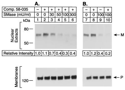
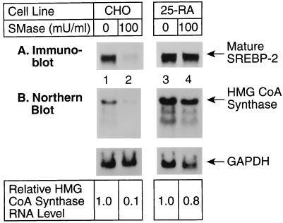
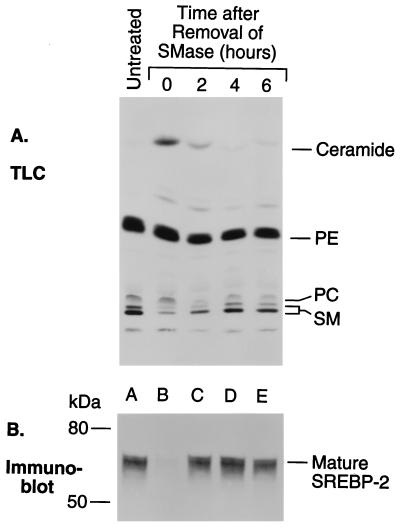
The current studies explore the mechanism by which the sphingomyelin content of mammalian cells regulates transcription of genes encoding enzymes of cholesterol synthesis. Previous studies by others have shown that depletion of sphingomyelin by treatment with neutral sphingomyelinase causes a fraction of cellular cholesterol to translocate from the plasma membrane to the endoplasmic reticulum where it expands a regulatory pool that leads to down-regulation of cholesterol synthesis and up-regulation of cholesterol esterification. Here we show that sphingomyelinase treatment of cultured Chinese hamster ovary cells prevents the nuclear entry of sterol regulatory element binding protein-2 (SREBP-2), a membrane-bound transcription factor required for transcription of several genes involved in the biosynthesis and uptake of cholesterol. Nuclear entry is blocked because sphingomyelinase treatment inhibits the proteolytic cleavage of SREBP-2 at site 1, thereby preventing release of the active NH2-terminal fragments from cell membranes. Sphingomyelinase treatment thus mimics the inhibitory effect on SREBP processing that occurs when exogenous sterols are added to cells. Sphingomyelinase treatment did not block site 1 proteolysis of SREBP-2 in 25-RA cells, a line of Chinese hamster ovary cells that is resistant to the suppressive effects of sterols, owing to an activating point mutation in the gene encoding SREBP cleavage-activating protein. In 25-RA cells, sphingomyelinase treatment also failed to down-regulate the mRNA for 3-hydroxy-3-methylglutaryl CoA synthase, a cholesterol biosynthetic enzyme whose transcription depends on the cleavage of SREBPs. Considered together with previous data, the current results indicate that cells regulate the balance between cholesterol and sphingomyelin content by regulating the proteolytic cleavage of SREBPs.


Figures

Figure 1
Figure 1
Proteolytic processing of SREBP-2 in CHO-7 cells treated with various amounts of neutral sphingomyelinase and ACAT inhibitor 58-035. On day 0, CHO-7 cells were set up for experiments as described. On day 2, the cells received 5 ml of medium B containing either 0.1% ethanol (lanes 1, 7, and 9) or 10 μg/ml of compound 58-035 dissolved in ethanol (final concentration 0.1%) (lanes 2–6, 8, and 10). After 30 min at 37°C, the indicated amount of sphingomyelinase (SMase) was added. After further incubation for 90 min at 37°C, the cells were harvested and fractionated as described. Aliquots of the nuclear extract (35 μg protein) and membranes (50 μg) were subjected to SDS/PAGE. Immunodetection of SREBP-2 was carried out with 5 μg/ml of mouse monoclonal IgG-7D4 antibody, and bound antibody was visualized by incubation with 4 × 10⁶ cpm/ml of 125I-labeled sheep anti-mouse antibody (Amersham) for 60 min at room temperature. The filters were washed three times (15 min per wash) with PBS/0.05% Tween 20 (Sigma P3563) and three times (15 min per wash) with PBS/0.05% Tween 20 containing 1% (vol/vol) Nonidet P-40, 0.1% (wt/vol) SDS, and 0.5% (wt/vol) deoxycholate. In A, the filters were exposed at −70°C to film with an intensifying screen for 48 h (nuclear extracts) and 14 h (membranes). In B, the filters were exposed for 33 h (nuclear extracts) and 14 h (membranes). Quantification was performed by exposing the filters to a Bas 1000 Fuji PhotoImager screen for 16 h. P and M denote the precursor and the mature cleaved forms of SREBP-2, respectively.
Figure 2
Figure 2
Immunoblot analysis of SREBP-2 in CHO-7 cells treated for various times with neutral sphingomyelinase in the presence of ACAT inhibitor 58-035. On day 0, CHO-7 cells were set up for experiments as described. On day 2, cells received 5 ml of medium B containing either 0.1% ethanol (lane 1) or 10 μg/ml compound 58-035 dissolved in ethanol (lanes 2–7). After 30 min at 37°C, additions of 100 milliunits/ml of sphingomyelinase (SMase) were made in a staggered fashion such that all the cells were harvested at the same time. After treatment with sphingomyelinase for the indicated time, the cells were harvested and fractionated as described. Aliquots of the nuclear extract (35 μg) and membranes (50 μg) were subjected to SDS/PAGE. Immunodetection of SREBP-2 was performed with 5 μg/ml of mouse monoclonal IgG-7D4 and 125I-labeled sheep anti-mouse antibody (4 × 10⁶ cpm/ml) as described in Fig. 1. The filters were exposed to film with an intensifying screen for 33 h (nuclear extracts) or 14 h (membranes) at −70°C, and the radioactivity in each protein band was quantified as described in Fig. 1. P and M denote the precursor and the mature cleaved forms of SREBP-2, respectively.
Figure 3
Figure 3
Mature form of SREBP-2 (A) and level of mRNA for HMG CoA synthase (B) in CHO-7 and 25-RA cells treated with neutral sphingomyelinase. (A) On day 0, the indicated cells were set up as described using medium A supplemented with 10 μg/ml of compound 58-035 (final concentration of ethanol, 0.1%). On day 2, the cells received 5 ml medium B containing 10 μg/ml of compound 58-035 and 100 milliunits/ml of sphingomyelinase (SMase) (lanes 2 and 4). After incubation for 4 h at 37°C, the medium was replaced by the above medium B containing fresh sphingomyelinase. After a further 2-h incubation, the cells from three dishes were harvested and fractionated as described. Aliquots (35 μg of protein) of the nuclear extract were subjected to SDS/PAGE and immunoblot analysis with 5 μg/ml of mouse monoclonal IgG-7D4 antibody directed against SREBP-2 and peroxidase-conjugated sheep anti-mouse IgG. The filters were exposed to film for 25 s. M denotes the mature form of SREBP-2. (B) Cells from the same experiment (three 100-mm dishes each) were used to prepare total RNA as described. Aliquots of total RNA (20 μg per lane) were subjected to electrophoresis and blot hybridization with the indicated 32P-labeled probe. The filters were exposed to film for 5 days (HMG CoA synthase) and 10 h (glyceraldehyde-3-phosphate dehydrogenase) at −70°C with an intensifying screen. The amount of radioactivity in each band was quantified with a Bio-Imaging analyzer as described after exposure for 12 h to a Fuji PhotoImager screen. The level of HMG CoA synthase mRNA relative to that of the zero-time values was calculated after correction for loading differences determined by the glyceraldehyde-3-phosphate dehydrogenase signal.
Figure 4
Figure 4
[3H]Serine-labeled lipids (A) and mature form of SREBP-2 (B) in CHO-7 cells during recovery period after treatment with neutral sphingomyelinase. (A) CHO-7 cells were set up and labeled with l-[3-3H]serine as described. On day 3, the medium was switched to 5 ml medium B containing 10 μg/ml compound 58-035. After incubation for 30 min at 37°C, 100 milliunits/ml sphingomyelinase (SMase) were added in a staggered fashion such that all cells were harvested at the same time. Cells were treated with sphingomyelinase for 2 h, washed twice with 5 ml of PBS, once with medium A, and incubated further for the indicated time in medium A supplemented with compound 58-035. Lipids were extracted and analyzed by thin-layer chromatography as described. (B) Unlabeled cells in the same experiment were incubated with the same additions as in (A) and harvested to prepare nuclear extract fractions as described. Aliquots of the nuclear extracts (30 μg) were subjected to SDS/PAGE. Immunoblot analysis was carried out with 5 μg/ml of mouse monoclonal IgG-7D4 antibody directed against SREBP-2 and peroxidase-conjugated sheep anti-mouse IgG. The filters were exposed to film for 15 s. PE, phosphatidylethanolamine; PC, phosphatidylcholine; SM, sphingomyelin.

References

    1. Brown D A, Rose J K. Cell. 1992;68:533–544. - PubMed
    1. Smart E J, Ying Y-S, Conrad P A, Anderson R G W. J Cell Biol. 1994;127:1185–1197. - PMC - PubMed
    1. Murata M, Peränen J, Schreiner R, Wieland F, Kurzchalia T, Simons K. Proc Natl Acad Sci USA. 1995;92:10339–10343. - PMC - PubMed
    1. Schuchman E H, Desnick R J. In: The Metabolic and Molecular Basis of Inherited Disease. Scriver C R, Beaudet A L, Sly W S, Valle D, editors; Scriver C R, Beaudet A L, Sly W S, Valle D, editors. New York: McGraw-Hill; 1995. pp. 2601–2624.
    1. Gatt S, Bierman E L. J Biol Chem. 1980;255:3371–3376. - PubMed

Publication types

MeSH terms

LinkOut - more resources