

doi: 10.1073/pnas.94.21.11301.
A functionally defined model for the M2 proton channel of influenza A virus suggests a mechanism for its ion selectivity
Affiliations
- PMID: 9326604
- PMCID: PMC23448
- DOI: 10.1073/pnas.94.21.11301
Item in Clipboard
A functionally defined model for the M2 proton channel of influenza A virus suggests a mechanism for its ion selectivity
Proc Natl Acad Sci U S A.
.
Abstract
The M2 protein from influenza A virus forms proton-selective channels that are essential to viral function and are the target of the drug amantadine. Cys scanning was used to generate a series of mutants with successive substitutions in the transmembrane segment of the protein, and the mutants were expressed in Xenopus laevis oocytes. The effect of the mutations on reversal potential, ion currents, and amantadine resistance were measured. Fourier analysis revealed a periodicity consistent with a four-stranded coiled coil or helical bundle. A three-dimensional model of this structure suggests a possible mechanism for the proton selectivity of the M2 channel of influenza virus.
Figures
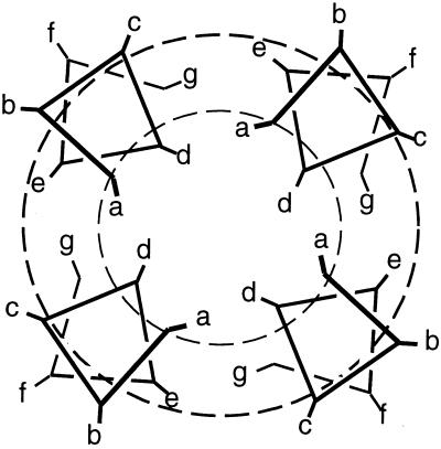
Helical wheel diagram showing one heptad of a parallel, four-stranded helical coiled coil (or four-helical bundle assuming a sequence repeat of 7.0 residues) viewed down the bundle axis. The dashed circles demarcate three regions that are predicted to show different perturbational effects (see text).
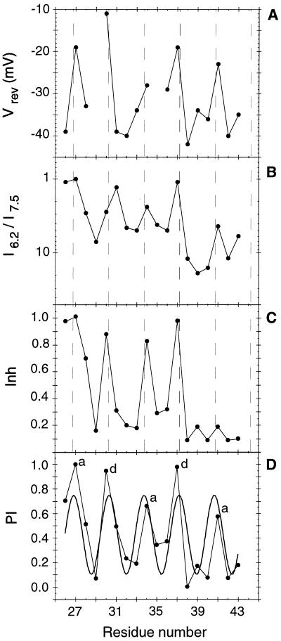
The variation in Vrev (A), I6.2/I7.5 (B), Inh (C), and PI(n) (D) as a function of position in the TM helix sequence. The smooth curve through the PI(n) data (D) represents the best fit to a cosine function (Eq. 2) generated using kaleidagraph with initial parameters A = 0.5, B = 1.0, C = 0, and P = 3.6. The dashed lines in A–C show peak positions expected for a 3.5-residue periodicity. Labels a and d in D represent heptad positions in the sequence. The perturbational index [PI(n)] is calculated from the three individual parameters. To combine these three parameters into PI(n), the individual values were normalized so their average values would be zero, then weighted according to their standard deviations. The values of the three parameters for each position were then averaged, and the combined PI(n) scale was normalized to range from 0 to 1. Because the periodic variation of Inh was limited to approximately the N-terminal half of the TM region, only residues 26–38 were included in calculating PI(n).
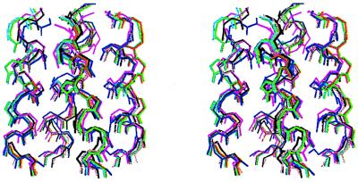
Stereoview of the seven representative models of the M2 proton channel using molscript (26). The backbone atoms of residues 26–41 are shown.
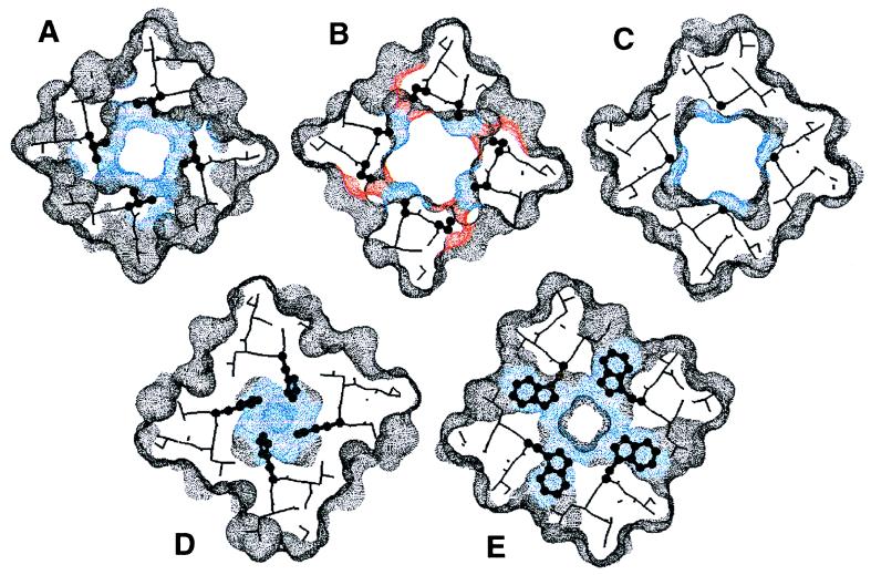
Axial view of the predicted structure of the M2 proton channel. The individual panels show successive slices (≈5 Å) through the structure. The residues that appear essential from mutagenic analysis—Val-27 (A), Ala-30 and Ser-31 (B), Gly-34 (C), His-37 (D), and Trp-41 (E)—-are shown as ball-and-stick representations. The dotted surface shows the parts of the model accessible to a probe with a 1.4-Å radius. Color designation: blue, Val-27, Ala-30, Gly-34, His-37, and Trp-41; red, Ser-31; gray, all other residues. This figure was generated using insightii .
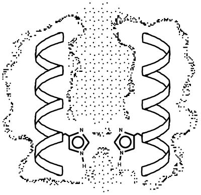
Side view of the M2 proton channel showing a slice through the structure (the other two helices are not shown for clarity).
References
-
- Hille B. Ionic Channels of Excitable Membranes. Sunderland, MA: Sinauer; 1992.
-
- Song L, Hobaugh M R, Shustak C, Cheley S, Bayley H, Gouaux J E. Science. 1996;274:1859–1866. - PubMed
-
- Schulz G E. Curr Opin Struct Biol. 1996;6:485–490. - PubMed
-
- Unwin N. J Mol Biol. 1996;257:586–596. - PubMed
-
- Montal M. Curr Opin Struct Biol. 1996;6:499–510. - PubMed
Publication types
MeSH terms
Substances
Grants and funding
LinkOut - more resources
Full Text Sources
Other Literature Sources
