Comparative Study
. 1997 Oct 14;94(21):11339-44.
doi: 10.1073/pnas.94.21.11339.

The signal recognition particle receptor of Escherichia coli (FtsY) has a nucleotide exchange factor built into the GTPase domain

Affiliations
Comparative Study

The signal recognition particle receptor of Escherichia coli (FtsY) has a nucleotide exchange factor built into the GTPase domain

C Moser et al. Proc Natl Acad Sci U S A. .

Abstract

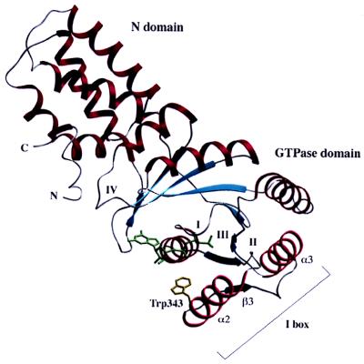
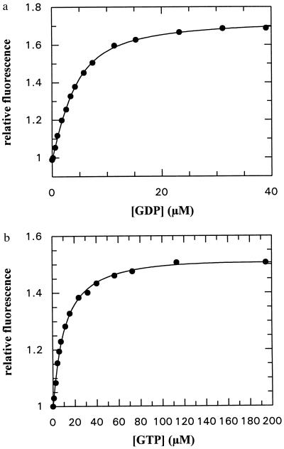
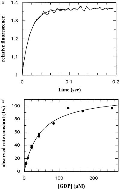
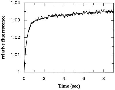
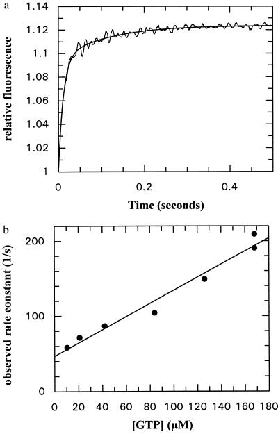
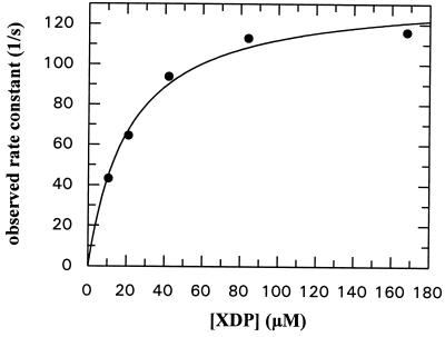
Targeting of many secretory and membrane proteins to the inner membrane in Escherichia coli is achieved by the signal recognition particle (SRP) and its receptor (FtsY). In E. coli SRP consists of only one polypeptide (Ffh), and a 4.5S RNA. Ffh and FtsY each contain a conserved GTPase domain (G domain) with an alpha-helical domain on its N terminus (N domain). The nucleotide binding kinetics of the NG domain of the SRP receptor FtsY have been investigated, using different fluorescence techniques. Methods to describe the reaction kinetically are presented. The kinetics of interaction of FtsY with guanine nucleotides are quantitatively different from those of other GTPases. The intrinsic guanine nucleotide dissociation rates of FtsY are about 10(5) times higher than in Ras, but similar to those seen in GTPases in the presence of an exchange factor. Therefore, the data presented here show that the NG domain of FtsY resembles a GTPase-nucleotide exchange factor complex not only in its structure but also kinetically. The I-box, an insertion present in all SRP-type GTPases, is likely to act as an intrinsic exchange factor. From this we conclude that the details of the GTPase cycle of FtsY and presumably other SRP-type GTPases are fundamentally different from those of other GTPases.


Figures

Figure 1
Figure 1
Cα ribbon representation of the structure of the NG domain of FtsY. The single tryptophan residue used for the fluorescence is shown with its side chain (Trp-343). The N and G domains are marked as well as the four consensus elements of GTP binding (I-IV) (14). The I-box (insertion consisting of α2-β3-α3) is indicated.
Figure 2
Figure 2
(a) Titration of GDP with the NG fragment of FtsY (3.6 μM), using tryptophan fluorescence as a signal of binding (excitation wavelength 297 nm, detection at 340 nm). The solid line shows the fit to the data obtained by using the quadratic equation describing the binding equilibrium. The fitted Kd value was 2.14 ± 0.22 μM. Conditions were as described in the text. (b) Titration of GTP with the NG fragment of FtsY (1 μM). Conditions as in a. Fitted Kd value = 10.7 ± 0.45 μM.
Figure 3
Figure 3
(a) Time dependence of the association between GDP (42 μM) and the NG fragment of FtsY (2 μM) monitored by tryptophan fluorescence in a stopped-flow apparatus. The smooth line shows a single exponential fit to the data and corresponds to a pseudo-first-order rate constant of 56.8 ± 0.42 s−1. (b) Concentration dependence of the pseudo-first-order rate constant for GDP association to the NG fragment of FtsY. The solid line shows the best fit to a hyperbolic curve that defines the apparent Kd of GDP in the monitored step as 44.9 ± 9.3 μM and the maximal rate constant of the fluorescence change as 119 s−1.
Figure 4
Figure 4
Dissociation kinetics of GDP from its complex with FtsY NG fragment. The complex between the NG fragment and GDP was generated by premixing 33.6 μM GDP with 2.2 μM NG fragment in one syringe of a stopped-flow apparatus and mixing with 500 μM mantGDP in the other syringe in the actual experiment. Excitation was at 295 nm, and fluorescence was detected above 389 nm. The continuous line shows a double-exponential fit to the data with rate constants of 3.74 ± 0.10 and 0.21 ± 0.02 s−1.
Figure 5
Figure 5
(a) Association kinetics of GTP and the NG fragment of FtsY. Conditions as in Fig. 3a. The smooth line shows a double-exponential fit to the data with rate constants of 87.6 ± 2.5 and 8.80 ± 0.45 s−1. (b) GTP concentration dependence of the pseudo-first-order rate constant for the first phase of the association reaction with NG fragment. The fitted straight line has a slope of 8.8 ± 0.7 × 10⁵ M−1⋅s−1 and an intercept of 46.63 ± 7.33 s−1.
Figure 6
Figure 6
Concentration dependence of the pseudo-first-order rate constant for the first phase of association of XTP with the Asp-449 → Asn mutant of the NG fragment of FtsY. The fitted hyperbola corresponds to an apparent Kd value of 21.3 ± 3.2 μM and a maximal rate of 135.9 ± 6.1 s−1. Conditions as in Fig. 3.

References

    1. Bukau B, Hesterkamp T, Luirink J. Trends Cell Biol. 1996;6:480–486. - PubMed
    1. Lütcke H. Eur J Biochem. 1995;228:531–550. - PubMed
    1. Poritz S, Bernstein H D, Strub K, Zopf D, Wilhelm H, Walter P. Science. 1990;250:1111–1117. - PubMed
    1. Ribes V, Roemisch K, Giner A, Dobberstein B, Tollervey D. Cell. 1990;63:591–600. - PubMed
    1. Luirink J, ten Hagen-Jongman C, van der Weijden C C, Oudega B, High S, Dobberstein B, Kusters R. EMBO J. 1994;13:2289–2296. - PMC - PubMed

Publication types

MeSH terms

LinkOut - more resources