Comparative Study
. 1997 Oct 14;94(21):11508-13.
doi: 10.1073/pnas.94.21.11508.

Molecular immune responses of the mosquito Anopheles gambiae to bacteria and malaria parasites

Affiliations
Comparative Study

Molecular immune responses of the mosquito Anopheles gambiae to bacteria and malaria parasites

G Dimopoulos et al. Proc Natl Acad Sci U S A. .

Abstract

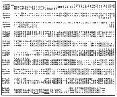
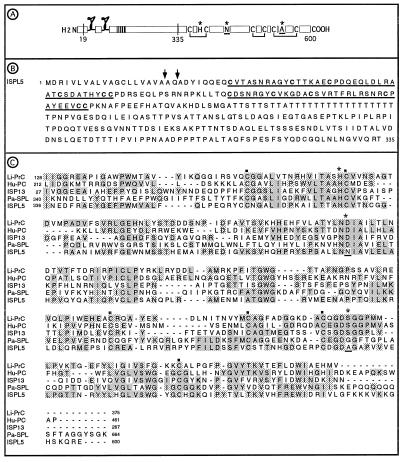
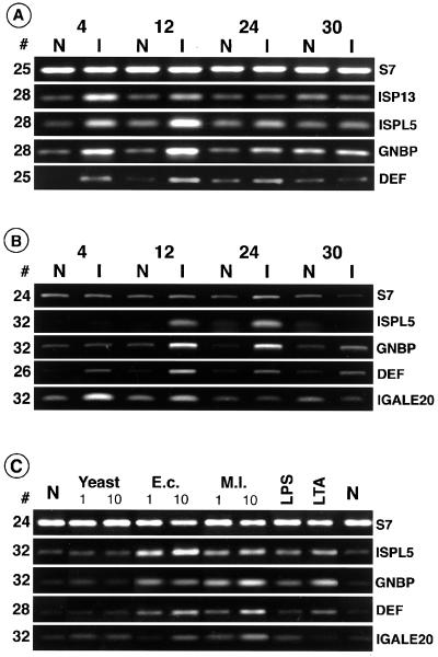
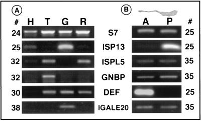
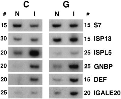
Immune responses of the malaria vector mosquito Anopheles gambiae were monitored systematically by the induced expression of five RNA markers after infection challenge. One newly isolated marker encodes a homologue of the moth Gram-negative bacteria-binding protein (GNBP), and another corresponds to a serine protease-like molecule. Additional previously described markers that respond to immune challenge encode the antimicrobial peptide defensin, a putative galactose lectin, and a putative serine protease. Specificity of the immune responses was indicated by differing temporal patterns of induction of specific markers in bacteria-challenged larvae and adults, and by variations in the effectiveness of different microorganisms and their components for marker induction in an immune-responsive cell line. The markers exhibit spatially distinct patterns of expression in the adult female mosquito. Two of them are highly expressed in different regions of the midgut, one in the anterior and the other in the posterior midgut. Marker induction indicates a significant role of the midgut in insect innate immunity. Immune responses to the penetration of the midgut epithelium by a malaria parasite occur both within the midgut itself and elsewhere in the body, suggesting an immune-related signaling process.


Figures

Figure 1
Figure 1
Full-length A. gambiae GNBP compared with B. mori GNBP (accession no. L38591) and B. circulans glucanase A1 (accession no. P23903). Residues matching in at least two of the sequences are shaded, and the highly conserved region of the putative B. circulans polysaccharide binding domain is indicated by ∗. The first 24 residues of AgGNBP show features suggestive of a signal peptide. The potential glycosylphosphatidylinositol anchor sequences of AgGNBP are underlined. Numbers indicate position of residues from the N terminus.
Figure 2
Figure 2
(A) Schematic sequence features of full-length ISPL5 showing the proposed signal peptide cleavage site (amino acid 19), the putative cysteine knots, the polythreonine stem region (vertical stripes), the putative activation site (amino acid 335), cysteines and disulfide bridges that are conserved in serine proteases, and the catalytic triad (∗) with two nonconserved residues underlined. (B) The amino terminal region of ISPL5 with the putative cysteine knots underlined. Two possible signal peptide cleavage sites are marked with arrows. (C) ISPL5 serine protease domain compared with ISP13 (accession no. Z69978), Limulus (Tachypleus tridentatus) clotting factor 2, which converts coagulen to insoluble coagulen gel (Li-PrC, accession no. P21902), human blood coagulation regulator protein C (Hu-PC, accession no. P04070), and Pacifastacus leniusculus (crayfish) hemocyte-specific serine protease-like protein (Pa-SPL, unpublished data; accession no. Y11145). The six conserved cysteines of serine proteases are marked with ▪; the residues of the serine protease catalytic triad are marked with ∗, with the nonconserved residues underlined as in A above. Aligned residues that match in two or more sequences are shaded. Numbers indicate position of residues from the N terminus.
Figure 3
Figure 3
Expression profiles of immune markers in bacterially challenged (I) larvae (A) and adults (B) at 4, 12, 24, and 30 hr after infection compared with unchallenged naive animals (N). Changes in expression levels of specific sequences are detected relative to the control ribosomal protein S7 transcript. In this and subsequent figures the PCR cycle number for each sequence is indicated to the side of the data panel. The induction profiles of ISP13 and defensin in larvae were reported previously (11, 12). [Part of A reproduced from ref. with permission (Copyright 1996, Royal Entomological Society) and part modified from figure 2 in ref. .] (C) Background levels of the markers in naive cells (N) and induced levels (I) after challenge with 10-fold differing concentrations of yeast, E. coli (E.c) and M. luteus (M.l.), or with LPS and LTA at concentrations as indicated in Materials and Methods.
Figure 4
Figure 4
(A) Regional expression profiles, determined by RT-PCR, in naive adult head (H), thorax (T), midgut (G), and remaining abdominal tissues (R; which includes hindgut, Malphigian tubules and ovaries). ISP13 is largely amplified from the gut cDNA but also shows lower level expression in the head and abdomen. ISPL5 is mainly amplified in the thorax and abdomen. GNBP is mainly amplified from the thorax, defensin from the thorax, midgut and abdomen, and IGALE20 from the midgut. (B) Specific expression of the markers in anterior (A) versus posterior (P) regions of the unchallenged midgut (shown in a photograph at the top). ISP13 is posterior midgut specific and defensin is limited to the anterior midgut. ISPL5, GNBP and IGALE20 amplify weakly, approximately equally from both posterior and anterior midguts.
Figure 5
Figure 5
Induction of immune markers after infection with P. berghei. Expression levels were assayed by radioactive RT-PCR in the midgut (G) and the remaining carcass (C) of mosquitoes fed 24 hr earlier on naive (N) or parasite-infected (I) mice. Note the prominent induction of IGALE20, defensin, and GNBP in the gut, as well as ISPL5, GNBP, and defensin in the carcass. Marginal levels of induction were observed for ISP13 in the infected gut and for IGALE20 in the carcass.

Comment in

  • Immune responsiveness in vector insects.
    Hoffmann JA. Hoffmann JA. Proc Natl Acad Sci U S A. 1997 Oct 14;94(21):11152-3. doi: 10.1073/pnas.94.21.11152. Proc Natl Acad Sci U S A. 1997. PMID: 9326575 Free PMC article. Review. No abstract available.

References

    1. Hoffman J A, Reichhard J-M, Hetru C. Curr Opin Immunol. 1996;8:8–13. - PubMed
    1. Hultmark D. Trends Genet. 1993;9:178–183. - PubMed
    1. Cociancich S, Ghazi A, Hetru C, Hoffman J, A, Letellier L. J Biol Chem. 1993;268:19239–19245. - PubMed
    1. Lemaitre B, Kromer-Metzger E, Michaut L, Nicolas E, Meister M, Georgel P, Reichhart M, Hoffman J A. Proc Natl Acad Sci USA. 1995;92:9465–9469. - PMC - PubMed
    1. Paskewitz S M, Christensen B M. In: The Biology of Disease Vectors. Beaty B J, Marquardt W C, editors; Beaty B J, Marquardt W C, editors. Boulder: Univer. Press of Colorado; 1997. pp. 371–392.

Publication types

MeSH terms

Associated data