. 1997 Oct 14;94(21):11514-9.
doi: 10.1073/pnas.94.21.11514.

MIC-1, a novel macrophage inhibitory cytokine, is a divergent member of the TGF-beta superfamily

Affiliations

MIC-1, a novel macrophage inhibitory cytokine, is a divergent member of the TGF-beta superfamily

M R Bootcov et al. Proc Natl Acad Sci U S A. .

Abstract

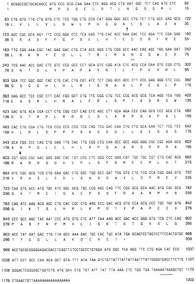
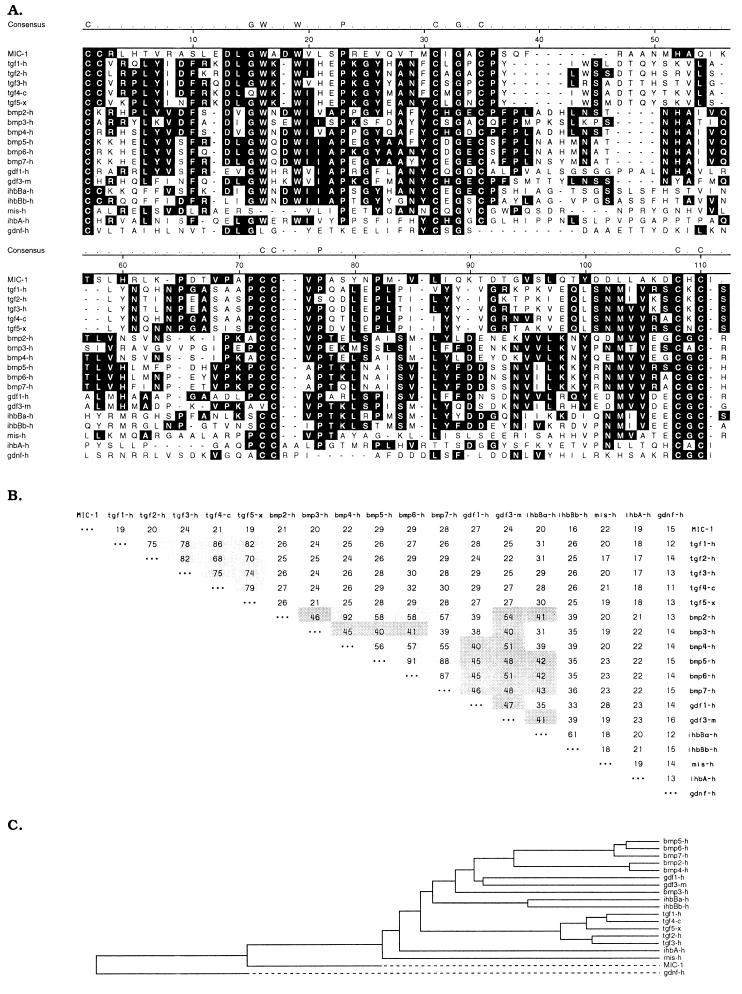
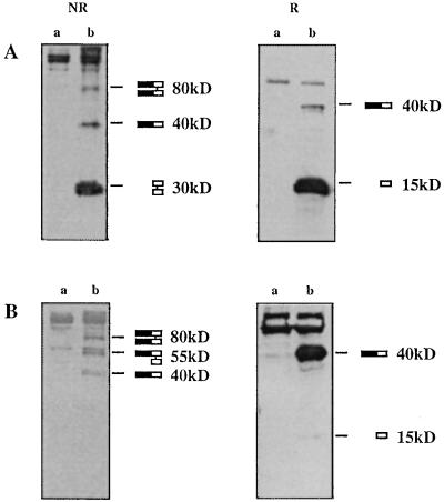
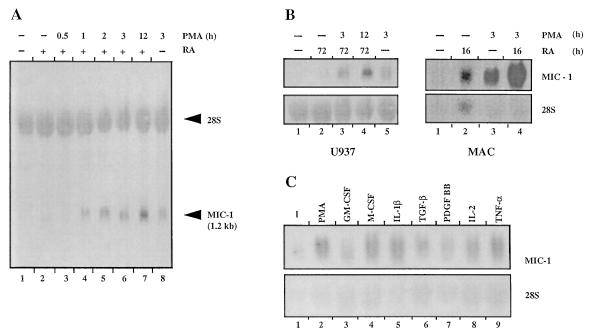
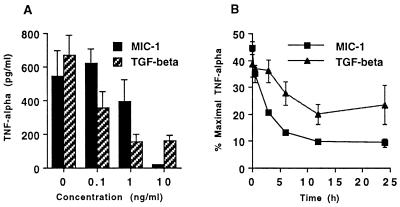
Macrophages play a key role in both normal and pathological processes involving immune and inflammatory responses, to a large extent through their capacity to secrete a wide range of biologically active molecules. To identify some of these as yet not characterized molecules, we have used a subtraction cloning approach designed to identify genes expressed in association with macrophage activation. One of these genes, designated macrophage inhibitory cytokine 1 (MIC-1), encodes a protein that bears the structural characteristics of a transforming growth factor beta (TGF-beta) superfamily cytokine. Although it belongs to this superfamily, it has no strong homology to existing families, indicating that it is a divergent member that may represent the first of a new family within this grouping. Expression of MIC-1 mRNA in monocytoid cells is up-regulated by a variety of stimuli associated with activation, including interleukin 1beta, tumor necrosis factor alpha (TNF-alpha), interleukin 2, and macrophage colony-stimulating factor but not interferon gamma, or lipopolysaccharide (LPS). Its expression is also increased by TGF-beta. Expression of MIC-1 in CHO cells results in the proteolytic cleavage of the propeptide and secretion of a cysteine-rich dimeric protein of Mr 25 kDa. Purified recombinant MIC-1 is able to inhibit lipopolysaccharide -induced macrophage TNF-alpha production, suggesting that MIC-1 acts in macrophages as an autocrine regulatory molecule. Its production in response to secreted proinflammatory cytokines and TGF-beta may serve to limit the later phases of macrophage activation.


Figures

Figure 1
Figure 1
Nucleotide and amino acid sequence of pre-pro MIC-1. Nucleotides and translated amino acids are numbered on both sides of the figure. The putative signal sequence cleavage site (↑), putative proteolytic cleavage site (↓), consensus N-linked glycosylation site (=), and stop codon (∗) are indicated. The polyadenylation signal (_) and mRNA instability sequences (..) are underlined.
Figure 2
Figure 2
Comparison of MIC-1 amino acid sequence with members of the TGF-β superfamily. (A) Alignment of the seven-cysteine domain of MIC-1 with corresponding regions of human TGF-β1 (28), human TGF-β2 (29), human TGF-β3 (30, 31), chicken TGF-β4 (32), Xenopus TGF-β5 (33), human BMP-2,3,4 (34), human MBP-5,6,7 (35), human growth and differentiation factor (GDF)-1 (36), murine GDF-3 (11), human inhibinβ-a, βb, and α (37), human mullerian-inhibiting substance (38), and human glial cell line-derived neurotopic factor (39). Multiple-sequence alignment was performed with the dnastar program (gap weight = 10, length weight = 10). The most conserved residues, including the seven highly conserved cysteines, are identified. Dashes denote gaps introduced to optimize alignment. (B) Percent amino acid identity between the seven-cysteine domains of members of the TGF-β superfamily calculated using the pairwise sequence comparison. Darkly shaded areas denote identity of 40–54%, and lightly shaded areas, identity of >55%. (C) Dendrogram of the relationship between TGF-β superfamily members. This has been produced using the dnastar program and is based on the percentage identity.
Figure 3
Figure 3
Expression of pre-pro-MIC-1 cDNA in 293-EBNA cells. Supernatant (A) and total cell lysate (B) from untransfected (a) and MIC-1 transfected (b) 293-EBNA cells were immunoprecipitated with anti-FLAG antibodies and analyzed by immunoblotting with anti-FLAG antibodies under both reducing (R) and nonreducing (NR) conditions. The various forms of MIC-1 corresponding to the indicated bands are depicted on the right, with the closed bar indicating the pro-domain and the open bar indicating the mature domain.
Figure 4
Figure 4
Expression of MIC-1 is induced by monocyte/macrophage differentiation and activation. Northern blot analysis was performed using total RNA (20 μg per lane) and hybridization with radiolabeled MIC-1 cDNA and 28S rRNA oligonucleotide probes. (A) MIC-1 expression in U937 cells after differentiation with RA and then activation with PMA. Lanes: 1, no treatment; 2, 1 μM RA for 3 days; 3–7, 1 μM RA for 3 days followed by 160 nM PMA for 30 min, 1 hr, 2 hr, 3 hr, and 12 hr, respectively; 8, 160 nM PMA for 3 hr. (B) MIC-1 expression in U937 and macrophages following treatment with 1 μM RA for either 3 days (U937) or 16 hr (macrophages) followed by activation with 160 nM PMA (U937) or 50 nM PMA (macrophages). (C) Cytokine regulation of MIC-1 expression in macrophages. Lanes: 1, no treatment; 2, 50 nM PMA; 3, 50 units/ml GM-CSF; 4, 100 units/ml M-CSF; 5, 100 units/ml IL-1β; 6, 10 ng/ml TGF-β; 7, 10 units/ml PDGF-BB; 8, 50 units/ml IL-2; 9, 100 units/ml TNF-α. All treatments were for 3 hr.
Figure 5
Figure 5
MIC-1 inhibits TNF-α production by macrophages. Macrophages were stimulated with LPS, and the effects on secretion of TNF-α were quantitated as the mean and standard deviation of triplicate cultures. Representative experiments showing the effect of MIC-1 or TGF-β1 (A) and of the preincubation period with MIC-1 or TGF-β1 (10 ng/ml) (B) on suppression of TNF-α secretion.

References

    1. Harris P, Uman R P. J Leukocyte Biol. 1985;37:407–422. - PubMed
    1. Valenzuela S M, Martin D K, Por S B, Robbins J M, Bootcov M, Schofield P R, Campbell T J, Breit S N. J Biol Chem. 1997;272:12575–12582. - PubMed
    1. Sanger F, Nicklen S, Coulson A. Proc Natl Acad Sci USA. 1977;74:5463–5467. - PMC - PubMed
    1. Bennett S, Por S B, Cooley M A, Breit S N. J Immunol. 1993;150:2364–2371. - PubMed
    1. Bennett S, Por S B, Stanley E R, Breit S N. J Immunol Methods. 1992;153:201–212. - PubMed

Publication types

MeSH terms

Associated data