. 1997 Oct 14;94(21):11520-5.
doi: 10.1073/pnas.94.21.11520.

Direct evidence to support the role of HLA-G in protecting the fetus from maternal uterine natural killer cytolysis

Affiliations

Direct evidence to support the role of HLA-G in protecting the fetus from maternal uterine natural killer cytolysis

N Rouas-Freiss et al. Proc Natl Acad Sci U S A. .

Abstract

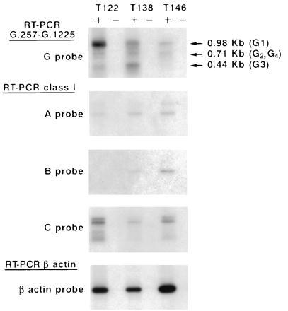
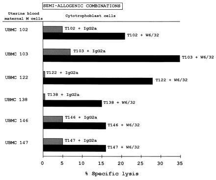
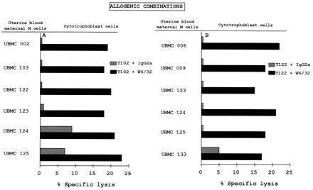
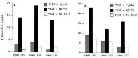
HLA-G is a nonclassical major histocompatibility complex class I molecule selectively expressed on cytotrophoblasts at the feto-maternal interface, where it may play an important role in maternal tolerance of the fetus. We provide direct evidence under physiological conditions that supports the role of HLA-G in protecting cytotrophoblasts against natural killer (NK) cytolysis in 6 semiallogenic combinations of maternal uterine NK cells and their own trophoblast counterparts, as well as in 20 allogenic combinations of maternal uterine NK cells and trophoblasts from different mothers. We show that, in all cases studied, this HLA-G-mediated protection was abolished by treatment of cytotrophoblasts with an HLA-G-specific mAb. The HLA class I-negative K562 cell line transfected with the predominant HLA-G1 isoform results in similar protection and abolition from maternal uterine NK lysis. Because maternal uterine NK cells express killer inhibitory receptors for HLA-G, we conclude that their interactions contribute to the survival of the fetal semiallograft by confering immunological tolerance to its tissues.


Figures

Figure 1
Figure 1
Detection of HLA class I transcripts in cytotrophoblast cells isolated in vitro from first trimester terminations of pregnancy. Reverse RNAs were amplified using either G.257 and G.1225 HLA-G-specific primers or pan class I-specific primers, and Southern blots were hybridized with either an HLA-G 32P-labeled probe (G probe), an HLA-A 32P-labeled probe (A probe), an HLA-B 32P-labeled probe (B probe), or an HLA-C 32P-labeled probe (C probe). Positive (+) and negative (−) lanes correspond to the RT+ and RT template, as described. To control the amount of RNA in the samples, RT-PCR amplification results obtained with β-actin-specific primers and Southern blots were hybridized with a β-actin 32P-labeled probe. Note that all the cytotrophoblasts present HLA-G1, -G2, -G3, and -G4 transcripts, but at various levels.
Figure 2
Figure 2
Protection of cytotrophoblasts from maternal uterine NK lysis in semi-allogenic combinations. Cytotoxicity tests were carried out using Percoll gradient-purified cytotrophoblasts as the targets (T): T102, T103, T122, T138, T146, and T147 and UBMC from mothers 102, 103, 122, 138, 146, and 147 as the effector cells (E) at a 25:1 E/T ratio. The results are expressed as the percentage of lysis recorded in a 4-hr 51Cr-release assay. The standard deviation of the mean of the triplicates was less than 5%. W6/32 and IgG2a isotype-matched control Abs were added to the assay at 10 μg/ml.
Figure 3
Figure 3
Protection of cytotrophoblasts from maternal uterine NK lysis in allogenic combinations. Cytotoxicity tests were carried out using the Percoll gradient-purified (A) T102 and (B) T122 cytotrophoblasts as targets and the indicated UBMC from different mothers as effector cells at a 25:1 E:T ratio. The tests were carried out as described in Fig. 2.
Figure 4
Figure 4
Protection of cytotrophoblasts from maternal uterine NK lysis in allogenic combinations. Cytotoxicity tests were carried out using the Percoll gradient-purified (A) T146 and (B) T138 cytotrophoblasts as targets and the indicated UBMC from different mothers as effector cells at a 25:1 E/T ratio. The tests were carried out as described in Fig. 2. W6/32, B1.23.2, and IgG2a isotype-matched control antibodies were added to the assay at 10 μg/ml.

References

    1. Kovats S, Main E K, Librach C, Stubblebine M, Fisher S J, DeMars R. Science. 1990;248:220–223. - PubMed
    1. King A, Boocock C, Sharkey A M, Gardner L, Beretta A, Siccardi A G, Loke Y W. J Immunol. 1996;156:2068–2076. - PubMed
    1. Carosella E D, Dausset J, Kirszenbaum M. Immunol Today. 1996;17:407–409. - PubMed
    1. Ishitani A, Geraghty D E. Proc Natl Acad Sci USA. 1992;89:3947–3951. - PMC - PubMed
    1. Kirszenbaum M, Moreau P, Gluckman E, Dausset J, Carosella E. Proc Natl Acad Sci USA. 1994;91:4209–4213. - PMC - PubMed

Publication types

MeSH terms