. 1997 Oct 14;94(21):11627-32.
doi: 10.1073/pnas.94.21.11627.

Ca2+-independent inhibition of inositol trisphosphate receptors by calmodulin: redistribution of calmodulin as a possible means of regulating Ca2+ mobilization

Affiliations

Ca2+-independent inhibition of inositol trisphosphate receptors by calmodulin: redistribution of calmodulin as a possible means of regulating Ca2+ mobilization

S Patel et al. Proc Natl Acad Sci U S A. .

Abstract

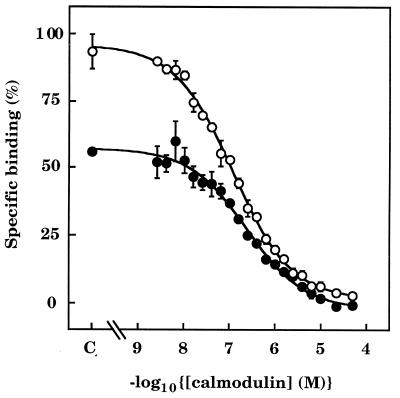
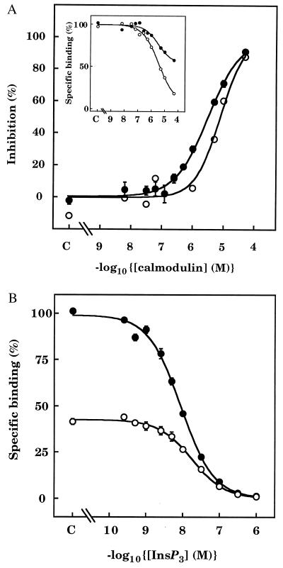
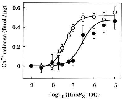
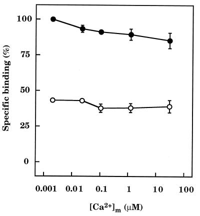
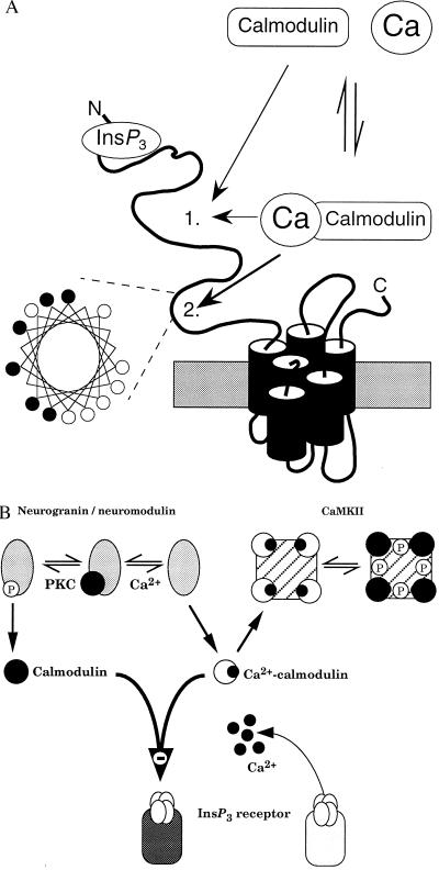
The interactions between calmodulin, inositol 1,4,5-trisphosphate (InsP3), and pure cerebellar InsP3 receptors were characterized by using a scintillation proximity assay. In the absence of Ca2+, 125I-labeled calmodulin reversibly bound to multiple sites on InsP3 receptors and Ca2+ increased the binding by 190% +/- 10%; the half-maximal effect occurred when the Ca2+ concentration was 184 +/- 14 nM. In the absence of Ca2+, calmodulin caused a reversible, concentration-dependent (IC50 = 3.1 +/- 0.2 microM) inhibition of [3H]InsP3 binding by decreasing the affinity of the receptor for InsP3. This effect was similar at all Ca2+ concentrations, indicating that the site through which calmodulin inhibits InsP3 binding has similar affinities for calmodulin and Ca2+-calmodulin. Calmodulin (10 microM) inhibited the Ca2+ release from cerebellar microsomes evoked by submaximal, but not by maximal, concentrations of InsP3. Tonic inhibition of InsP3 receptors by the high concentrations of calmodulin within cerebellar Purkinje cells may account for their relative insensitivity to InsP3 and limit spontaneous activation of InsP3 receptors in the dendritic spines. Inhibition of InsP3 receptors by calmodulin at all cytosolic Ca2+ concentrations, together with the known redistribution of neuronal calmodulin evoked by protein kinases and Ca2+, suggests that calmodulin may also allow both feedback control of InsP3 receptors and integration of inputs from other signaling pathways.


Figures

Figure 1
Figure 1
Reversible binding of 125I-calmodulin to pure InsP3 receptors in the absence and presence of Ca2+. (A) The time course of specific binding of 125I-calmodulin (3 nM) to SPA-InsP3 receptor beads is shown, first in nominally Ca2+-free medium ([Ca2+]m ≈ 2 nM), then after addition of Ca2+ ([Ca2+]m ≈ 30 μM), and finally after addition of EGTA (40 mM) to restore [Ca2+]m to ≈2 nM. The main panel illustrates results from a single experiment, representative of three. Equilibrium binding of 125I-calmodulin in Ca2+-free medium (n = 12), Ca2+-containing medium (n = 12), and after addition of EGTA (n = 3) are shown in the histogram (means ± SEM). (B) The stimulatory effect of [Ca2+]m on specific 125I-calmodulin binding is plotted as a percentage of the maximal effect, which was obtained when [Ca2+]m was ≈30 μM. The 0 and 100% values were both derived by extrapolation of the curves. Results are means ± SEM of three independent experiments. (C) Dissociation of 125I-calmodulin from InsP3 receptors is shown after addition of calmodulin (50 μM) to SPA-InsP3 receptor beads that had equilibrated (3 h) with 125I-calmodulin (3 nM) in the absence (•, [Ca2+]m ≈ 2 nM) or presence (○, [Ca2+]m ≈ 30 μM) of Ca2+. Results are plotted on a semilogarithmic scale and are representative of three independent experiments. In Ca2+-free medium, 45% ± 5% of the 125I-calmodulin dissociated with a half-time (t½) = 474 ± 156 s, and the remainder with a t½ = 143 ± 29 min; the comparable numbers in Ca2+-containing medium were t½ = 180 ± 48 s (44% ± 2%) and 74 ± 9 min.
Figure 2
Figure 2
Equilibrium binding of 125I-calmodulin to pure InsP3 receptors in the absence and presence of Ca2+. SPA-InsP3 receptor beads were incubated with 125I-calmodulin (3 nM) in the presence of the indicated concentrations of unlabeled calmodulin, first in Ca2+-free medium (•, [Ca2+]m ≈ 2 nM) and then after addition of Ca2+ (○, [Ca2+]m ≈ 30 μM). C denotes the control. Results (means ± SEM) are from three independent experiments.
Figure 3
Figure 3
Calmodulin inhibits [3H]InsP3 binding to InsP3 receptors. (A) Effects of calmodulin in Ca2+-free medium on equilibrium binding of [3H]InsP3 (3 nM) to cerebellar membranes (○, typical results from one of three independent preparations) or to pure InsP3 receptors on SPA beads (•, means ± SEM of three independent receptor purifications). Results are shown as percentages of the maximal inhibition obtained in the presence of a saturating concentration of calmodulin (derived by extrapolation of the binding curve to infinite calmodulin concentration). (Inset) Typical effects of calmodulin on specific binding of [3H]InsP3 to pure InsP3 receptors from two preparations, showing the differences in the maximal inhibition caused by calmodulin. (B) Equilibrium competition binding curves are shown for specific [3H]InsP3 binding to InsP3 receptors in Ca2+-free medium in the absence of calmodulin (•) and then after addition of a submaximal concentration of calmodulin (3 μM) (○). Results are the means ± SEM of three independent experiments.
Figure 4
Figure 4
Calmodulin inhibits InsP3-evoked Ca2+ release from cerebellar microsomes. Cerebellar microsomes were loaded with 45Ca2+ in the absence (○) or presence (•) of 10 μM calmodulin before addition of the indicated concentrations of InsP3 in the continued presence or absence of calmodulin. The results (means ± SEM of 3–17 independent experiments, each performed in triplicate) show the amount of Ca2+ released during the 45-s incubation with InsP3.
Figure 5
Figure 5
Calmodulin inhibition of [3H]InsP3 binding is independent of Ca2+. Results (means ± SEM of three independent experiments) show the effect of varying [Ca2+]m on the specific binding of [3H]InsP3 (3 nM) to receptor beads in the absence (•) and presence (○) of a submaximal concentration of calmodulin (3 μM).
Figure 6
Figure 6
Interactions between calmodulin and neuronal InsP3 receptors. (A) Predicted structure of a single subunit of the type 1 InsP3 receptor. Our results establish that both calmodulin and Ca2+–calmodulin bind with the same affinity to a site on the InsP3 receptor to decrease its affinity for InsP3; the exact location of this site 1 is unknown. Another calmodulin-binding site (site 2) within the modulatory domain of the receptor binds only Ca2+–calmodulin (27) and, as the helical wheel representation demonstrates, that site has the basic amphipathic helical structure found in other Ca2+–calmodulin-binding proteins (42). Within the helical wheel, basic residues (Arg, His, Lys) are denoted by •, and hydrophobic residues (Ile, Ala, Trp, Val, Leu) by ○. (B) Both neurogranin (postsynaptic) and neuromodulin (presynaptic) are exclusively neuronal and release their bound calmodulin after either an increase in cytosolic Ca2+ concentration or phosphorylation by protein kinase C (PKC). CaMKII binds Ca2+–calmodulin, which triggers autophosphorylation causing the calmodulin to remain bound after the Ca2+ concentration has returned to its resting level. The ensuing changes in cytosolic calmodulin concentration will regulate binding of InsP3 to its receptor irrespective of the prevailing cytosolic Ca2+ concentration.

References

    1. Gnegy M E. Annu Rev Pharmacol Toxicol. 1993;33:45–70. - PubMed
    1. Carafoli E. J Biol Chem. 1992;267:2115–2118. - PubMed
    1. Buratti R, Prestipino G, Menegazzi P, Treves S, Zorzato F. Biochem Biophys Res Commun. 1995;213:1082–1090. - PubMed
    1. Liu M, Chen T-Y, Ahamed B, Li J, Yau K-W. Science. 1994;266:1348–1354. - PubMed
    1. Lan L, Bawden M J, Auld A M, Barritt G J. Biochem J. 1996;316:793–803. - PMC - PubMed

Publication types

MeSH terms

LinkOut - more resources