. 1997 Oct 14;94(21):11657-62.
doi: 10.1073/pnas.94.21.11657.

Activation of a caspase 3-related cysteine protease is required for glutamate-mediated apoptosis of cultured cerebellar granule neurons

Affiliations

Activation of a caspase 3-related cysteine protease is required for glutamate-mediated apoptosis of cultured cerebellar granule neurons

Y Du et al. Proc Natl Acad Sci U S A. .

Abstract

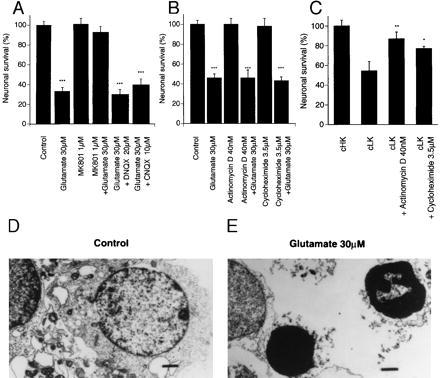
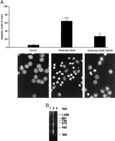
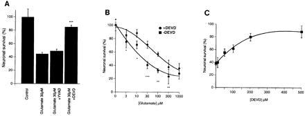
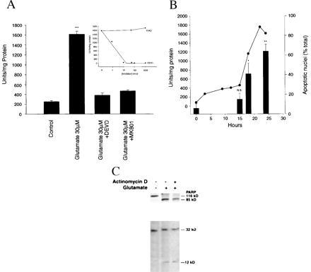
Neurotoxicity induced by overstimulation of N-methyl-D-aspartate (NMDA) receptors is due, in part, to a sustained rise in intracellular Ca2+; however, little is known about the ensuing intracellular events that ultimately result in cell death. Here we show that overstimulation of NMDA receptors by relatively low concentrations of glutamate induces apoptosis of cultured cerebellar granule neurons (CGNs) and that CGNs do not require new RNA or protein synthesis. Glutamate-induced apoptosis of CGNs is, however, associated with a concentration- and time-dependent activation of the interleukin 1beta-converting enzyme (ICE)/CED-3-related protease, CPP32/Yama/apopain (now designated caspase 3). Further, the time course of caspase 3 activation after glutamate exposure of CGNs parallels the development of apoptosis. Moreover, glutamate-induced apoptosis of CGNs is almost completely blocked by the selective cell permeable tetrapeptide inhibitor of caspase 3, Ac-DEVD-CHO but not by the ICE (caspase 1) inhibitor, Ac-YVAD-CHO. Western blots of cytosolic extracts from glutamate-exposed CGNs reveal both cleavage of the caspase 3 substrate, poly(ADP-ribose) polymerase, as well as proteolytic processing of pro-caspase 3 to active subunits. Our data demonstrate that glutamate-induced apoptosis of CGNs is mediated by a posttranslational activation of the ICE/CED-3-related cysteine protease caspase 3.


Figures

Figure 1
Figure 1
(A) Glutamate-induced neurotoxicity (excitotoxicity) is mediated by the NMDA receptor. Cultured CGNs were pretreated with 1 μM dizocilpine (MK-801), 10 μM CNQX (6-cyano-7-nitroquinoxaline-2,3-dione), or 20 μM DNQX (6,7-dinitroquinoxaline-2,3-dione) for 5 min, then exposed to 30 μM glutamate for 24 hr. (B) Glutamate-induced apoptosis is not dependent on de novo gene transcription or translation because neither 40 nM actinomycin D nor 3.5 μM cycloheximide attenuates glutamate-induced neurotoxicity (∗∗∗, P < 0.001 treated vs. control by Student’s t test). (C) Apoptosis of CGNs induced by switching from depolarizing (high K+, cHK) to nondepolarizing (low K+, cLK) media is greatly attenuated by 40 nM actinomycin D and 3.5 μM cycloheximide (cLK = 5 mM KCl, cHK = 20 mM KCl) (∗, P < 0.05, ∗∗, P < 0.01, cLK treated vs. cLK alone by Student’s t test). Neuronal viability was determined as described in Materials and Methods and calculated as a % of control (untreated cultures). Values are expressed as the mean ± standard error of triplicate determinations from at least three separate experiments (∗∗∗, P < 0.001 treated vs. control by Student’s t test). Ultrastructural analysis (×8,400) of CGNs after glutamate exposure reveals morphological features typical of apoptosis including condensed chromatin (bar = 1 μm) (E), as compared with control (D). Note markedly condensed chromatin and small pyknotic nuclei. Lower power electron microscopy photomicrographs (×4,400) were used to quantify number (%) apoptotic CGNs after glutamate exposure. Data is presented in text.
Figure 4
Figure 4
(A) The caspase 3 protease inhibitor Ac-DEVD-CHO attenuates the morphological features of apoptosis induced by glutamate in cultured CGNs. CGNs were treated with 30 μM glutamate for 24 hr in the presence or absence of 200 μM Ac-DEVD-CHO and then stained with 5 μg/ml Hoechst 33258. The number of apoptotic nuclei (small with condensed chromatin) were counted from representative photomicrographs (×1,250) and are represented as a % of the total number of nuclei counted (n = 400) (∗∗∗, P < 0.001 glutamate vs. control; ∗, P < 0.05 glutamate vs. glutamate + Ac-DEVD-CHO by Student’s t test). (B) Internucleosomal DNA fragmentation (DNA laddering) induced in CGNs after exposure to glutamate (30 μM, 24 hr) is greatly attenuated by 200 μM Ac-DEVD-CHO. CGNs (control, lane 1) were exposed to glutamate (30 μM, 24 hr) in the absence (lane 2) or presence of 200 μM Ac-DEVD-CHO (lane 3).
Figure 2
Figure 2
(AH) Effects of the cell-permeable caspase 3 (CPP32) and caspase 1 (ICE) protease inhibitors, Ac-DEVD-CHO and Ac-YVAD-CHO, respectively, on glutamate-induced apoptosis of cultured CGNs. Representative fields of CGNs were photographed under phase contrast microscopy (AD) and after double staining with FDA and PI (EH) as described in Materials and Methods. Compare untreated control cultures (A and E) to glutamate (30 μm; 24 hr)-treated (B and F) vs. glutamate + Ac-DEVD-CHO (C and G) to glutamate + Ac-YVAD-CHO (D and H). Notice that the CPP32/apopain protease inhibitor, Ac-DEVD-CHO, but not the ICE-specific protease inhibitor Ac-YVAD-CHO, greatly attenuates glutamate-induced toxicity of CGN as measured by phase contrast microscopy or double staining with FDA and PI. (×100)
Figure 3
Figure 3
(A) Quantification of the effects of Ac-DEVD-CHO and Ac-YVAD-CHO on glutamate-treated (30 μm, 24 hr) CGNs also shows almost complete protection by Ac-DEVD-CHO but not by Ac-YVAD-CHO. (B) The CPP32 inhibitor Ac-DEVD-CHO significantly attenuates glutamate-induced apoptosis of CGNs (ANOVA, P < 0.001). CGNs were exposed to increasing concentrations of glutamate (3–1,000 μM) for 24 hr in the presence or absence of 200 μM Ac-DEVD-CHO (∗∗∗, P < 0.001, ∗, P < 0.05 by Student’s t test). (C) CGNs were exposed to 30 μM glutamate for 24 hr in the presence of increasing concentrations of the CPP32 protease inhibitor, Ac-DEVD-CHO. Neuronal viability was assessed by FDA and PI staining and visual counting of viable neurons from representative photomicrographs. Values in A, B, and C are expressed as a % of control cultures for each experiment, and the data represent the mean ± standard error of triplicate determinations from a representative experiment repeated at least three times with similar results.
Figure 5
Figure 5
Caspase 3 protease activity in cultured cerebellar granule neurons is induced by glutamate. (A) The caspase 3-specific color substrate Ac-DEVD-pNA was incubated for 60 min at 37°C with 30 μg cytosolic protein collected from CGNs treated with glutamate (30 μM, 24 hr) in the presence or absence of Ac-DEVD-CHO (200 μM, 24 hr) or dizocilpine (MK-801), (1 μM, 24 h). Measurements were taken at 405 nm (see Materials and Methods for details). One unit of enzyme is defined as the amount of enzyme required to release 0.22 pmol of p-nitroaniline per min at 37°C. Note that both Ac-DEVD-CHO and dizocilpine (MK-801) completely inhibit glutamate-induced activation of caspase 3 activity. (B) Caspase 3 protease activity is induced significantly at 15 hr after glutamate exposure and increases 8-fold over the ensuing 9 hr (solid dark line), just before the measurement of apoptosis (bars). Note increase in caspase 3 activity that is temporally associated with an increase in the number (%) apoptotic nuclei measured by Hoechst 33258 staining (see Materials and Methods for details). Caspase 1 protease activity was not detected at any time point after glutamate exposure (30 μM, 24 hr). Protease activity was determined as described above. Glutamate-induced caspase 3 protease activity measured in cytosolic extracts from CGNs exposed to glutamate is potently inhibited in vitro by Ac-DEVD-CHO (IC50 < 10nM), but not by the caspase 1-specific inhibitor Ac-YVAD-CHO (A, Inset). (C) PARP and caspase 3 proenzyme from cell extracts of CGNs exposed to glutamate (30 μM, 24 hr) are proteolytically processed to their 85-kDa and 12-kDa subunits, respectively. Protein from CGNs exposed to glutamate (30 μM, 24 hr) were size-fractionated on a 10–20% gradient gel, transferred to nitrocellulose, and detected with polyclonal antibodies directed to PARP or the 12-kDa subunit of caspase 3 using enhanced chemiluminescence (see Materials and Methods for details).

References

    1. Choi D W. Neuron. 1988;1:623–634. - PubMed
    1. Meldrum B, Garthwaite J. Trends Pharmacol Sci. 1990;11:379–387. - PubMed
    1. Coyle J T, Puttfarcken P. Science. 1993;262:689–695. - PubMed
    1. Lipton S A, Rosenberg P A. N Engl J Med. 1994;330:613–622. - PubMed
    1. Manev H, Favaron A, Guidotti A, Costa E. Mol Pharmacol. 1989;36:106–112. - PubMed

MeSH terms