. 1997 Oct 1;11(19):2456-67.
doi: 10.1101/gad.11.19.2456.

The PAG gene product, a stress-induced protein with antioxidant properties, is an Abl SH3-binding protein and a physiological inhibitor of c-Abl tyrosine kinase activity

Affiliations

The PAG gene product, a stress-induced protein with antioxidant properties, is an Abl SH3-binding protein and a physiological inhibitor of c-Abl tyrosine kinase activity

S T Wen et al. Genes Dev. .

Abstract

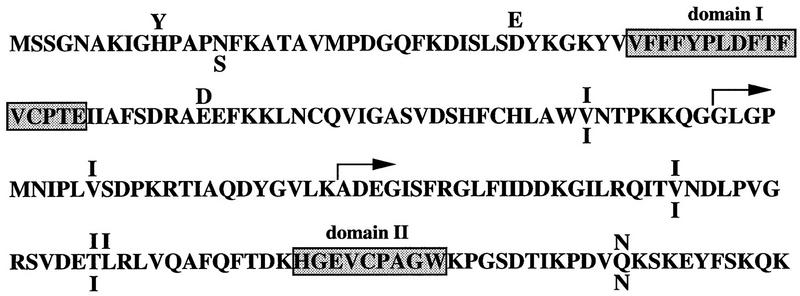
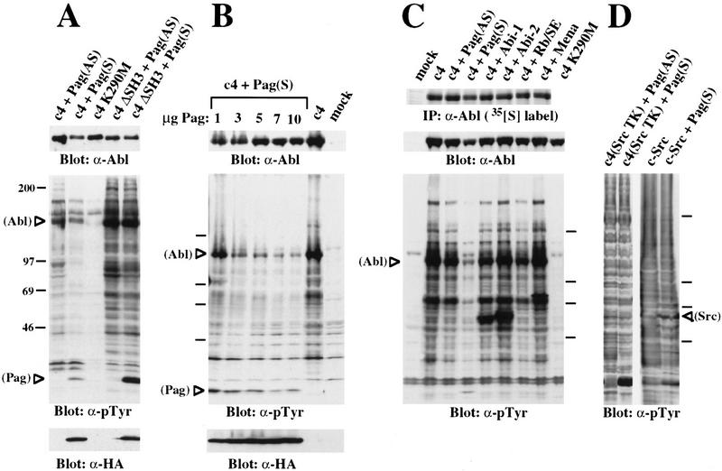
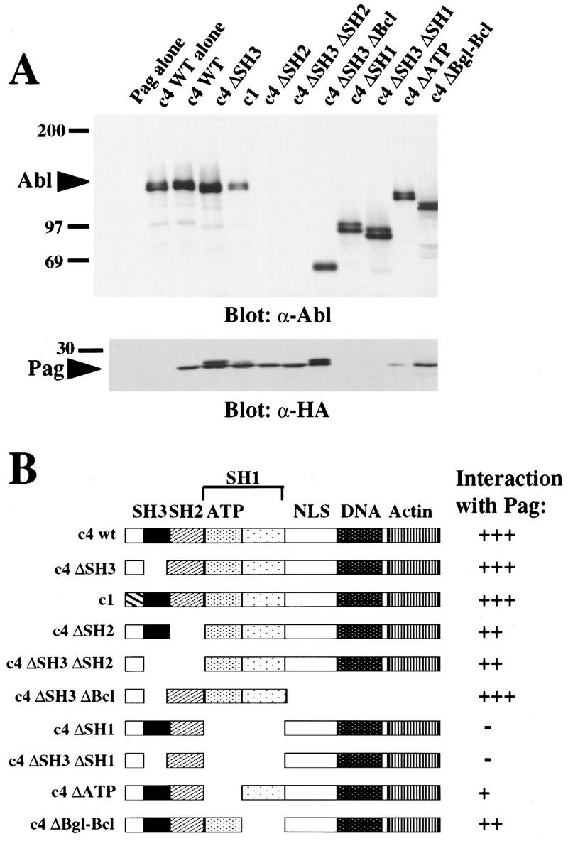
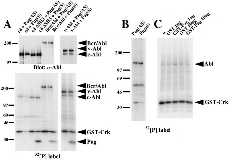
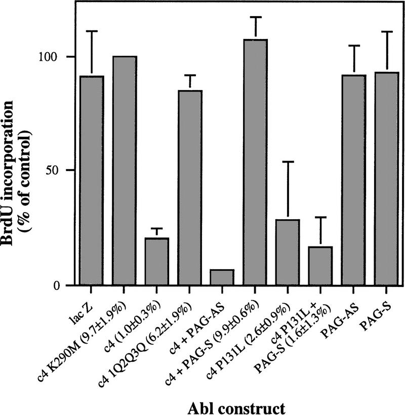
Biochemical and genetic evidence suggests that the tyrosine kinase activity of c-Abl is tightly regulated in vivo by a cellular factor binding to the Src homology 3 (SH3) domain of Abl. We used the yeast two-hybrid system to identify a gene, PAG, whose protein product (Pag) interacts specifically with the Abl SH3 domain. Pag, also known as macrophage 23-kD stress protein (MSP23), is a member of a novel family of proteins with antioxidant activity implicated in the cellular response to oxidative stress and in control of cell proliferation and differentiation. In a co-expression assay, Pag associates with c-Abl in vivo and inhibits tyrosine phosphorylation induced by overexpression of c-Abl. Inhibition requires the Abl SH3 and kinase domains and is not observed with other Abl SH3-binding proteins. Expression of Pag also inhibits the in vitro kinase activity of c-Abl, but not SH3-mutated Abl or v-Abl. When transfected in NIH-3T3 cells, Pag is localized to nucleus and cytoplasm and rescues the cytostatic effect induced by c-Abl. These observations suggest Pag is a physiological inhibitor of c-Abl in vivo.


Figures

Figure 1
Figure 1
Amino acid sequence of Pag and its murine and rat homologs. The sequence of human Pag (Prosperi et al. 1993) is given according to the single letter amino acid code. Letters above denote substitutions present in the murine homolog MSP23 (Ishii et al. 1993); letters below indicate substitutions in the rat homolog (HBP23) (Iwahara et al. 1995). Domains I and II are regions containing cysteine residues that are conserved among the family of peroxide reductases (Chae et al. 1994a). The arrows indicate the locations of the point of fusion of the two cDNAs recovered in the yeast two-hybrid screen with the B42 transactivation domain.
Figure 2
Figure 2
Pag interacts specifically with the Abl SH3 domain in the yeast two-hybrid system. Yeast strain EGY48, containing Pag fused to the B42 transactivation domain, was transformed with different LexA baits and β-galactosidase activity detected on galactose X-gal plates as blue color (dark). I3 and I7 are positive controls (Gyuris et al. 1993) with I3 demonstrating strong transactivation and I7 weaker transactivation. (A) Pag interacts with the Abl SH3 domain, but not with a mutant SH3 domain containing an activating point mutation (P131L) or the Abl kinase domain (SH1). (B) Pag fails to interact with SH3 domains from Grb2, PI3K p85 subunit, and c-Src.
Figure 3
Figure 3
Pag inhibits the tyrosine phosphorylation induced by c-Abl in vivo. The indicated proteins were expressed by transient transfection of 293T cells, and lysates containing equal amounts of protein compared for their phosphotyrosine levels by immunoblotting with anti-phosphotyrosine antibody 4G10. Where indicated, the blot was then stripped and reprobed with anti-Abl mAb 8E9 to detect c-Abl or anti-hemagglutinin mAb 12CA5 to detect epitope-tagged Pag. (A) Pag inhibits c-Abl but not SH3-deleted Abl. Pag, in either sense (S) or antisense (AS) orientation, was cotransfected with wild-type c-Abl (c4) or SH3-deleted Abl into 293T cells. (B) Inhibition of Abl by Pag is dose-dependent. 293T cells were cotransfected with 5 ug of c-Abl (c4) and increasing amounts (1, 3, 5, 7, and 10 μg) of Pag. (C) Other SH3-binding proteins and pRb fail to inhibit c-Abl. 293T cells were cotransfected with Abl and different Abl SH3 domain binding proteins (Pag, Mena, Abi-1, Abi-2) or Rb SE, the C pocket of retinoblastoma gene product. Expression of these proteins at levels similar to that of Pag was confirmed by immunoblotting with anti-Mena or anti-epitope antibodies (data not shown). (D) Inhibition of Abl by Pag requires the Abl kinase domain. 293T cells were cotransfected with Pag in either sense (S) or antisense (AS) orientation and with the Abl c4-SrcTK chimera or c-Src.
Figure 4
Figure 4
Pag interacts with Abl in vivo. (A) Epitope-tagged Pag and the indicated Abl proteins were coexpressed in 293T cells, cell extracts immunoprecipitated with anti-Abl antisera α-GEX4 (directed against the carboxyl terminus of Abl) (Jackson et al. 1993) or α-type Ib Abl antisera (directed against the Abl amino terminus, for the c4 ΔSH3 ΔBcl protein), and immunoblotted with anti-HA mAb 12CA5 for the detection of Pag or anti-Abl Ab 8E9 (directed against the SH2 domain of Abl). The ΔSH2 proteins were detected with a different anti-Abl Ab and found to be expressed at equivalent levels (data not shown). Coimmunoprecipitation of Abl with Pag was also observed when the anti-HA mAb was used for immunoprecipitation (data not shown). (B) Schematic representation of Abl deletion constructs and their interaction with Pag as measured by coimmunoprecipitation. The location of the SH3, SH2, and SH1 domains, the ATP-binding lobe of the kinase domain, the pentalysine nuclear localization signal, and the DNA-binding and actin-binding domains are indicated.
Figure 5
Figure 5
Pag is localized to nucleus and cytoplasm. NIH-3T3 cells were transfected with epitope-tagged Pag and type IV c-Abl and the expressed proteins were localized with indirect immunofluorescence with rabbit anti-Abl antibodies (A) and anti-HA mAb 12CA5 (B). Background staining levels are indicated by several nontransfected cells in the field. Cells transfected with Pag or Abl alone and stained with the opposite secondary antibody gave similar background staining (data not shown). Bar, 10 μm.
Figure 6
Figure 6
Pag inhibits Abl kinase activity in vitro. (A) 293T cells cotransfected with Pag and wild-type c-Abl, SH3-deleted Abl (c4 ΔSH3), Bcr/Abl, or p160 v-Abl were immunoprecipitated with anti-Abl antibody and immune complex kinase assays performed with GST–Crk as a substrate. (Top) Western blot with anti-Abl antibody showing equivalent amounts of Abl proteins were immunoprecipitated. (Bottom) Autoradiograph showing32P incorporation. (B) NIH-3T3 cells were transfected with Pag in either sense (S) or antisense (AS) orientation and huICAM-1 as a cell-surface marker, selected with magnetic beads, immunoprecipitated with anti-Abl Ab, and immune complex kinase assays performed with GST–Crk as a substrate. (C) (His)6-purified Abl protein was tested for its in vitro kinase activity with GST–Crk as a substrate and increasing amounts of parental GST or GST–Pag fusion protein added.
Figure 7
Figure 7
Pag rescues the cytostatic and cytotoxic effects induced by c-Abl. NIH-3T3 cells were transfected with the indicated Abl constructs with or without PAG in the sense (S) or antisense (AS) orientation, and entry into the S phase of the cell cycle was measured as the percentage of transfected cells positive for BrdU incorporation relative to kinase-inactive Abl. (LacZ) Transfection of the β-galactosidase reporter construct alone; (c4) c-Abl type IV; (K290M) kinase-inactivating point mutant; (1Q2Q3Q) site-specific mutations in the first, second, and third Abl nuclear localization signals; (P131L) activated SH3 point mutant of c-Abl; (PAG-S and PAG-AS) cotransfection of a PAG expression vector in the sense or antisense orientation, respectively. The transfection efficiency (absolute percentage of β-galactosidase positive cells) is shown in parentheses after the construct name.

References

    1. Adzhubei AA, Sternberg MJ. Left-handed poly-proline II helices commonly occur in globular proteins. J Mol Biol. 1993;229:472–493. - PubMed
    1. Andreotti AH, Bunnell SC, Feng S, Berg LJ, Schreiber SL. Regulatory intramolecular association in a tyrosine kinase of the Tec family. Nature. 1997;385:93–97. - PubMed
    1. Chae HZ, Robison K, Poole LB, Church G, Storz G, Rhee SG. Cloning and sequencing of thiol-specific antioxidant from mammalian brain: Alkyl hydroperoxide reductase and thiol-specific antioxidant define a large family of antioxidant enzymes. Proc Natl Acad Sci. 1994a;91:7017–7021. - PMC - PubMed
    1. Chae HZ, Uhm TB, Rhee SG. Dimerization of thiol-specific antioxidant and the essential role of cystein 47. Proc Natl Acad Sci. 1994b;91:7022–7026. - PMC - PubMed
    1. Chien C-T, Bartel PL, Sternglanz R, Fields S. The two-hybrid system: A method to identify and clone genes for proteins that interact with a protein of interest. Proc Natl Acad Sci. 1991;88:9578–9582. - PMC - PubMed

Publication types

MeSH terms