Comparative Study
. 1997 Oct 15;11(20):2679-90.
doi: 10.1101/gad.11.20.2679.

The DAF-3 Smad protein antagonizes TGF-beta-related receptor signaling in the Caenorhabditis elegans dauer pathway

Affiliations
Comparative Study

The DAF-3 Smad protein antagonizes TGF-beta-related receptor signaling in the Caenorhabditis elegans dauer pathway

G I Patterson et al. Genes Dev. .

Abstract

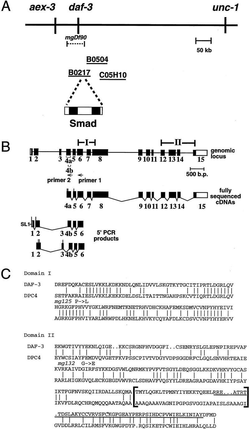
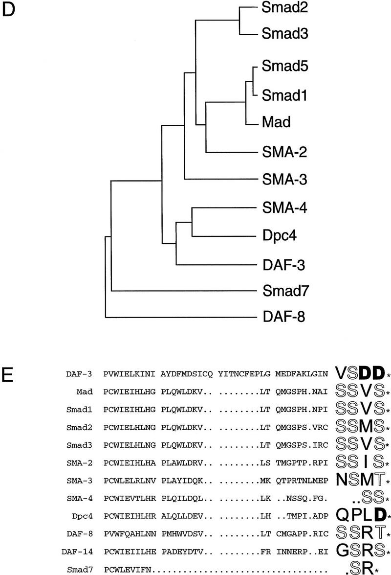
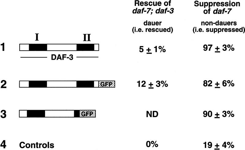
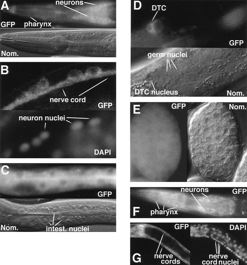
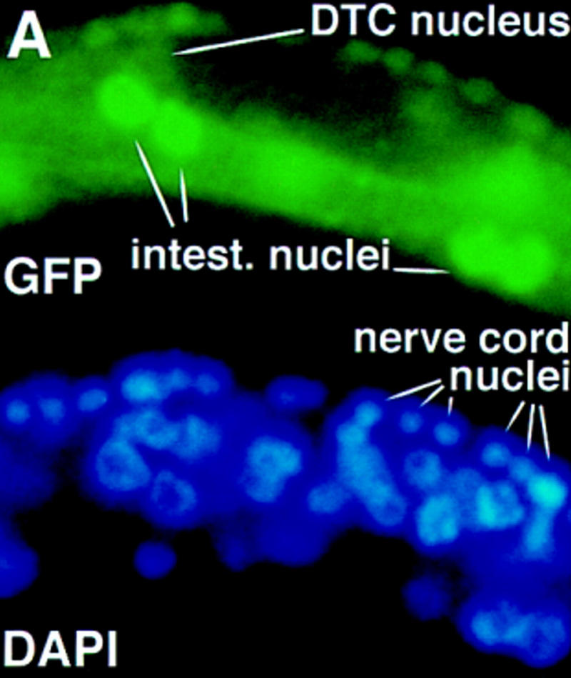
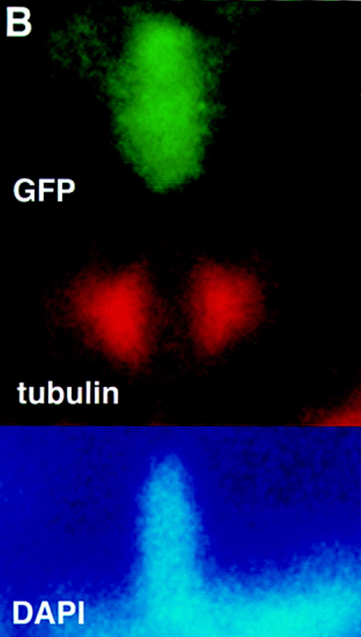
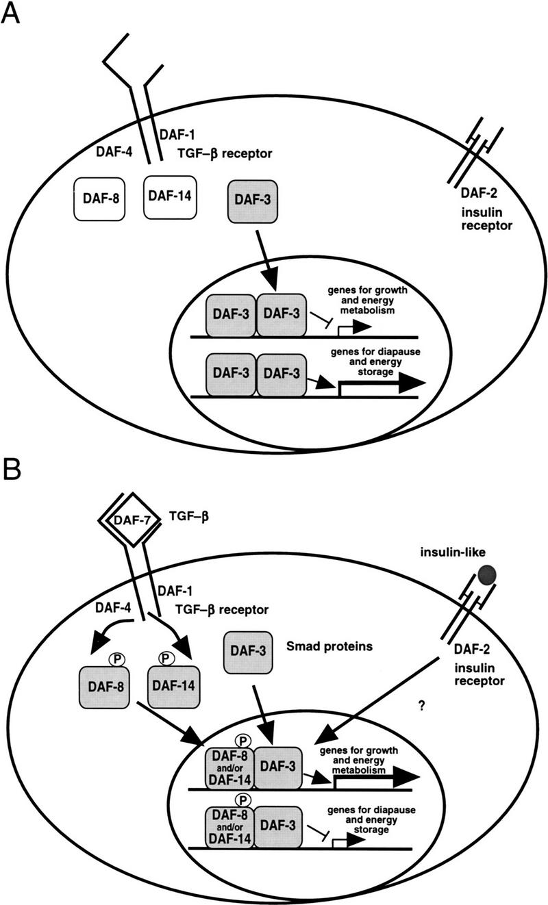
Signals from TGF-beta superfamily receptors are transduced to the nucleus by Smad proteins, which transcriptionally activate target genes. In Caenorhabditis elegans, defects in a TGF-beta-related pathway cause a reversible developmental arrest and metabolic shift at the dauer larval stage. Null mutations in daf-3 suppress mutations in genes encoding this TGF-beta signal, its receptors, and associated Smad signal transduction proteins. daf-3 encodes a Smad protein that is most closely related to mammalian DPC4, and is expressed throughout development in many of the tissues that are remodeled during dauer development. DAF-4, the type II TGF-beta receptor in this pathway, is also expressed in remodeled tissues. These data suggest that the DAF-7 signal from sensory neurons acts as a neuroendocrine signal throughout the body to directly regulate developmental and metabolic shifts in tissues that are remodeled during dauer formation. A full-length functional DAF-3/GFP fusion protein is predominantly cytoplasmic, and this localization is independent of activity of the upstream TGF-beta-related pathway. However, this fusion protein is associated with chromosomes in mitotic cells, suggesting that DAF-3 binds DNA directly or indirectly. DAF-3 transgenes also interfere with dauer formation, perhaps attributable to a dosage effect. A truncated DAF-3/GFP fusion protein that is predominantly nuclear interferes with dauer formation, implying a role for DAF-3 in the nucleus. These data suggest that DAF-7 signal transduction antagonizes or modifies DAF-3 Smad activity in the nucleus to induce reproductive development; when DAF-7 signals are disabled, unmodified DAF-3 Smad activity mediates dauer arrest and its associated metabolic shift. Therefore, daf-3 is unique in that it is antagonized, rather than activated, by a TGF-beta pathway.


Figures

Figure 1
Figure 1
daf-3 encodes a Smad protein most closely related to DPC4. (A) daf-3 cloning. daf-3 was genetically mapped to a region on the X chromosome between aex-3 and unc-1 (A. Koweek and G. Patterson, unpubl.). B0217 complements daf-3 mutants in transformation rescue experiments; B0504 and C05H10 do not. mgDf90 is a deletion that removes all of daf-3. (B) Structure of daf-3-coding region. At the top is the exon/intron structure of daf-3; coding exons are solid boxes, noncoding regions are open boxes, and lines are introns. Conserved Smad domains I and II are indicated. The three classes of cDNA are shown, and 5′ ends are indicated by vertical lines. The accession numbers for the cDNAs shown are, top to bottom: AF005205, AF005206, and AF005207. (C) Protein sequence alignment of C. elegans DAF-3 and its closest homolog, human DPC4, in the Smad conserved domains I and II. Dots indicate gaps introduced to maximize alignment. The Smad mutational hot spot is underscored, and the insertion in DAF-3 and DPC4 relative to other Smads is bracketed. In addition to mg125 and mg132, seven other daf-3 alleles were sequenced in the hot spot; none of them contains a mutation. Alleles sequenced were mg91, mg93, mg105, mg121, mg126, mg133 (isolated by A. Koweek and G. Patterson, unpubl.) and sa205 (Thomas et al. 1993). (D) Relationship of DAF-3 domain I to other Smads. Lineup was performed with the program pileup (Genetics Computer Group 1994) using amino acids 137–245 of DAF-3 (GenBank accession no. AF005205) and corresponding residues of the other Smads. (E) Comparison of carboxyl termini of Smads. The final 28–44 residues are shown. Residues that are phosphorylated by receptor (in Smad1 and Smad2) or similar residues in similar positions (in other Smads) are shown in outline. Aspartates in similar positions are shown in boldface type.
Figure 1
Figure 1
daf-3 encodes a Smad protein most closely related to DPC4. (A) daf-3 cloning. daf-3 was genetically mapped to a region on the X chromosome between aex-3 and unc-1 (A. Koweek and G. Patterson, unpubl.). B0217 complements daf-3 mutants in transformation rescue experiments; B0504 and C05H10 do not. mgDf90 is a deletion that removes all of daf-3. (B) Structure of daf-3-coding region. At the top is the exon/intron structure of daf-3; coding exons are solid boxes, noncoding regions are open boxes, and lines are introns. Conserved Smad domains I and II are indicated. The three classes of cDNA are shown, and 5′ ends are indicated by vertical lines. The accession numbers for the cDNAs shown are, top to bottom: AF005205, AF005206, and AF005207. (C) Protein sequence alignment of C. elegans DAF-3 and its closest homolog, human DPC4, in the Smad conserved domains I and II. Dots indicate gaps introduced to maximize alignment. The Smad mutational hot spot is underscored, and the insertion in DAF-3 and DPC4 relative to other Smads is bracketed. In addition to mg125 and mg132, seven other daf-3 alleles were sequenced in the hot spot; none of them contains a mutation. Alleles sequenced were mg91, mg93, mg105, mg121, mg126, mg133 (isolated by A. Koweek and G. Patterson, unpubl.) and sa205 (Thomas et al. 1993). (D) Relationship of DAF-3 domain I to other Smads. Lineup was performed with the program pileup (Genetics Computer Group 1994) using amino acids 137–245 of DAF-3 (GenBank accession no. AF005205) and corresponding residues of the other Smads. (E) Comparison of carboxyl termini of Smads. The final 28–44 residues are shown. Residues that are phosphorylated by receptor (in Smad1 and Smad2) or similar residues in similar positions (in other Smads) are shown in outline. Aspartates in similar positions are shown in boldface type.
Figure 3
Figure 3
Rescuing ability and suppression of daf-7 by daf-3 plasmids. The solid boxes represent the Smad conserved domains I and II of daf-3; the stippled boxes represent GFP. For all experiments shown, daf-3 plasmids were injected at a concentration of 10 ng/μl and the pRF4 injection marker was injected at a concentration of 90 ng/μl. To score dauer formation, synchronous broods of transgenic animals were scored after two days at 25°C. The rescue experiment shows the rescue of daf-7(m62); daf-3(e1376) by each of the fusion proteins. The control is a transgenic array with the pRF4 transformation marker and a nonrescuing cosmid. For each construct, four or more lines were measured in two separate experiments. To measure suppression of daf-7, transgenic arrays were crossed to a daf-7 strain (for plasmids 1 and 3), or produced by injecting directly into daf-7 (plasmid 2). The controls are two transgenic strains with the pRF4 marker and an unrelated GFP-expressing transgene.
Figure 2
Figure 2
DAF-3 and DAF-4 expression. All DAF-3 photographs show animals with the full-length functional DAF-3/GFP fusion gene shown in Fig. 3. In A and C–F, intrinsic fluorescence is used to visualize GFP; in B and G, anti-GFP antibodies are used. (A) DAF-3/GFP head expression in an L1 animal. Weak DAF-3/GFP expression in the pharynx impedes cell identification, but the main body of the pharynx is filled, implying expression in pharyngeal muscle. (B) DAF-3/GFP expression in the ventral nerve cord of an adult animal. L1 animals look similar. (C) DAF-3/GFP expression in the intestine of an L1 animal. (D) DAF-3/GFP expression in the distal tip cell of an L4 animal. (E) DAF-3/GFP expression in an embryo with ∼200 nuclei. (F) DAF-4/GFP expression in the head of an L1 animal. (G) DAF-4/GFP expression in the dorsal nerve cord and ventral nerve cord of an L4 animal. Although the intestine can be seen in this picture, the DAF-4/GFP is not visible, because in this focal plane, the intestinal cell membrane is obscured by the bright fluorescence in the nerve cords.
Figure 4
Figure 4
(A) A truncated DAF-3/GFP protein is predominantly nuclear. Wild-type animals were injected with the truncated construct shown in Fig. 3 at a concentration of 10 ng/ml. The pRF4 transformation marker was injected at 100 ng/ml. The photograph shows an early L2 animal, and DAF-3 is predominantly nuclear. The clear spot in the center of some of the nuclei is the nucleolus. All cells in these animals have predominantly nuclear DAF-3/GFP, including the ventral cord neurons, intestinal cells and distal tip cell (all shown), as well as head and tail neurons and hypodermal cells. (B) DAF-3/GFP is associated with metaphase chromosomes. Fixed L1 animals were immunostained with anti-GFP antibody (Clontech) and anti-α-tubulin antibody (Amersham). DNA was visualized with DAPI.
Figure 4
Figure 4
(A) A truncated DAF-3/GFP protein is predominantly nuclear. Wild-type animals were injected with the truncated construct shown in Fig. 3 at a concentration of 10 ng/ml. The pRF4 transformation marker was injected at 100 ng/ml. The photograph shows an early L2 animal, and DAF-3 is predominantly nuclear. The clear spot in the center of some of the nuclei is the nucleolus. All cells in these animals have predominantly nuclear DAF-3/GFP, including the ventral cord neurons, intestinal cells and distal tip cell (all shown), as well as head and tail neurons and hypodermal cells. (B) DAF-3/GFP is associated with metaphase chromosomes. Fixed L1 animals were immunostained with anti-GFP antibody (Clontech) and anti-α-tubulin antibody (Amersham). DNA was visualized with DAPI.
Figure 5
Figure 5
A model for the role of the DAF-3/DAF-8/DAF-14 Smads in dauer formation, as described in text. (A) Dauer growth induction; (B) reproductive growth induction.

References

    1. Attisano L, Wrana JL, Montalvo E, Massague J. TGF-β receptors and actions. Biochim Biophys Acta. 1994;1222:71–80. - PubMed
    1. Baker JC, Harland RM. A novel mesoderm inducer, Madr2, functions in the activin signal transduction pathway. Genes & Dev. 1996;10:1880–1889. - PubMed
    1. Bargmann CI, Horvitz HR. Control of larval development by chemosensory neurons in Caenorhabditis elegans. Science. 1991;251:1243–1246. - PubMed
    1. Chalfie M, Tu Y, Euskirchen G, Ward WW, Prasher DC. Green fluorescent protein as a marker for gene expression. Science. 1994;263:802–805. - PubMed
    1. Chen X, Rubrock M, Whitman M. A transcriptional partner for MAD proteins in TGF-β signaling. Nature. 1996;383:691–696. - PubMed

Publication types

MeSH terms

LinkOut - more resources