. 1997 Oct 15;11(20):2691-700.
doi: 10.1101/gad.11.20.2691.

The Drosophila neuregulin homolog Vein mediates inductive interactions between myotubes and their epidermal attachment cells

Affiliations

The Drosophila neuregulin homolog Vein mediates inductive interactions between myotubes and their epidermal attachment cells

T Yarnitzky et al. Genes Dev. .

Abstract

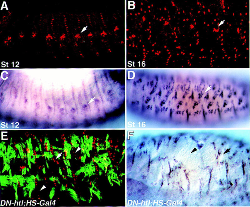
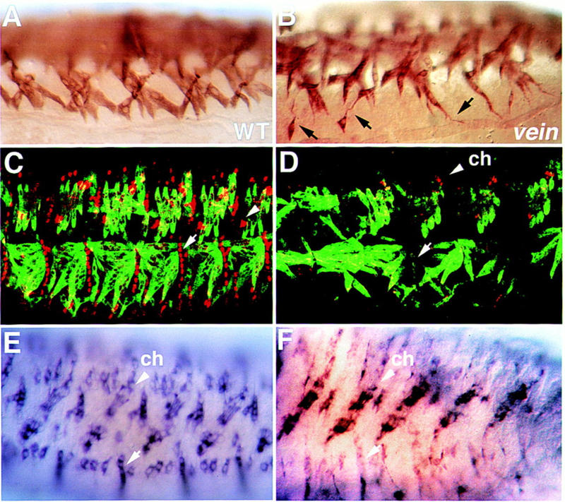
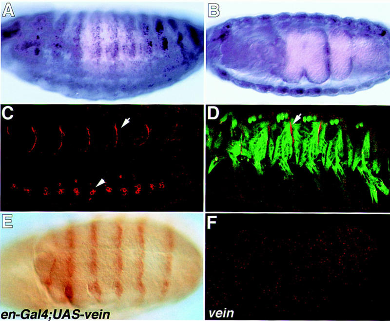
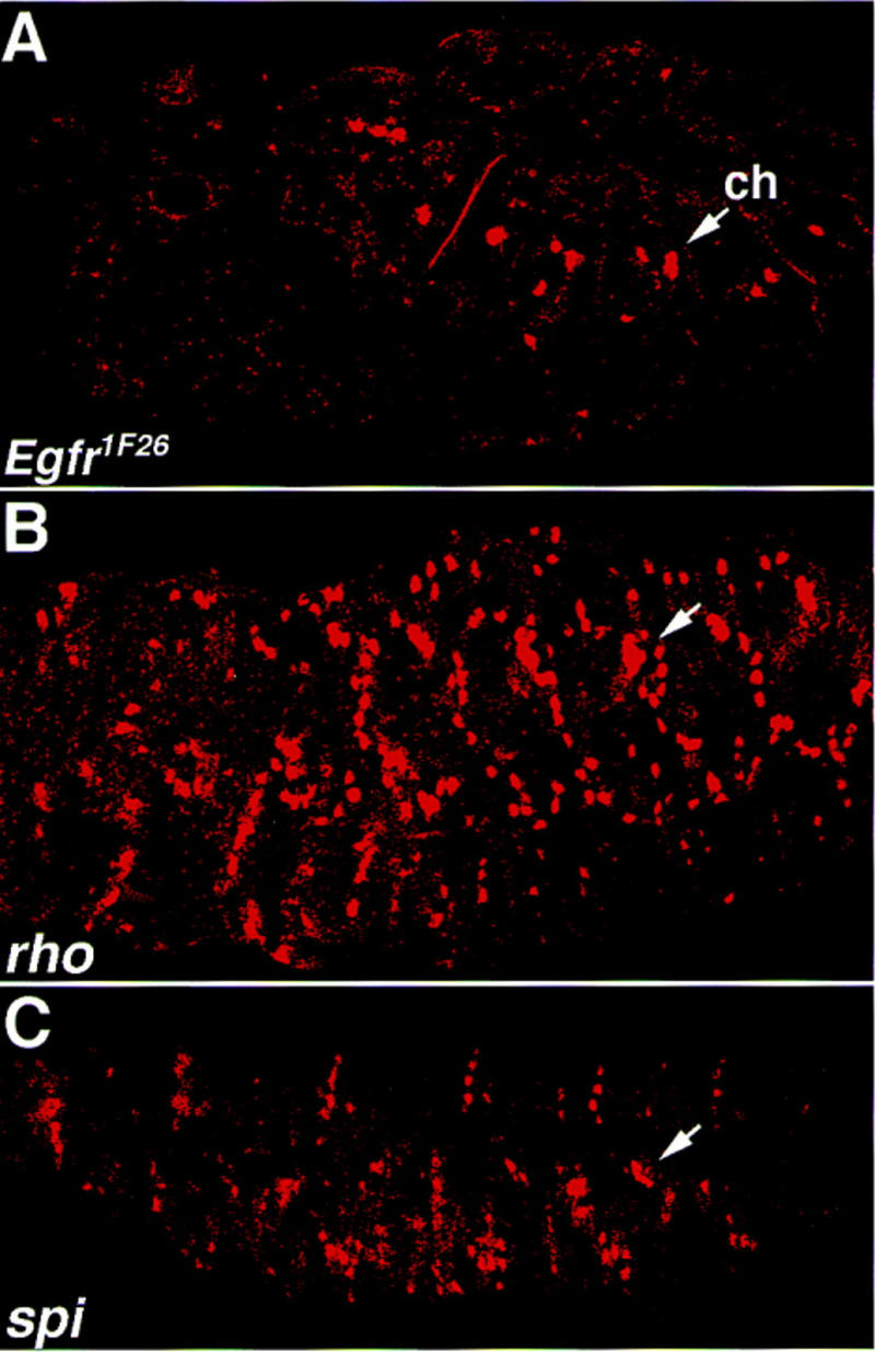
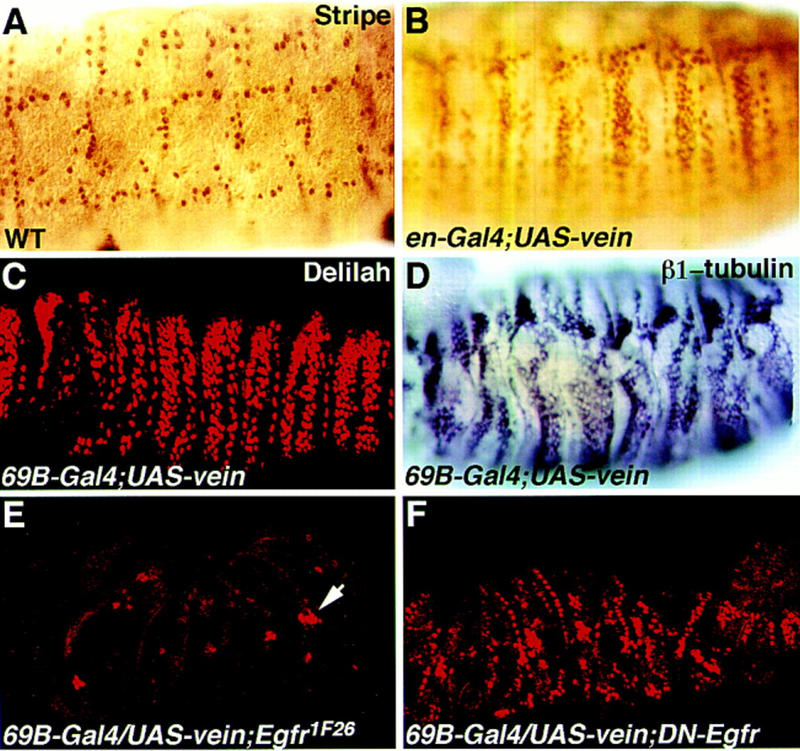
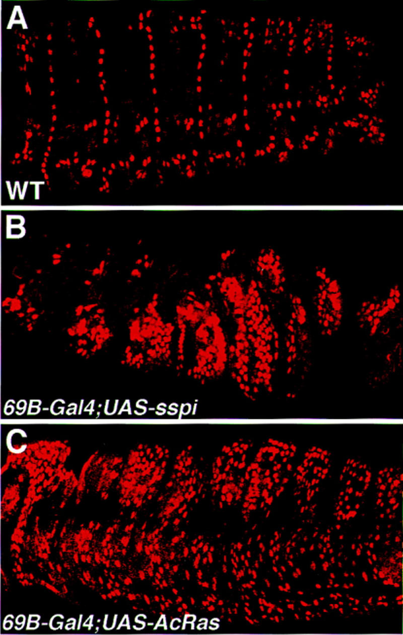
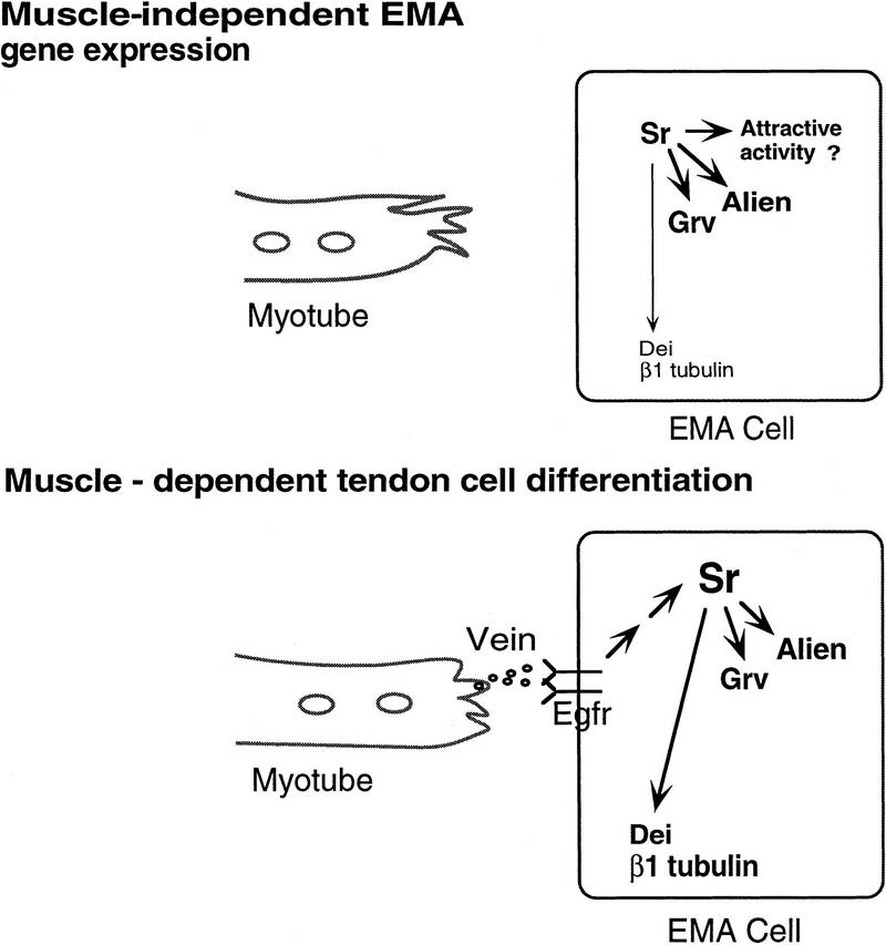
Inductive interactions between cells of distinct fates underlie the basis for morphogenesis and organogenesis across species. In the Drosophila embryo, somatic myotubes form specific interactions with their epidermal muscle attachment (EMA) cells. The establishment of these interactions is a first step toward further differentiation of the EMA cells into elongated tendon cells containing an organized array of microtubules and microfilaments. Here we show that the molecular signal for terminal differentiation of tendon cells is the secreted Drosophila neuregulin-like growth factor Vein produced by the myotubes. Although vein mRNA is produced by all of the myotubes, Vein protein is secreted and accumulates specifically at the muscle-tendon cell junctional site. In loss-of-function vein mutant embryos, muscle-dependent differentiation of tendon cells, measured by the level of expression of specific markers (Delilah and beta1 tubulin) is blocked. When Vein is expressed in ectopic ectodermal cells, it induces the ectopic expression of these genes. Our results favor the possibility that the Drosophila EGF receptor DER/Egfr expressed by the EMA cells functions as a receptor for Vein. We show that Vein/Egfr binding activates the Ras pathway in the EMA cells leading to the transcription of the tendon-specific genes, stripe, delilah, and beta1 tubulin. In Egfr1F26 mutant embryos that lack functional Egfr expression, the levels of Delilah and beta1 tubulin are very low. In addition, the ability of ectopic Vein to induce the expression of Delilah and beta1 tubulin depends on the presence of functional Egfrs. Finally, activation of the Egfr signaling pathway by either ectopically secreted Spitz, or activated Ras, leads to the ectopic expression of Delilah. These results suggest that inductive interactions between myotubes and their epidermal muscle attachment cells are initiated by the binding of Vein, to the Egfr on the surface of EMA cells.


Figures

Figure 1
Figure 1
Muscle-independent, and muscle-dependent expression of Delilah and β1 tubulin. Wild-type embryos at stage 12 (A,C) or stage 16 (B,D) were stained with anti-Delilah antibody (A,B) or hybridized with a β1 tubulin antisense probe (C,D). At stage 12 of embryonic development, somatic muscles are not yet connected to their EMA cells, and the expression of Delilah and β1 tubulin is weak, relative to the strong expression of both markers in embryos at stage 16 when binding of somatic myotubes has been established. In mutant embryos carrying UAS–DN–Htl, and HS–Gal4 (Gal4 under a heat shock promoter), which were heat-shocked at 4–5 hr after egg laying, some of the muscles are missing (E,F). Embryos were double-labeled for Delilah and Myosin (E), or hybridized with a β1 tubulin probe (F). Note that the absence of somatic muscles leads to reduced expression of Delilah and β1 tubulin. Arrows mark chordotonals; arrowheads in E and F mark regions in which Delilah or β1 tubulin expression is abnormal.
Figure 2
Figure 2
Somatic muscles and tendon cells are defective in vein mutant embryos. Wild-type (A,C,E) and vein mutant (B,D,F) embryos were stained with anti-Myosin antibody (A,B), double-labeled with anti-Myosin (green), and anti-Delilah (red) antibody (C,D), and hybridized with a β1 tubulin antisense probe (E,F). Notice that the somatic myotubes in the vein mutant embryo send elongated random filopodia (black arrows in B) and that the expression of Delilah or β1 tubulin in the tendon cells is significantly reduced (cf. white arrows in D and F to C and E, respectively). Arrowheads in C, D, E, and F show a high level of Delilah and β1 tubulin expression in the chordotonal organs (ch).
Figure 3
Figure 3
(A) Schematic illustration of the molecular structure of Vein. A signal peptide (SP) is present at its amino-terminal domain, followed by PEST sequences, a single immunoglobulin-like (Ig) domain, and an EGF domain at its carboxyl terminus. (B) Localization of the P1749 construct within the first non-coding exon of the vein gene. Solid boxes represent noncoding sequences; open boxes represent coding exons.
Figure 3
Figure 3
(A) Schematic illustration of the molecular structure of Vein. A signal peptide (SP) is present at its amino-terminal domain, followed by PEST sequences, a single immunoglobulin-like (Ig) domain, and an EGF domain at its carboxyl terminus. (B) Localization of the P1749 construct within the first non-coding exon of the vein gene. Solid boxes represent noncoding sequences; open boxes represent coding exons.
Figure 4
Figure 4
The mRNA and protein expression pattern of Vein. Wild-type embryos (A–D) were hybridized with Vein antisense probe (A,B) or with anti-Vein antibody (red in C and D). The embryo in D is double labeled for Myosin (green) and vein (red). Note that although vein mRNA is expressed throughout the somatic and visceral muscles, Vein protein is concentrated in the muscle–tendon junctional site (arrows in C and D). Vein protein staining is also noted in a cluster of cells along the CNS (arrowhead in C). The embryo in E labeled with anti-Vein antibody, carries gal4 under the engrailed promoter and UAS–vein. The engrailed pattern is prominent. The embryo in F is a veinΔ25/DfXAS96 mutant embryo labeled with anti-Vein antibody and shows no positive staining.
Figure 5
Figure 5
The expression of Delilah in spitz group mutant embryos. Egfr1F26 mutant embryos (A), rho mutant embryos (B), or spitz (spi) mutant embryos were labeled with anti-Delilah antibody. While in rho and spitz mutants, Delilah expression in the tendon cells is maintained, in the Egfr1F26 mutant, Delilah expression is significantly reduced. Compare the expression of Delilah in the EMA cells to that in the chordotonal organs (arrows).
Figure 6
Figure 6
Ectopic expression of Vein induces ectopic expression of tendon-specific genes. Embryos carrying the UAS–vein construct (B–F) in combination with either the 69B–gal4 construct (C–F) or the engrailed–gal4 construct (B) were stained with anti-Stripe antibody (B) or with anti-Delilah antibody, or with a β1 tubulin antisense probe. Note that ectopic Vein induces the ectopic expression of Stripe (cf. embryo in B to a wild-type embryo stained for Stripe in A). Ectopic expression of Vein also induces Delilah (C) and β1 tubulin (D). The embryo in E carries a UAS–vein construct in combination with flb1F26 (null mutation for Egfr) and the 69B–gal4 inducer, and was stained with anti-Delilah antibody. The ectopic expression of Delilah in this embryo is eliminated (the arrow marks the chordotonal organs). The embryo in F carries a UAS–vein construct in combination with the UAS–DN–Egfr construct and the 69B–gal4 inducer, and is stained with anti-Delilah antibody. The ectopic expression of Delilah is significantly reduced.
Figure 7
Figure 7
Activation of the Ras pathway is sufficient to induce ectopic expression of Delilah. Embryos carrying a UAS-secreted spitz construct (B), or UAS-activated ras1 construct (C) in combination with the whole ectoderm gal4 inducer 69B were collected and stained with anti-Delilah antibody. Note that in both genetic combinations the induction of ectopic Delilah expression is prominent (cf. to the wild-type expression of Delilah shown in A).
Figure 8
Figure 8
Summary of the genetic circuitry in the EMA cell before and following muscle binding. In the first phase of EMA gene expression, Stripe (Sr) activates the expression of Alien, Groovin (Grv), and low levels of Delilah (Dei) and β1 tubulin. In the second muscle-dependent phase of gene expression, Vein/Egfr interactions activate high levels of Stripe, Groovin, Alien, Delilah, and β1 tubulin.

References

    1. Abmayr SM, Erickson MS, Bour BA. Embryonic development of the larval body wall musculature of Drosophila melanogaster. Trends Genet. 1995;11:153–159. - PubMed
    1. Armand P, Knapp AC, Hirsch AJ, Wieschaus EF, Cole MD. A novel basic helix-loop-helix protein is expressed in muscle attachment sites of the Drosophila epidermis. Mol Cell Biol. 1994;14:4145–4154. - PMC - PubMed
    1. Ashburner M. Drosophila: A laboratory manual. Cold Spring Harbor, NY: Cold Spring Harbor Laboratory Press; 1989. pp. 44–49.
    1. Azpiazu N, Lawrence PA, Vincent JP, Frasch M. Segmentation and specification of the Drosophila mesoderm. Genes & Dev. 1996;10:3183–3194. - PubMed
    1. Baker R, Schubiger G. Ectoderm induces muscle-specific gene expression in Drosophila embryos. Development. 1995;121:1387–1398. - PubMed

Publication types

MeSH terms

LinkOut - more resources