. 1997 Oct 20;139(2):435-47.
doi: 10.1083/jcb.139.2.435.

The microtubule-dependent motor centromere-associated protein E (CENP-E) is an integral component of kinetochore corona fibers that link centromeres to spindle microtubules

Affiliations

The microtubule-dependent motor centromere-associated protein E (CENP-E) is an integral component of kinetochore corona fibers that link centromeres to spindle microtubules

X Yao et al. J Cell Biol. .

Abstract

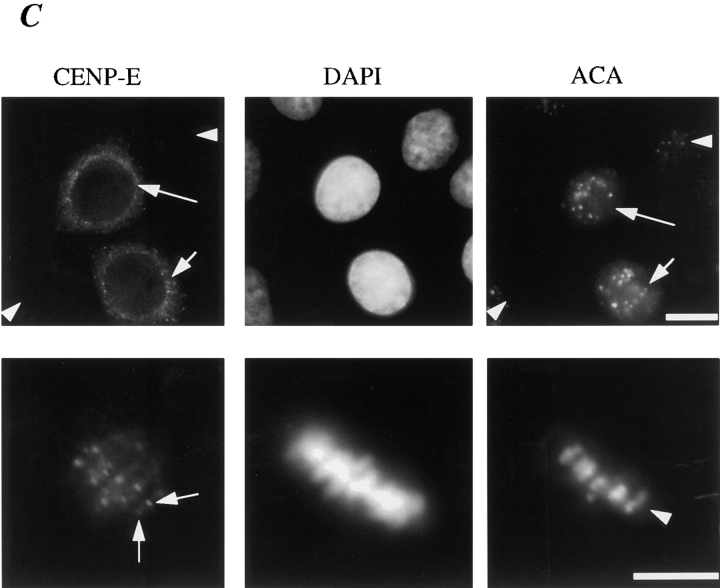
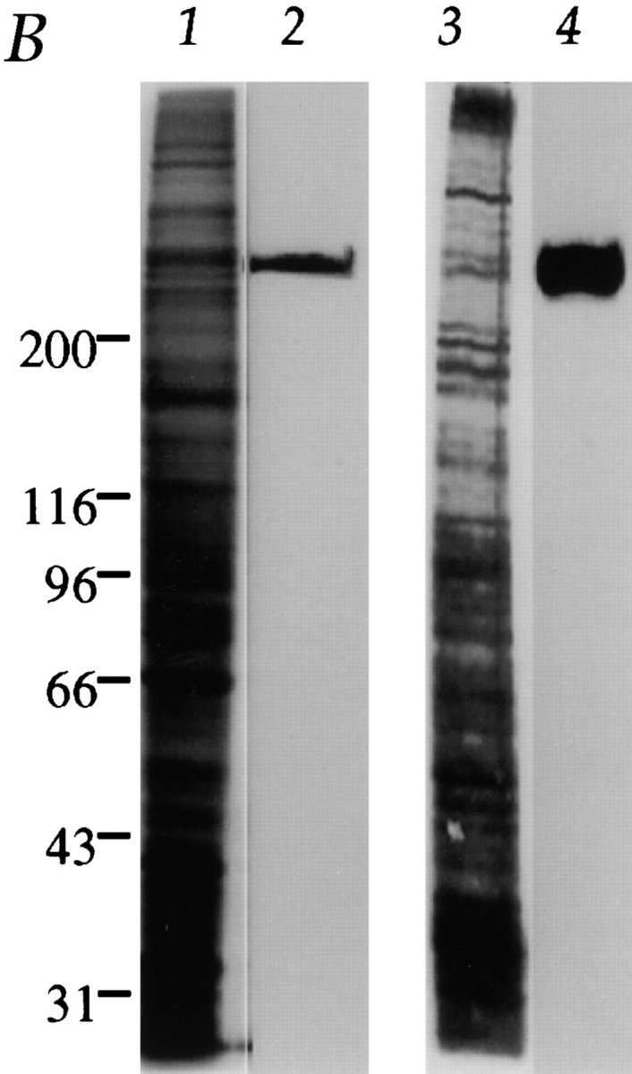
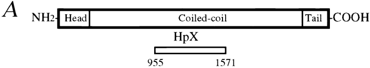
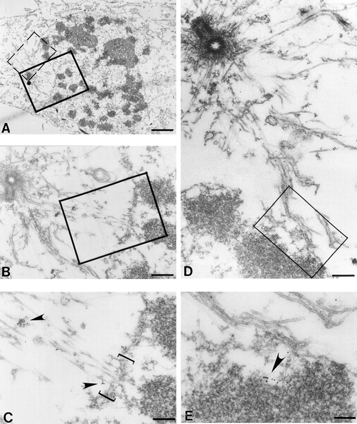
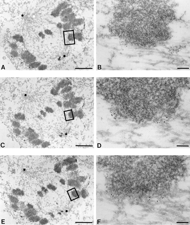
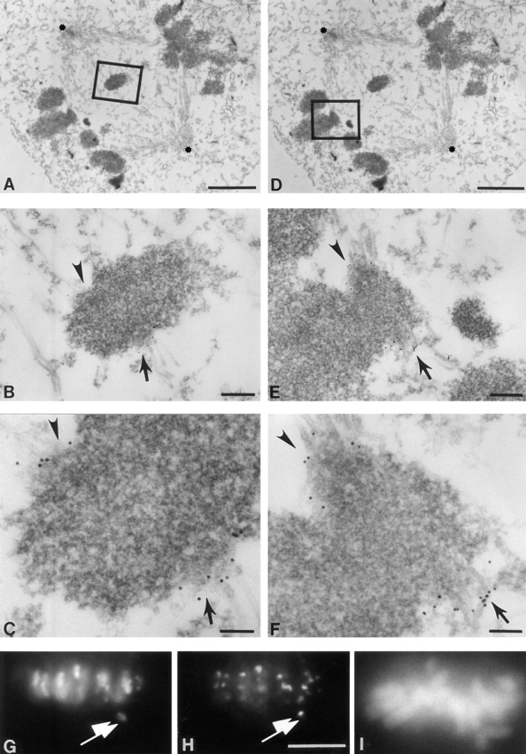
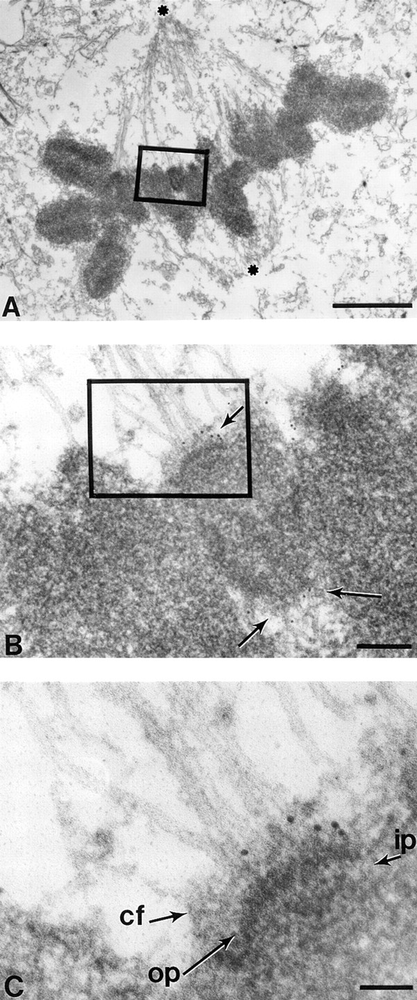
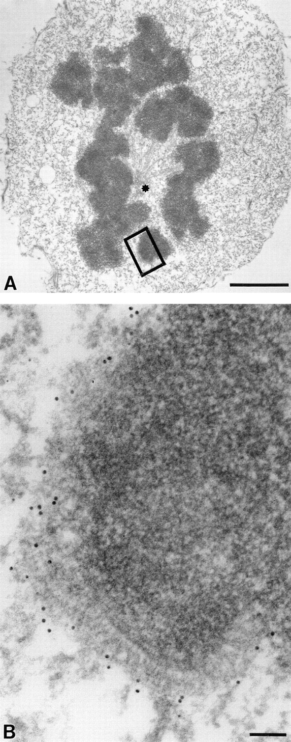
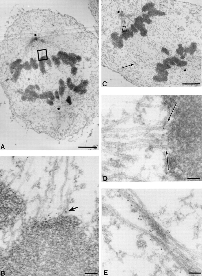
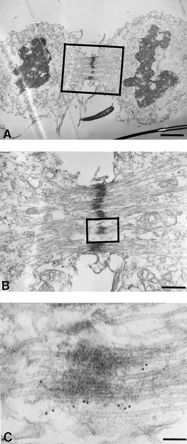
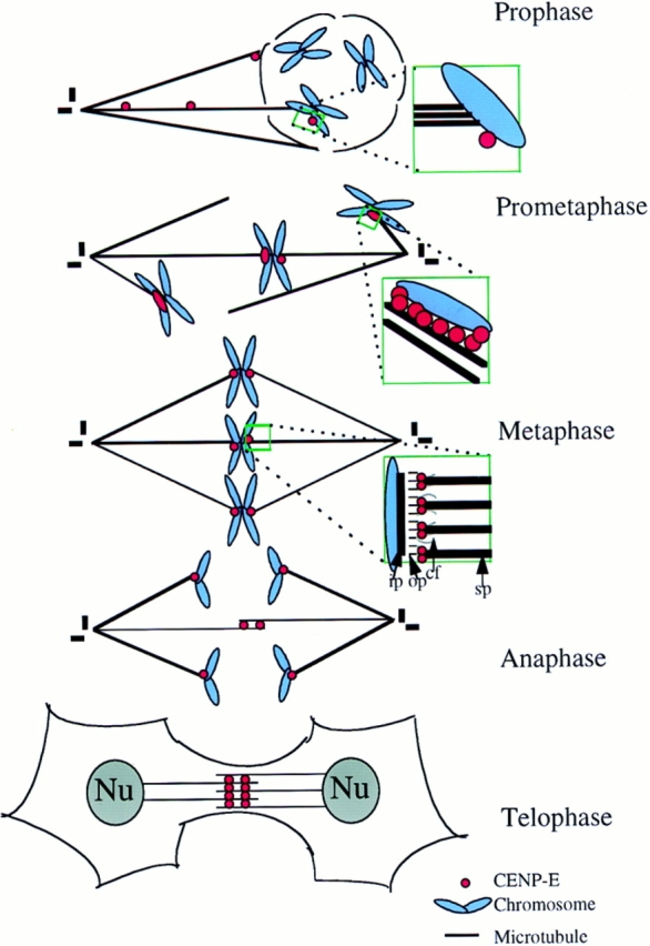
Centromere-associated protein E (CENP-E) is a kinesin-related microtubule motor protein that is essential for chromosome congression during mitosis. Using immunoelectron microscopy, CENP-E is shown to be an integral component of the kinetochore corona fibers that tether centromeres to the spindle. Immediately upon nuclear envelope fragmentation, an associated plus end motor trafficks cytoplasmic CENP-E toward chromosomes along astral microtubules that enter the nuclear volume. Before or concurrently with initial lateral attachment of spindle microtubules, CENP-E targets to the outermost region of the developing kinetochores. After stable attachment, throughout chromosome congression, at metaphase, and throughout anaphase A, CENP-E is a constituent of the corona fibers, extending at least 50 nm away from the kinetochore outer plate and intertwining with spindle microtubules. In congressing chromosomes, CENP-E is preferentially associated with (or accessible at) the stretched, leading kinetochore known to provide the primary power for chromosome movement. Taken together, this evidence strongly supports a model in which CENP-E functions in congression to tether kinetochores to the disassembling microtubule plus ends.


Figures

Figure 1
Figure 1
Characterization of the affinity-purified CENP-E antibody HpX. (A) Schematic drawing of CENP-E denoting the region used to generate HpX, a fragment of 70 kD recombinant polypeptide expressed in bacteria. (B) Specificity of affinity-purified HpX antibody. Immunoblots of mitotic whole cell lysates (lane 2, 50 μg) and isolated chromosome scaffold (lane 4, 25 μg). The same materials were separated in SDS-PAGE and stained with Coomassie blue (lanes 1 and 3). (C) Upper panels: CENP-E is accumulated in the cytoplasm just before nuclear envelope breakdown. Indirect immunofluorescence image of HeLa cells stained with HpX antibody (upper left), DAPI (upper middle) and human CREST sera (upper right). CREST sera stained centromeres in both early interphase cell (arrowheads) and late interphase cells (arrows). CENP-E signal appeared only in the late interphase cells (upper left, arrows). Interphase nuclei lack CENP-E staining (arrowheads). Lower panels: CENP-E is located to kinetochores as pairs of clearly resolved double dots (lower left, arrows), while CREST sera mark centromeres as unresolved dots (lower right, arrowhead). Bars: (upper panels) 20 μm; (lower panels) 10 μm.
Figure 1
Figure 1
Characterization of the affinity-purified CENP-E antibody HpX. (A) Schematic drawing of CENP-E denoting the region used to generate HpX, a fragment of 70 kD recombinant polypeptide expressed in bacteria. (B) Specificity of affinity-purified HpX antibody. Immunoblots of mitotic whole cell lysates (lane 2, 50 μg) and isolated chromosome scaffold (lane 4, 25 μg). The same materials were separated in SDS-PAGE and stained with Coomassie blue (lanes 1 and 3). (C) Upper panels: CENP-E is accumulated in the cytoplasm just before nuclear envelope breakdown. Indirect immunofluorescence image of HeLa cells stained with HpX antibody (upper left), DAPI (upper middle) and human CREST sera (upper right). CREST sera stained centromeres in both early interphase cell (arrowheads) and late interphase cells (arrows). CENP-E signal appeared only in the late interphase cells (upper left, arrows). Interphase nuclei lack CENP-E staining (arrowheads). Lower panels: CENP-E is located to kinetochores as pairs of clearly resolved double dots (lower left, arrows), while CREST sera mark centromeres as unresolved dots (lower right, arrowhead). Bars: (upper panels) 20 μm; (lower panels) 10 μm.
Figure 2
Figure 2
An associated plus end motor activity trafficks CENP-E along newly assembled astral microtubules into the nuclear domain after nuclear envelope fragmentation. HeLa cells were processed as described in Materials and Methods. (A) Low magnification view of a prophase/prometaphase HeLa cell bearing condensed chromosomes and a partially fragmented nuclear envelope. One spindle pole is readily apparent (asterisk). Examination of serial sections did not reveal another pole, consistent with a prophase cell before centriole separation. (B) Magnified view of boxed region in A shows that astral microtubules emanating from the centriole come in close proximity to the nuclear envelope. (C) Higher magnification view reveals that CENP-E is microtubule-associated along astral microtubules adjacent to the remaining nuclear envelope (bracket). Arrowheads point to microtubule-bound gold particles reporting CENP-E location. (D) Magnified view of the dashed box in A and highlighting astral microtubules passing through the fragmented lamina and lying in close proximity to a chromosome. (E) Higher magnification of the area boxed in D, revealing that some CENP-E is found along the microtubules, but additional CENP-E is associated with a localized domain on the chromosome, presumably the developing kinetochore. Note the chromosome is not yet attached to microtubules. Bars: (A) 2 μm; (B) 400 nm; (C) 200 nm; (D) 800 nm; (E) 140 nm.
Figure 3
Figure 3
At early prometaphase, CENP-E binds all long the outermost surface of monooriented kinetochores attached laterally to spindle microtubules. HeLa cells grown on coverslips were preextracted and fixed. The visualization of CENP-E was achieved by 10-nm gold–conjugated goat anti–rabbit IgG. (A, C, and E) Low magnification serial sections of an early prometaphase HeLa cell. Asterisks denote the two poles of the developing bipolar spindle. An apparently monooriented chromosome is boxed, and higher power views are shown in B, D, and F. 10-nm gold particles representing CENP-E position decorate the interface between immature kinetochore and the laterally attached spindle microtubules. Note the labeling of CENP-E on the kinetochore appears as a crescent (C) shape. Bars: (A, C, and E) 2 μm; (B) 160 nm; (D and F) 110 nm.
Figure 4
Figure 4
The leading kinetochore of a congressing chromosomes pair has increased level or accessibility of CENP-E. HeLa cells were processed as described in Fig. 2. (A and D) Low magnification views of a prometaphase HeLa cell (poles of the bipolar spindle are labeled with asterisks). (B and E) Intermediate magnification of two examples of a bioriented chromosome. (B) Boxed area of A showing chromosomes pair partially congressed from spindle poles toward the equator of spindle poles, but not yet aligned at the equator. (C and F) High magnification of the two bioriented chromosomes in B and E. 10-nm gold particles representing CENP-E decorate the outer kinetochore surface. A trilaminar structure of the kinetochore is not yet apparent, indicating that these kinetochores are not fully mature. (G–I) Double immunofluorescence demonstrating preferential CENP-E staining on the kinetochore closest to the midzone on a chromosome not yet congressed to the metaphase plate. (G) CENP-B, (H) CENP-E, and (I) DAPI to display chromosome positioning. Bars: (A and D) 2 μm; (B and E) 230 nm; (C) 90 nm; (F) 110 nm; (H–I) 10 μm.
Figure 5
Figure 5
At metaphase CENP-E extends from the kinetochore outer plate at least 50 nm along spindle microtubules. Low magnification view of a metaphase HeLa cell with chromosomes aligned at the equator between the spindle poles (asterisks). (B) Magnified view of one metaphase chromosome showing that spindle microtubules indeed associate with a kinetochore with a trilaminar structure. Five 10-nm gold particles are located to each sister kinetochore (arrows). Five additional gold particles just to the right of the boxed area represent CENP-E associated with the kinetochore of another chromosome (more clearly seen in adjacent sections). (C) Higher magnification view shows that CENP-E is located to the corona fibers of the kinetochore. op, outer plate; ip, inner plate; cf, corona fibers. Bars: (A) 2 μm; (B) 170 nm; (C) 70 nm.
Figure 6
Figure 6
CENP-E is a integral component of kinetochore corona fibers extending 50 nm from the outer plate. HeLa cells were initially treated with a low dose of nocodazole for 12 h to disassemble microtubules and processed as described in Fig. 2. Nocodazole treatment bulges the kinetochore outward from the surface of the chromosome. (A) Low magnification view of a nocodazole-treated prometaphase HeLa cell. The asterisk denotes one spindle pole. Note that not all spindle microtubules are depolymerized. Microtubule-free kinetochores appeared swollen and crescent shaped. (B) Magnified view of boxed area in A shows that 33 gold particles representing CENP-E position are located to the tip of the enlarged kinetochore outer plate, which appears to consist of tightly packed fibers. Bars: (A) 2 μm; (B) 85 nm.
Figure 7
Figure 7
CENP-E remains a component of the kinetochore fibers as sister chromatids move toward the poles in anaphase. HeLa cells were processed as described in Fig. 2. (A) Low magnification view of an early anaphase HeLa cell. The two spindle pole positions are marked with asterisks (one is apparent while another is in a different section). (B) Magnified view of a kinetochore–microtubule interface shows that CENP-E is located between the kinetochore outer plate and the spindle microtubules (arrow). No gold particles are seen in other regions on chromosomes or on microtubules. (C) Low magnification view of a late anaphase HeLa cell bearing elongated spindle poles, labeled with asterisks; one is apparent while another is in a different section. Interzonal microtubules are readily seen (arrow). (D) Magnified view of the upper boxed region in C, showing that CENP-E is located between a kinetochore and its associated spindle microtubules (arrow). Examination of the number of particles revealed a reduction relative to metaphase. (E) Magnified view of area pointed by the arrow in C. Some CENP-E is now localized to the interzonal microtubules. Bars: (A) 2 μm; (B) 120 nm; (C) 2 μm; (D) 70 nm; (E) 90 nm.
Figure 8
Figure 8
CENP-E cross-links the interzonal microtubules during telophase. HeLa cells were processed as described in Fig. 2. (A) Low magnification view of a late telophase HeLa cell. Lamin deposition to reform nuclei is partially complete. (B) Magnified view of boxed area in A shows that CENP-E is located along and/ or between the interzonal microtubules (boxed). (C) Higher magnification of interzonal microtubules shows that gold particles are primarily located between the microtubule bundles. Bars: (A) 2 μm; (B) 500 nm; (C) 90 nm.
Figure 9
Figure 9
Model for CENP-E function in chromosome movements. CENP-E is recruited to the immature kinetochore as soon as the nuclear envelope disassembles. CENP-E localizes to kinetochores before stable microtubule attachment, apparently by trafficking to the kinetochore by movement over astral microtubules. CENP-E is situated on the outermost surface of the kinetochore during initial lateral microtubule attachment. After biorientation, CENP-E remains on the corona fibers that link kinetochores in an apparent end-on interaction with spindle microtubules. CENP-E abundance, conformation, or accessibility is increased on the leading kinetochore of a congressing pair of chromosomes. Upon the sister chromatid separation, CENP-E remains kinetochore-associated and leads the poleward-moving chromosome. Once the chromosomes have reached the poles, CENP-E releases and redistributes to midzone where it may stabilize interzonal microtubules or power microtubule sliding that leads to pole-to-pole separation. op, outer plate; ip, inner plate; cf, corona fibers; sp, spindle microtubules.

Similar articles

Cited by

References

    1. Brinkley BR, Stubblefield E. The fine structure of the kinetochore of a mammalian cell in vitro. . Chromosoma (Berl) 1966;19:28–43. - PubMed
    1. Brown KD, Coulson RM, Yen TJ, Cleveland DW. Cyclin-like accumulation and loss of the putative kinetochore motor CENP-E results from coupling continuous synthesis with specific degradation at the end of mitosis. J Cell Biol. 1994;125:1303–1312. - PMC - PubMed
    1. Brown KD, Wood KW, Cleveland DW. The kinesin-like protein CENP-E is kinetochore-associated thoughout poleward chromosome segregation during anaphase-A. J Cell Sci. 1996;109:961–969. - PubMed
    1. Chen R-H, Waters JC, Salmon ED, Murray AW. Association of spindle assembly checkpoint component XMAD2 with unattached kinetochores. Science (Wash DC) 1996;274:242–246. - PubMed
    1. Comings DE, Okada TA. Holocentric chromosomes in oncopettus kinetochore plates are present in mitosis but absent in meiosis. Chromosoma (Berl) 1973;37:177–192. - PubMed

Publication types