. 1997 Oct 20;139(2):497-506.
doi: 10.1083/jcb.139.2.497.

ATP- and gap junction-dependent intercellular calcium signaling in osteoblastic cells

Affiliations

ATP- and gap junction-dependent intercellular calcium signaling in osteoblastic cells

N R Jorgensen et al. J Cell Biol. .

Abstract

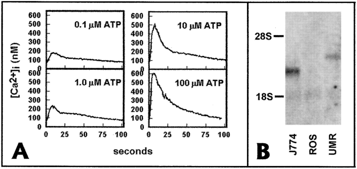
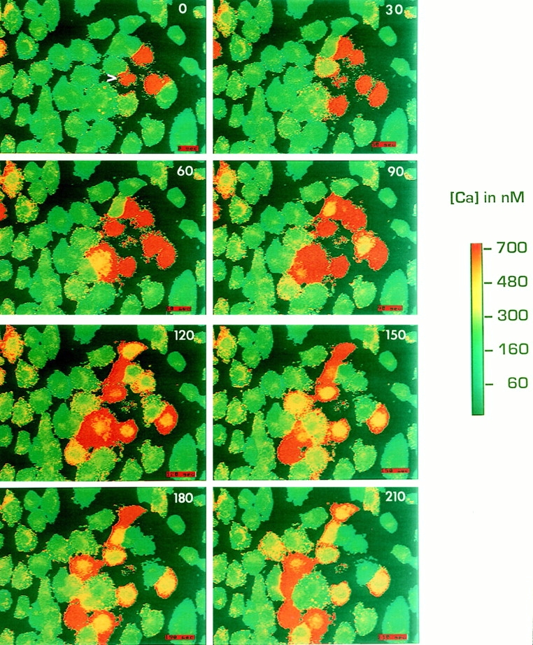
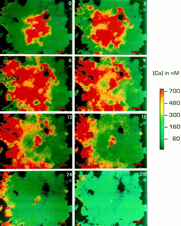
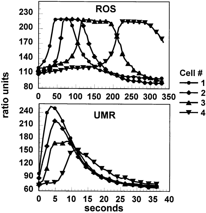
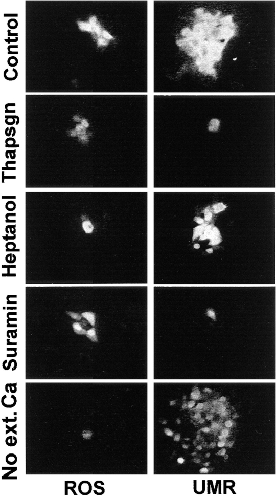
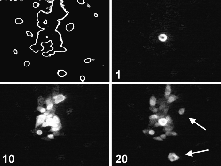
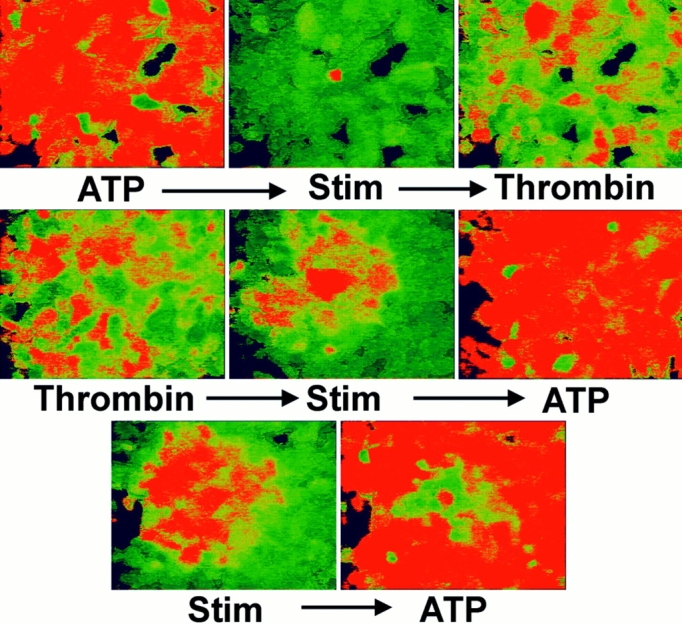
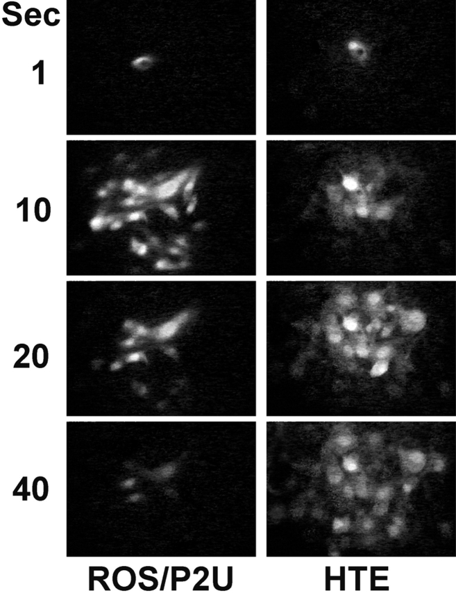
Many cells coordinate their activities by transmitting rises in intracellular calcium from cell to cell. In nonexcitable cells, there are currently two models for intercellular calcium wave propagation, both of which involve release of inositol trisphosphate (IP3)- sensitive intracellular calcium stores. In one model, IP3 traverses gap junctions and initiates the release of intracellular calcium stores in neighboring cells. Alternatively, calcium waves may be mediated not by gap junctional communication, but rather by autocrine activity of secreted ATP on P2 purinergic receptors. We studied mechanically induced calcium waves in two rat osteosarcoma cell lines that differ in the gap junction proteins they express, in their ability to pass microinjected dye from cell to cell, and in their expression of P2Y2 (P2U) purinergic receptors. ROS 17/2.8 cells, which express the gap junction protein connexin43 (Cx43), are well dye coupled, and lack P2U receptors, transmitted slow gap junction-dependent calcium waves that did not require release of intracellular calcium stores. UMR 106-01 cells predominantly express the gap junction protein connexin 45 (Cx45), are poorly dye coupled, and express P2U receptors; they propagated fast calcium waves that required release of intracellular calcium stores and activation of P2U purinergic receptors, but not gap junctional communication. ROS/P2U transfectants and UMR/Cx43 transfectants expressed both types of calcium waves. Gap junction-independent, ATP-dependent intercellular calcium waves were also seen in hamster tracheal epithelia cells. These studies demonstrate that activation of P2U purinergic receptors can propagate intercellular calcium, and describe a novel Cx43-dependent mechanism for calcium wave propagation that does not require release of intracellular calcium stores by IP3. These studies suggest that gap junction communication mediated by either Cx43 or Cx45 does not allow passage of IP3 well enough to elicit release of intracellular calcium stores in neighboring cells.


Figures

Figure 1
Figure 1
UMR cells, but not ROS cells express P2U receptor mRNA. (A) Calcium transients elicited by different concentrations of ATP in UMR cells. Fura-2–loaded UMR cells were placed in a stirred cuvette at 37°C, and 340:380 ratios were obtained. (B) Total cell RNA (10 μg) from ROS, UMR, and the mouse macrophage cell line J774 was electrophoresed on agarose-formaldehyde gel, transferred to nylon, and hybridized with a radiolabeled cDNA probe for the human P2U receptor. Both J774 cells and UMR cells expressed a hybridizing band of the appropriate size for mouse and rat P2U, respectively, but no band was detected in ROS cell RNA.
Figure 2
Figure 2
ROS cells propagate slow intercellular calcium waves. Monolayers of ROS cells were loaded with fura-2, and a single cell (arrowhead) was mechanically stimulated during fluorescence ratio imaging. Time after stimulation in seconds is indicated on each panel. The pseudocolor map represents the estimated [Ca2+]i.
Figure 3
Figure 3
UMR cells propagate fast intercellular calcium waves. Methods as in Fig. 2.
Figure 4
Figure 4
Kinetics of calcium wave propagation in ROS and UMR cells. Four cells in a row from the stimulated cells were analyzed from the sequences shown in Figs. 2 and 3. Data are presented as relative fluorescence ratio.
Figure 5
Figure 5
Inhibition of calcium waves in ROS and UMR cells. Adherent cells were loaded with fluo-3, and thapsigargin, heptanol, or suramin was added, or extracellular calcium was removed, as described in the text. Intercellular calcium waves were initiated by mechanical stimulation. Panels were obtained by subtracting a frame taken immediately before mechanical stimulation from a frame taken at the time of maximal wave propagation. Note that, as described in the text, heptanol blocks calcium wave propagation in UMR cells in many cases, and this occurs when heptanol also inhibits ATP-induced calcium transients. The panel shown is the typical response in experiments where ATP-mediated calcium transients remain intact after heptanol treatment.
Figure 6
Figure 6
Calcium waves are propagated among islands of UMR cells. Calcium waves were induced in a subconfluent monolayer of fluo-3–loaded UMR cells. The outline of cells in the field of view is shown in the upper left panel. A cell in the middle of the cell island was stimulated, and images were taken at intervals. Cells that propagated the calcium wave but were not in physical contact with the stimulated cell are indicated by arrows. Time after stimulation in s is shown on each panel.
Figure 7
Figure 7
Desensitization of P2U receptors inhibits calcium waves in UMR cells. Ratio imaging was performed on fura-2–loaded UMR cell monolayers. Top panels: left, 1 mM ATP-elicited calcium transients; center, subsequent mechanical stimulation failed to induce intercellular calcium waves; right, subsequent challenge with 20 U/ml thrombin revealed that calcium stores were not depleted. Middle panels: left, thrombin-induced calcium transients; center, subsequent mechanical stimulation induced a calcium wave; right, subsequent addition of ATP induced calcium transients. Bottom panels: propagated calcium waves (left) frequently desensitized a local area to subsequent addition of ATP (right).
Figure 8
Figure 8
Calcium waves in ROS/P2U transfectants and HTE cells. ROS/P2U transfectants (left) and HTE cells (right) were loaded with fluo-3 and images were taken after a mechanical stimulation of a single cell. Panels are images taken at the indicated time after subtraction of the prestimulation fluo-3 image.

References

    1. Babich M, Choi H, Johnson RM, King KL, Alford GE, Nissenson RA. Thrombin and parathyroid hormone mobilize intracellular calcium in rat osteosarcoma cells by distinct pathways. Endocrinology. 1991;129:1463–1470. - PubMed
    1. Boitano S, Dirksen ER, Sanderson MJ. Intercellular propagation of calcium waves mediated by inositol trisphosphate. Science (Wash DC) 1992;258:292–295. - PubMed
    1. Cao D, Lin G, Westphale EM, Beyer EC, Steinberg TH. Mechanisms for the coordination of intercellular calcium signaling in insulin-secreting cells. J Cell Sci. 1997;110:497–504. - PubMed
    1. Charles AC, Merrill JE, Dirksen ER, Sanderson MJ. Intercellular signaling in glial cells: calcium waves and oscillations in response to mechanical stimulation and glutamate. Neuron. 1991;6:983–992. - PubMed
    1. Charles AC, Naus CCG, Zhu D, Kidder GM, Dirksen ER, Sanderson MJ. Intercellular calcium signaling via gap junctions in glioma cells. J Cell Biol. 1992;118:195–201. - PMC - PubMed

Publication types

MeSH terms