. 1997 Oct 20;139(2):541-52.
doi: 10.1083/jcb.139.2.541.

Expression of a truncated, kinase-defective TGF-beta type II receptor in mouse skeletal tissue promotes terminal chondrocyte differentiation and osteoarthritis

Affiliations

Expression of a truncated, kinase-defective TGF-beta type II receptor in mouse skeletal tissue promotes terminal chondrocyte differentiation and osteoarthritis

R Serra et al. J Cell Biol. .

Abstract

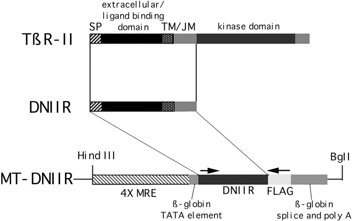
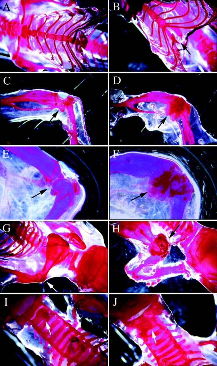
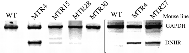
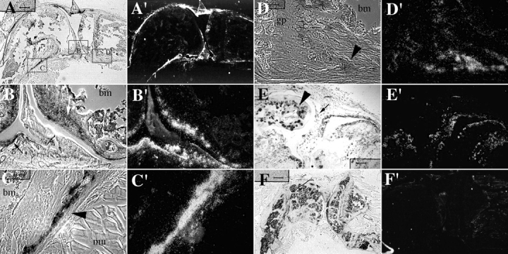
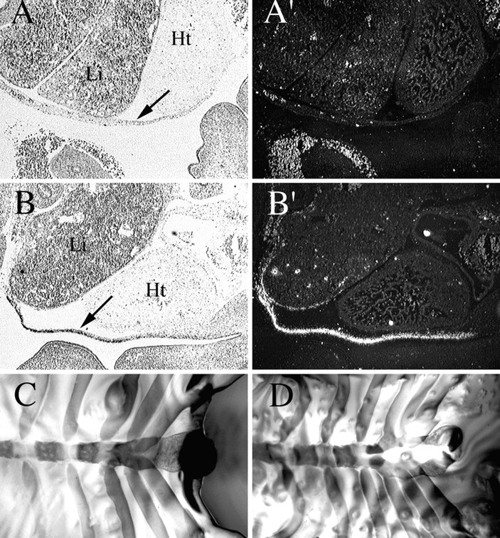
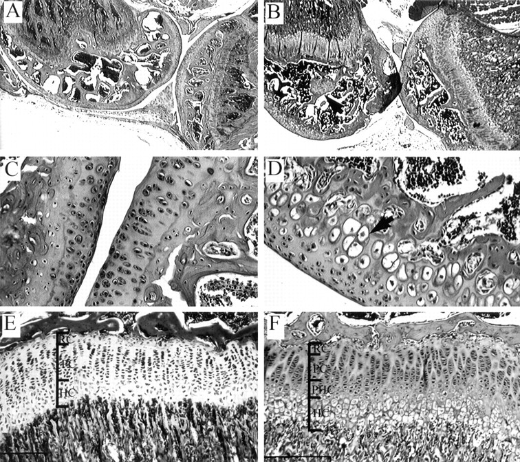
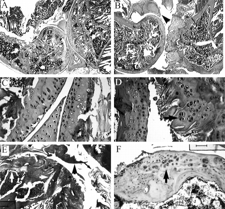
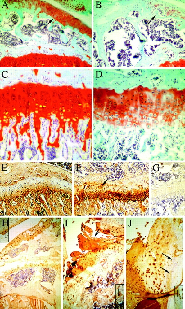
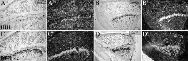
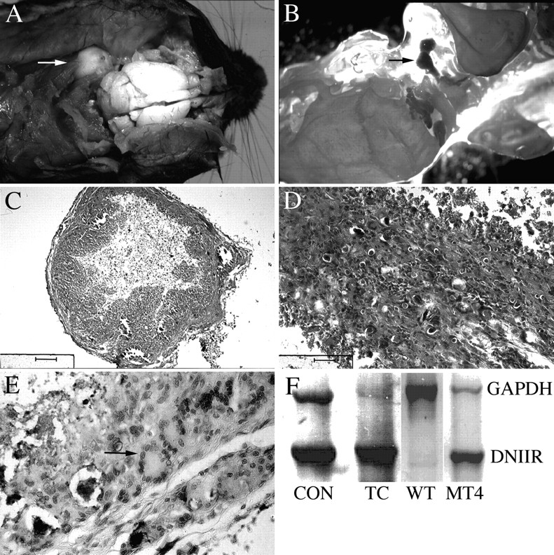
Members of the TGF-beta superfamily are important regulators of skeletal development. TGF-betas signal through heteromeric type I and type II receptor serine/threonine kinases. When over-expressed, a cytoplasmically truncated type II receptor can compete with the endogenous receptors for complex formation, thereby acting as a dominant-negative mutant (DNIIR). To determine the role of TGF-betas in the development and maintenance of the skeleton, we have generated transgenic mice (MT-DNIIR-4 and -27) that express the DNIIR in skeletal tissue. DNIIR mRNA expression was localized to the periosteum/perichondrium, syno-vium, and articular cartilage. Lower levels of DNIIR mRNA were detected in growth plate cartilage. Transgenic mice frequently showed bifurcation of the xiphoid process and sternum. They also developed progressive skeletal degeneration, resulting by 4 to 8 mo of age in kyphoscoliosis and stiff and torqued joints. The histology of affected joints strongly resembled human osteo-arthritis. The articular surface was replaced by bone or hypertrophic cartilage as judged by the expression of type X collagen, a marker of hypertrophic cartilage normally absent from articular cartilage. The synovium was hyperplastic, and cartilaginous metaplasia was observed in the joint space. We then tested the hypothesis that TGF-beta is required for normal differentiation of cartilage in vivo. By 4 and 8 wk of age, the level of type X collagen was increased in growth plate cartilage of transgenic mice relative to wild-type controls. Less proteoglycan staining was detected in the growth plate and articular cartilage matrix of transgenic mice. Mice that express DNIIR in skeletal tissue also demonstrated increased Indian hedgehog (IHH) expression. IHH is a secreted protein that is expressed in chondrocytes that are committed to becoming hypertrophic. It is thought to be involved in a feedback loop that signals through the periosteum/ perichondrium to inhibit cartilage differentiation. The data suggest that TGF-beta may be critical for multifaceted maintenance of synovial joints. Loss of responsiveness to TGF-beta promotes chondrocyte terminal differentiation and results in development of degenerative joint disease resembling osteoarthritis in humans.


Figures

Figure 1
Figure 1
Map of the MT-DNIIR expression plasmid. The EcoRI/XbaI fragment of the human TGF-β type II receptor from the plasmid p102 containing a FLAG epitope tag and the signal sequence (SP), ligand binding, transmembrane (TM), and juxtamembrane (JM) domains of the receptor was inserted into the BamHI site of the MT-β metallothionein expression vector by blunt end ligation. The MT-β vector contains four metal responsive elements and the β globin TATA element, splice sites, and polyadenylation signal (69). The HindIII/BglI fragment was injected into single cell embryos. Arrows mark the location of primer sequences used for PCR and RT-PCR analysis.
Figure 2
Figure 2
Skeletal defects in MT-DNIIR transgenic mice. Photographs of alizarin red whole mount skeletal preparations from adult wild-type (A, C, E, G, and I) MTR-DNIIR-4 (B, D, H, and J), and MT-DNIIR-27 (F) mice. Arrows point to xiphoid process (A and B), knee (C–F), shoulder (G and H) joints, and cervical vertebrae (I and J).
Figure 3
Figure 3
RT-PCR analysis of DNIIR mRNA expression in MT-DNIIR transgenic mouse lines. RNA isolated and pooled from the hind limbs of two to four wild-type (WT) or transgenic (MT-DNIIR-4, -15, -27, -28, -30) mice maintained on normal food and tap water was analyzed by RT-PCR. To specifically amplify the truncated DNIIR cDNA, primers targeted to FLAG epitope sequences were used (Fig. 1). Amplifications of GAPDH was used as an internal control. Two separate assays are shown separated by the black bar.
Figure 4
Figure 4
Localization of DNIIR mRNA in skeletal tissue. Sections of knee joints from 8-wk MT-DNIIR transgenic mice were hybridized to an 35S-labeled antisense DNIIR riboprobe (A–D). Boxes shown in A delineate the approximate locations on the joints shown in (B–D). DNIIR expression was detected in the articular cartilage (B, white arrow), synovium (B, black arrow), and periosteum/perichondrium (C and D, black arrowhead) of transgenic mice. Representative images from analysis of two different mice are shown. In 6-mo-old transgenic mice, DNIIR expression was detected in hyperplastic synovium (E, small, black arrow) surrounding areas of cartilage metaplasia (E, large, black arrowhead). A representative image from two separate mice is shown. No hybridization was detected in wild-type tissue (F) or in transgenic mice with an 35S-labeled sense riboprobe (data not shown). Toluidine blue–stained bright field (A–F) and dark field (A′–F′) images are shown. Bars: (A and F) 400 μm; (B–E) 100 μm.
Figure 5
Figure 5
Expression of DNIIR mRNA in embryos. Sections of 12.5-d post-coital wild-type (A) and MTR-DNIIR transgenic (B) embryos were hybridized to an 35S-labeled DNIIR riboprobe. DNIIR nRNA expression was detected in the mesenchyme of the thoracic body wall (A and B, arrow) in MT-DNIIR transgenic but not wild-type embryos at the time the sternum begins to develop. Toluidine blue–stained bright field (A and B) and dark field (A′ and B′) images are shown. Li, liver, Ht, heart. Bifurcated sternum in 17.5-d post-coital MT-DNIIR-4 mice. Alizarin red/alcian blue-stained skeletal preparations of wild-type (C) and MT-DNIIR transgenic (D) embryos at 17.5 d post coitus. Bar, 200 μm.
Figure 6
Figure 6
Knee joint histology in young MT-DNIIR mice. Images of hematoxylin- and eosin-stained sections from wild-type (A, C, and E) and MT-DNIIR (B, D, and F) knee joints at 4 wk of age. Disorganized cartilage islands were often observed in the transgenic epiphysis (B, black arrowhead). Hypertrophic cells were located in the deep zones of the articular cartilage in transgenic (D) but not wild-type (C) mice. Resting (RC), proliferating (PC), and hypertrophic (HC) cells were easily detectable in the wild-type growth plate (E). In transgenic mice, the histology of the growth plate was altered. The hypertrophic zone was thicker, and two distinct cell populations were observed, hypertrophic cells (HC) and smaller, round prehypertrophic cells (PHC). Bars: (A and B) 400 μm; (C and D) 50 μm; (E and F) 77 μm.
Figure 7
Figure 7
Knee joint histology in older MT-DNIIR mice. Joints from 6-mo-old transgenic mice with joint damage (B and D–F) and from wild-type mice (A and C) are shown. Cartilage was observed in the joint space (B and E, arrowheads) and the synovium was hyperplastic (E, arrow) in transgenic mice. The articular surface of wild-type mice was smooth and organized (C). In transgenic mice, the articular surface was fibrillated and chondrocytes were grouped into clusters (D, arrow). Early osteophytes were also present on the articular surface (F). The growth plate was often undetectable or highly disorganized (E, white arrows) relative to the wild-type growth plate (A). Bars: (A and B) 400 μm; (E) 270 μm; (C, D, and F) 50 μm.
Figure 8
Figure 8
Localization of proteoglycans and type X collagen in the knee joint. Sections from 8-wk-old wild-type (A and C) and MT-DNIIR (B and D) knee joints stained with safranine O (A–D). Images at 150× (A and B) show staining in the articular surface (arrows). Images C and D focus on staining in the growth plate. There was intense proteoglycan staining in the articular cartilage of wild-type mice (A) while staining was less intense and patchy on the articular surface of transgenic mice (B). Staining was also less intense in the transgenic growth plate (D) relative to the wild-type growth plate (C). Sections from 8 wk (E–G) and 6-mo-old (H–J) wild-type (E and H) and MT-DNIIR (F, G, I, and J) were used for immunohistochemical staining of type X collagen (E–J). There was increased type X collagen staining in the transgenic growth plate at 8 wk (F) relative to wild-type controls (E). Intracellular staining was detected in transgenic chondrocytes in the upper zones of the growth plate (F, arrow). Type X collagen immunoreactivity was not readily detectable in articular cartilage from wild-type mice at 6 mo of age (H) but was detected in fibrillated cartilage (I, arrow) from older (6 mo) transgenic mice. Chondrocytes in osteophytes also stained for type X collagen (J). Arrows represent the original joint lining (J). No staining was detected in the absence of primary anitbody (G). Bars: (A and B) 66 μm; (C and D) 25 μm; (E and F) 66 μm; (G) 200 μm; (H and I) 100 μm; (J) 50 μm.
Figure 9
Figure 9
IHH and PTH receptor expression. Sections of knee joints from 8 wk wild-type (A and C) and MT-DNIIR transgenic (B and D) mice were hybridized to 35S-labeled antisense IHH (A and B) and PTH receptor (C and D) riboprobes. Bars, 100 μm.
Figure 10
Figure 10
Tumoral calcinosis-like lesions in MT-DNIIR transgenic mice. Photograph of a large tumoral calcinosis-like lesion (arrow) from the cervical vertebrae of an MT-DNIIR transgenic mouse (A). Whole mount skeletal preparation showing alizarin red-stained tumoral carcinosis-like lesion (arrow) from the cervical vertebrae of an MT-DNIIR transgenic mouse (B). Images of hematoxylin- and eosin-stained sections from tumoral calcinosis lesion (C–E). The tumors were encapsulated and necrotic in the center (C). The tumors consisted primarily of poorly differentiated mesencyme (B) and large multinucleated giant cells (E). Expression of DNIIR mRNA in tumoral calcinosis lesions (F). RNA from Mv1Lu cells that expressed the truncated receptor (CON), a tumoral calcinosis lesion (TC), and the hind limb of a wild-type (WT) or MT-DNIIR-4 (MT4) transgenic mice was used in RT-PCR analysis. Amplification of GAPDH cDNA was used as an internal control. The tumoral calcinosis lesion expressed high levels of the DNIIR mRNA. Bars: (C) 200 μm; (D) 100 μm; (E) 50 μm.

References

    1. Allen JB, Manthey CL, Hand AR, Ohura K, Ellingsworth L, Wahl SM. Rapid onset of synovial inflammation and hyperplasia induced by transforming growth factor β. J Exp Med. 1990;171:231–247. - PMC - PubMed
    1. Amizuka N, Warshawsky H, Henderson JE, Goltzman D, Karaplis AC. Parathyroid hormone-related peptide-depleted mice show abnormal epiphyseal cartilage development and altered endochondral bone formation. J Cell Biol. 1994;126:1611–1623. - PMC - PubMed
    1. Amling M, Neff L, Tanaka S, Inoue D, Kuida K, Weir E, Philbrick WM, Broadus AE, Baron R. Bcl-2 lies downstream of parathyroid hormone-related peptide in a signaling pathway that regulates chondrocyte maturation during skeletal development. J Cell Biol. 1997;136:205–213. - PMC - PubMed
    1. Ballock RT, Heydemann A, Wakefield LM, Flanders KC, Roberts AB, Sporn MB. TGF-β1 prevents hypertrophy of epiphyseal chondrocytes: regulation of gene expression for cartilage matrix proteins and metalloproteases. Dev Biol. 1993;158:414–429. - PubMed
    1. Bohme K, Winterhalter KH, Bruckner P. Terminal differentiation of chondrocytes in culture is a spontaneous process and is arrested by TGF-β2 and basic fibroblast growth factor in synergy. Exp Cell Res. 1995;216:191–198. - PubMed

Publication types

MeSH terms

Substances