. 1997 Oct 20;139(2):553-62.
doi: 10.1083/jcb.139.2.553.

Laminin alpha1 chain synthesis in the mouse developing lung: requirement for epithelial-mesenchymal contact and possible role in bronchial smooth muscle development

Affiliations

Laminin alpha1 chain synthesis in the mouse developing lung: requirement for epithelial-mesenchymal contact and possible role in bronchial smooth muscle development

L Schuger et al. J Cell Biol. .

Abstract

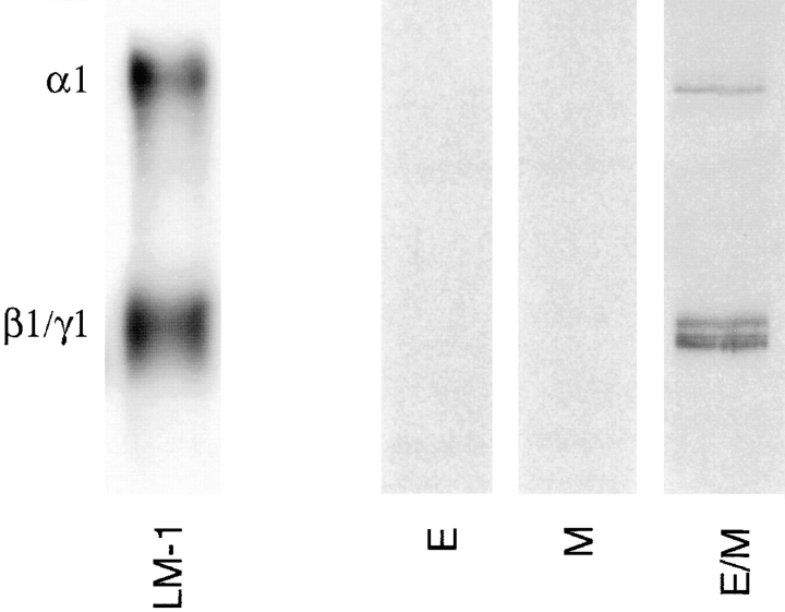
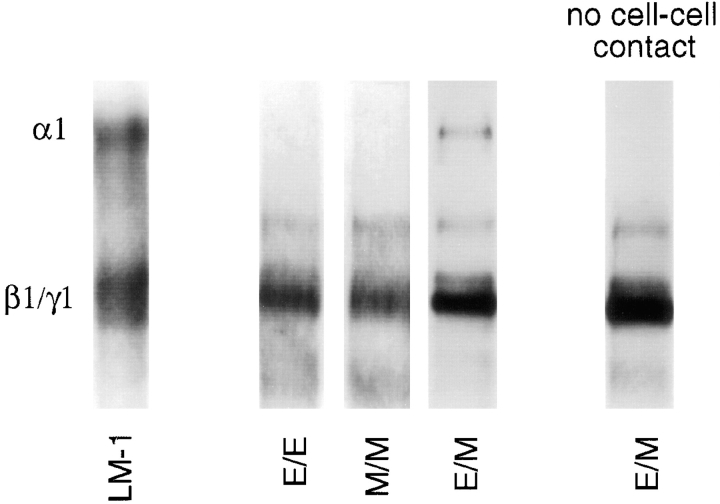
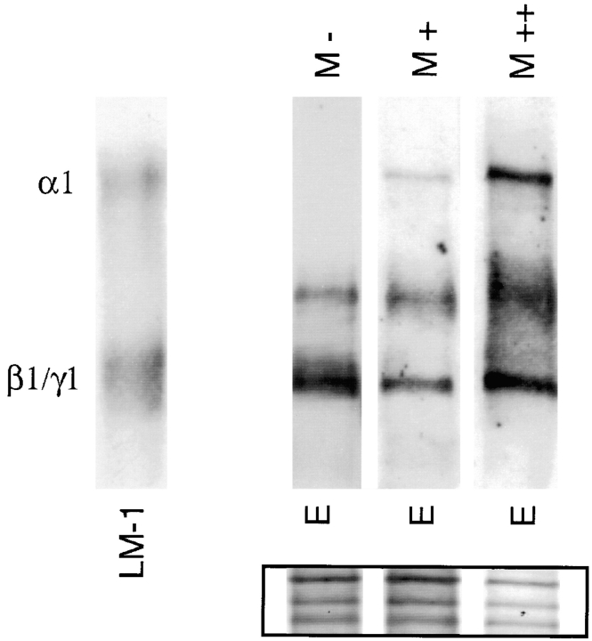
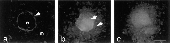
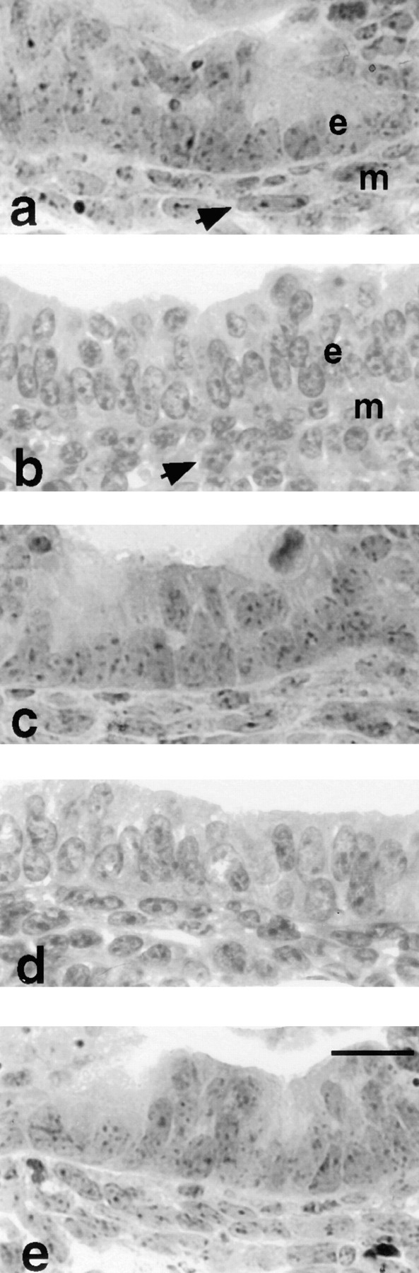
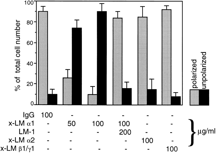
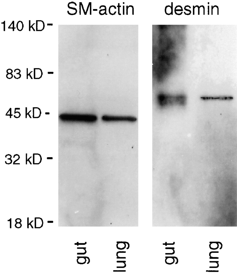
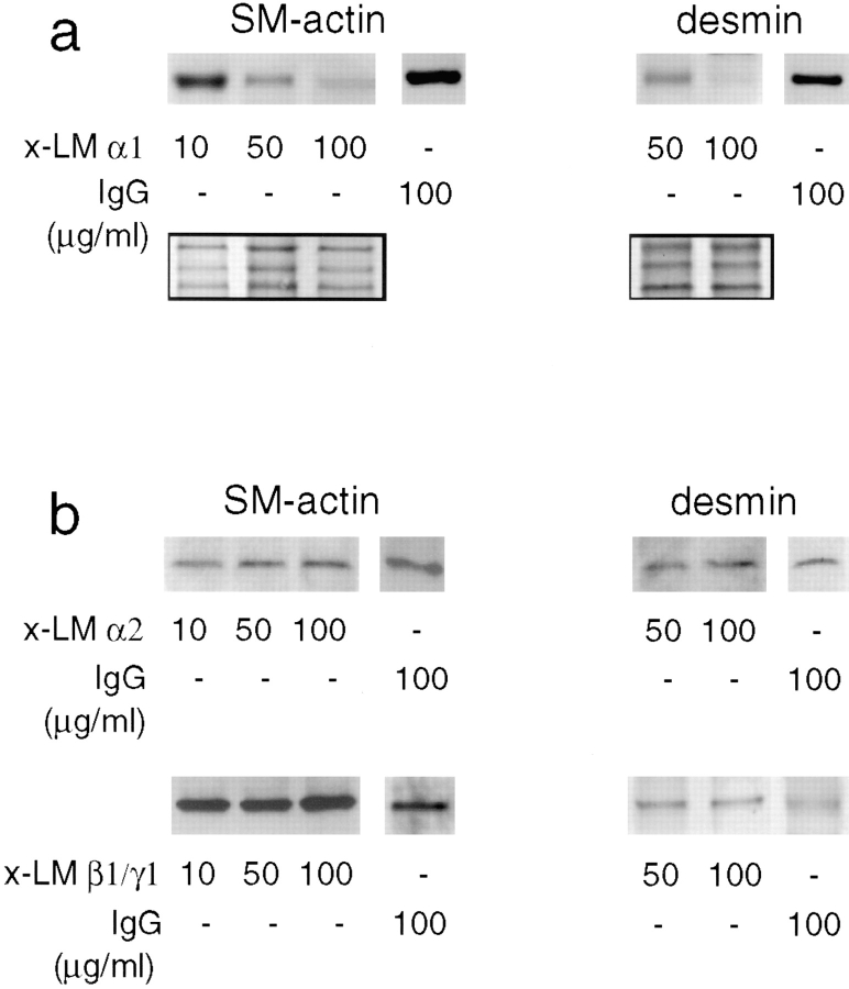
Laminins, the main components of basement membranes, are heterotrimers consisting of alpha, beta, and gamma polypeptide chains linked together by disulfide bonds. Laminins-1 and -2 are both composed of beta1 and gamma1 chains and differ from each other on their alpha chain, which is alpha1 and alpha2 for laminin-1 and -2, respectively. The present study shows that whereas laminins-1 and -2 are synthesized in the mouse developing lung and in epithelial-mesenchymal cocultures derived from it, epithelial and mesenchymal monocultures lose their ability to synthesize the laminin alpha1 chain. Synthesis of laminin alpha1 chain however returns upon re-establishment of epithelial-mesenchymal contact. Cell-cell contact is critical, since laminin alpha1 chain is not detected in monocultures exposed to coculture-conditioned medium or in epithelial-mesenchymal cocultures in which heterotypic cell-cell contact is prevented by an interposing filter. Immunohistochemical studies on cocultures treated with brefeldin A, an inhibitor of protein secretion, indicated both epithelial and mesenchymal cells synthesize laminin alpha1 chain upon heterotypic cell- cell contact. In a set of functional studies, embryonic lung explants were cultured in the presence of monoclonal antibodies to laminin alpha1, alpha2, and beta/gamma chains. Lung explants exposed to monoclonal antibodies to laminin alpha1 chain exhibited alterations in peribronchial cell shape and decreased smooth muscle development, as indicated by low levels of smooth muscle alpha actin and desmin. Taken together, our studies suggest that laminin alpha1 chain synthesis is regulated by epithelial-mesenchymal interaction and may play a role in airway smooth muscle development.


Figures

Figure 1
Figure 1
Immunoblots demonstrating the presence of LM α1, α2, β1, and γ1 chains in the developing lung (day 15 of gestation). A polyclonal antibody against EHS LM that recognizes α1, α2, β1, and γ1 chains (x-LM), a monoclonal antibody to the LM α1 chain (AL-4, x-LM α1), and a monoclonal antibody to LM α2 chain (4H8-2, x-LM α2) were used in these studies. LM-1 from the EHS tumor and embryonic heart (rich in LM-2) served as controls. Immunoblots with normal rabbit IgG or rat IgG did not detect any protein bands.
Figure 2
Figure 2
LM α1, α2, β1, and γ1 chains produced by epithelial– epithelial (E/E), mesenchymal–mesenchymal (M/M), and epithelial–mesenchymal (E/M) cocultures. A polyclonal antibody against EHS LM that recognizes α1, α2, β1, and γ1 chains was used for immunobloting. LM α1 chain is observed only in epithelial–mesenchymal cocultures. As controls, the first lane contained LM-1 from the EHS tumor and the second lane contained lung extract. Immunoblots with normal rabbit IgG or rat IgG did not detect any protein bands.
Figure 3
Figure 3
ELISA showing different levels of LM α1 chain synthesis in different cocultures. (Column 1) Epithelial–mesenchymal coculture established from mixed cell populations directly isolated from the lung. (Column 2) Epithelial–mesenchymal coculture established with cells from monocultures mixed in a 1:3 epithelial/mesenchymal ratio. (Column 3) Epithelial–mesenchymal coculture established by adding epithelial cells to a mesenchymal monolayer (both confluent at the time of determining LM-1 production). (Column 4) Epithelial–mesenchymal coculture established by adding mesenchymal cells to an epithelial monolayer (both confluent at the time of determining LM-1 production). The bars represent SD. The means and SD are based on quadruplicate examples in a single experiment. These were repeated three times with similar results.
Figure 4
Figure 4
Epithelial–mesenchymal organotypic coculture double-stained with anti-keratin antibodies by immunoperoxidase (a) and with anti-LM α1 chain antibody by immunofluorescence (b). The epithelial cells are recognized because they form round clusters and stain light brown with anti-keratin antibodies (not clearly appreciated in a black and white photograph). LM-1 is exclusively deposited at the epithelial–mesenchymal interface (arrows). (Inset) Day 12 lung stained with anti-LM α1 chain antibody shows LM-1 deposition restricted to the basement membrane alongside the bronchial tree. Bar: (a and b) 20 μm; (inset) 0.2 mm.
Figure 5
Figure 5
Immunoprecipitation of 35S-metabolically labeled LM-1 synthesized by epithelial (E) and mesenchymal (M) monocultures and epithelial–mesenchymal cocultures (E/M) using a monoclonal antibody to the LM α1 chain. Only epithelial–mesenchymal cocultures produced detectable levels of LM-1.
Figure 6
Figure 6
Immunoblots using a polyclonal antibody to LM-1 that recognizes α1, α2, β1, and γ1 chains. No LM α1 chain was detected in epithelial–epithelial (E/E) or mesenchymal–mesenchymal (M/M) cocultures, whereas LM α1 chain was seen in epithelial–mesenchymal (E/M) cocultures. LM α1 chain was not detected in the absence of epithelial–mesenchymal contact (monocultures separated by a filter), whereas α2, β1, and γ1 chains remained constant. The first lane is LM-1 from the EHS tumor, used as control.
Figure 7
Figure 7
Addition of mesenchymal cells to epithelial monocultures stimulates LM α1 chain synthesis. (M−) No mesenchymal cells; (M+) 1:2 epithelial/mesenchymal cell ratio; (M++) 1:4 epithelial/mesenchymal cell ratio. The first lane is LM-1 from the EHS tumor, used as control. The inset shows staining of the nitrocellulose membrane with 0.2% amido black after immunobloting to visualize the amount of protein loaded per lane.
Figure 8
Figure 8
Epithelial–mesenchymal organotypic cocultures exposed to 0 (a), 5 (b), and 10 (c) μg/ml of brefeldin A for 3 h, followed by immunostaining with anti-LM α1 chain antibody. e, Epithelial cell; m, mesenchymal cells. In untreated cocultures (a), LM α1 chain accumulates at the epithelial–mesenchymal interface (arrow). In brefeldin A-treated cocultures (b and c), LM α1 chain can be detected in the epithelial cells as well as in the mesenchymal cells surrounding them (b, arrows). Bar, 20 μm.
Figure 9
Figure 9
Photomicrographs of a main bronchus including peribronchial mesenchymal cells in lung explants cultured for 3 d in the presence of 100 μg/ml of normal mouse IgG (a), anti-LM α1 chain antibody (b), anti-LM α1 chain antibody preincubated with 200 μg/ml of LM-1 (c), anti-LM α2 antibody (d), and anti-LM β1/γ1 antibody (e). Note that in the control (a) the peribronchial mesenchymal cells (m) are polarized (elongated and oriented concentrically to the bronchus), whereas the peribronchial mesenchymal cells in explants exposed to anti-LM α1 chain antibody (b) are round. This effect was corrected by preincubation of the antibody with LM-1 (c) and was not observed in explants exposed to the other antibodies (d and e). The epithelial cells (e) showed no morphological alterations. Bar, 20 μm.
Figure 10
Figure 10
Histogram showing the percentage of polarized (elongated) and unpolarized (round) peribronchial cells in day 12 embryonic lung explants exposed for 3 d to various anti-LM antibodies and control immunoglobulin. The number of polarized (elongated) and unpolarized (round) peribronchial mesenchymal cells was determined on histological sections of the explants. These were cut longitudinally to and including the full main bronchus. Only the cells closest to the BM of the main bronchus and its first order branches were quantitated. The bars represent SD. The means and SD are based on five examples of each treatment.
Figure 11
Figure 11
Immunoblots using monoclonal antibodies to smooth muscle α actin and desmin on mouse adult gut (rich in smooth muscle) and fetal lung tissue lysates, the latter on day 15 of gestation. A single band migrating at an Mr of ∼42,000, consistent with smooth muscle α actin, and a single band migrating at an Mr of ∼52,000, consistent with desmin, are visualized in both adult gut and fetal lung.
Figure 12
Figure 12
Detection of smooth muscle α actin (SM-actin) and desmin in lung explants cultured for 3 d in the presence of monoclonal antibodies against LM chains or control IgG. There is a decrease in smooth muscle α actin and desmin proportional to the concentration of anti-α1 chain antibody added to the lung organ cultures (a). The inset shows a portion of the nitrocellulose membrane stained with 0.2% amido black after immunobloting to visualize the amount of protein loaded per lane. No change in smooth muscle α actin or desmin synthesis is observed in lung explants cultured for 3 d in the presence of a mAb to LM β1/γ1 chains, a monoclonal antibody to LM-α2 chain, or normal mouse IgG (b).

References

    1. Anklesaria P, Teixidó J, Laiho M, Pierce JH, Greenberger JS, Massagué J. Cell-cell adhesion mediated by binding of membrane-anchored transforming growth factor α to epidermal growth factor receptors promotes cell proliferation. Proc Natl Acad Sci USA. 1990;87:3289–3293. - PMC - PubMed
    1. Boström H, Willetts K, Pekny M, Leveen P, Lindahl P, Hedstrand H, Pekna M, Hellström M, Gebre-Medhin S, Schalling M, et al. PDGF-A signaling is a critical event in lung alveolar myofibroblast development and alveogenesis. Cell. 1996;85:863–873. - PubMed
    1. Burgeson RE, Chiquet M, Deutzmann R, Ekblom P, Engel J, Kleinmann H, Martin GR, Meneguzzi G, Paulsson M, Sanes J, et al. A new nomenclature for the laminins. Matrix Biol. 1994;263:16536–16544. - PubMed
    1. Carlin BE, Durkin ME, Bender B, Jaffe R, Chung AE. Synthesis of laminin and entactin by F9 cells induced with retinoic acid and dibutyryl cyclic AMP. J Biol Chem. 1983;25:7729–7737. - PubMed
    1. Cunha GR, Battle E, Young P, Brody J, Donjacour A, Hayashi N, Kinbara H. Role of epithelial-mesenchymal interactions in the differentiation and spatial organization of visceral smooth muscle. Epithelial Cell Biol. 1992;2:76–83. - PubMed

Publication types

MeSH terms