. 1997 Oct 20;139(2):563-71.
doi: 10.1083/jcb.139.2.563.

Presentation of integrins on leukocyte microvilli: a role for the extracellular domain in determining membrane localization

Affiliations

Presentation of integrins on leukocyte microvilli: a role for the extracellular domain in determining membrane localization

M A Abitorabi et al. J Cell Biol. .

Abstract

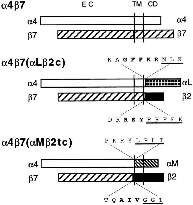
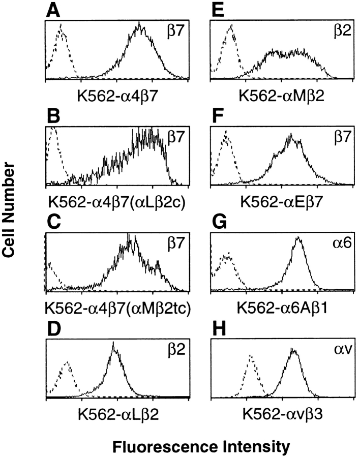
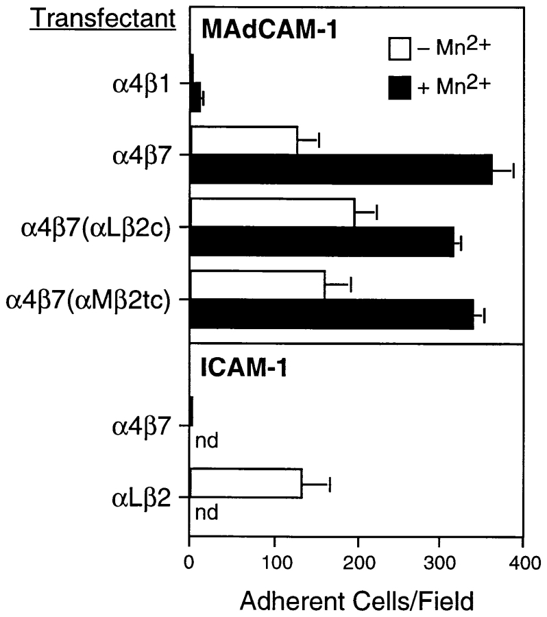
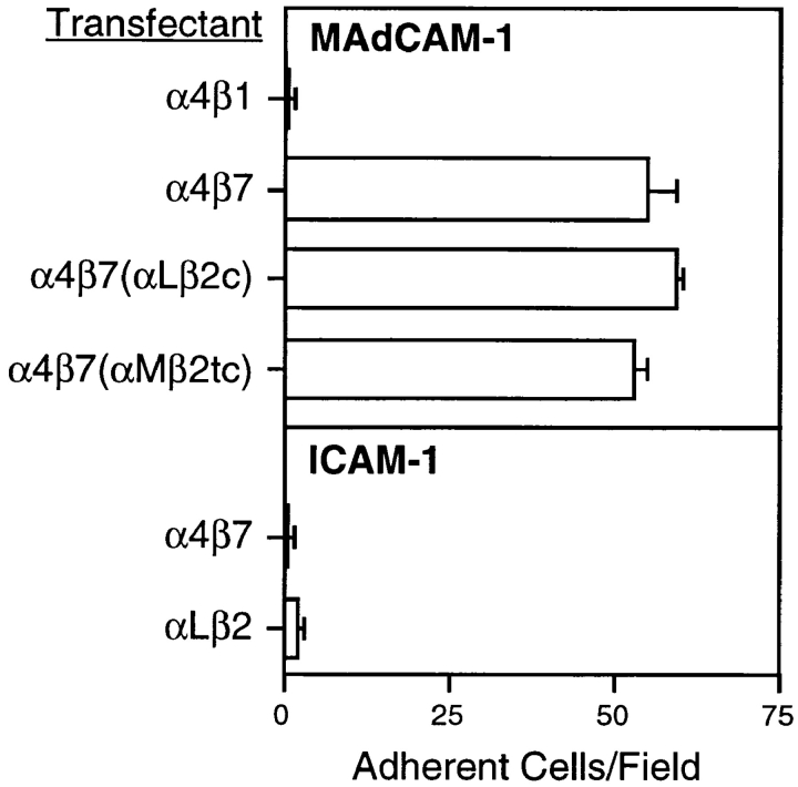
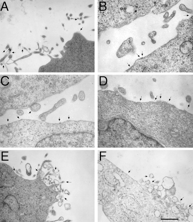
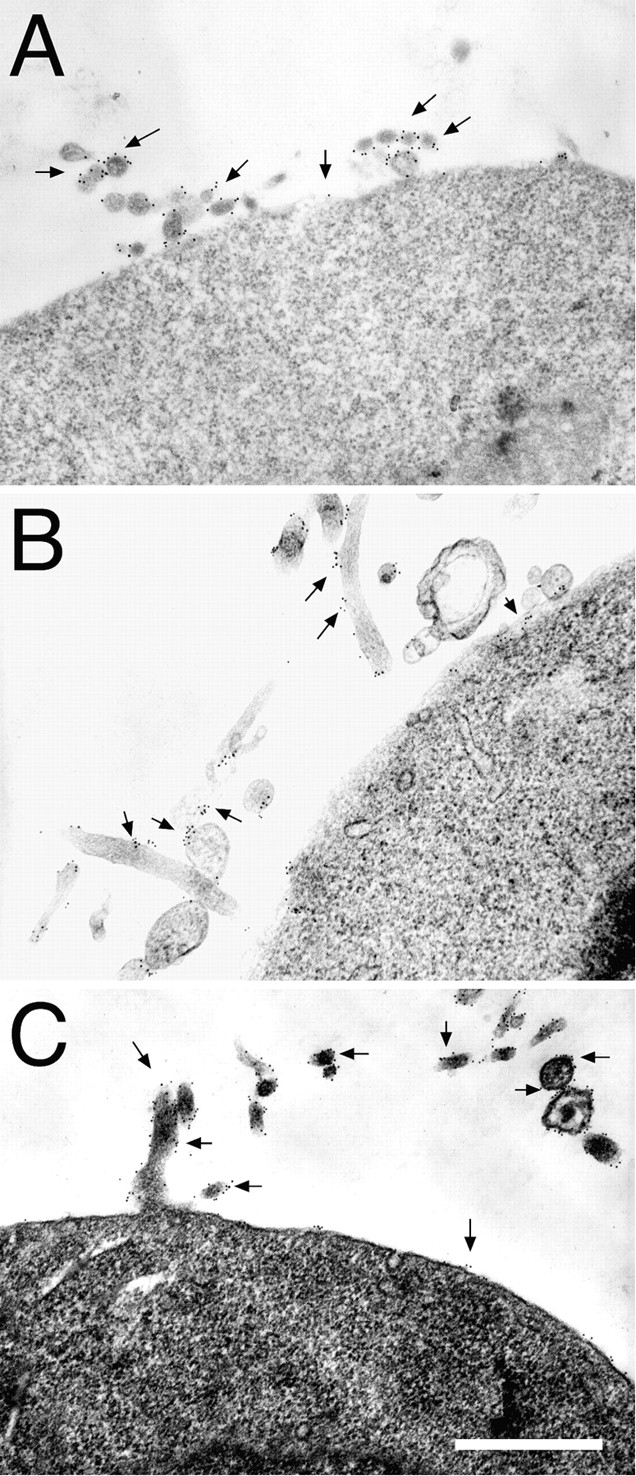
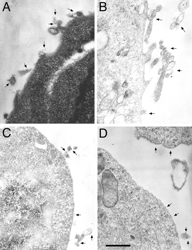
Adhesion of blood leukocytes to the endothelium involves multiple steps including initial attachment (tethering), rolling, and firm arrest. Presentation of adhesion molecules on leukocyte microvilli can substantially enhance tethering. Localization of L-selectin to microvilli and of CD44 to the planar cell body have been shown to depend upon their transmembrane and cytoplasmic domains. We investigated the role of leukocyte integrin transmembrane and cytoplasmic domains in initiating adhesion under flow and in microvillous localization. Integrins alpha4beta7, alphaLbeta2, and alphaMbeta2 were heterologously expressed in K562 cells. alpha4beta7 initiated adhesion under flow and localized to microvilli, whereas beta2 integrins did not initiate adhesion and localized to the cell body. Chimeric integrins were produced by replacing the alpha4beta7 cytoplasmic and/or transmembrane domains with the homologous domains of alphaLbeta2 or alphaMbeta2. Unexpectedly, these chimeras efficiently mediated adhesion to the alpha4beta7 ligand mucosal addressin cell adhesion molecule-1 under flow and localized to microvilli. Therefore, differences between the transmembrane and cytoplasmic domains of alpha4 and beta2 integrins do not account for differences in ability to support attachment under flow or in membrane localization. Integrins alpha4beta1, alpha5beta1, alpha6Abeta1, alphavbeta3, and alphaEbeta7 also localized to microvilli. Transmembrane proteins known or suspected to associate with extracellular domains of microvillous integrins, including tetraspans and CD47, were concentrated on microvilli as well. These findings suggest that interactions between the extracellular domains of integrins and associated proteins could direct the assembly of multimolecular complexes on leukocyte microvilli.


Figures

Figure 1
Figure 1
Schematic representation of wild-type and chimeric α4β7 integrins. The amino acid sequence at the splice site is shown with conserved regions in bold, and the αLβ2 or αMβ2 sequences underlined.
Figure 2
Figure 2
Cell surface expression of wild-type and chimeric integrins. K562-α4β7 (A), K562-α4β7 (αLβ2c) (B), and K562-α4β7 (αMβ2tc) (C) cells were stained with the anti-β7 antibody, Fib 504, as shown. Each of these three transfectants was also recognized by other antibodies specific for the α4 subunit or the α4β7 heterodimer, but were not recognized by anti-αE antibodies (not shown). K562-αLβ2 were recognized by antibodies to β2 (D) and αL (not shown). K562-αMβ2 cells were recognized by antibodies to β2 (E) and αM (not shown). K562-αEβ7 cells stained with antibodies to β7 (F) and αE, but not with anti-α4 antibodies (not shown). K562-α6Aβ1 cells were stained with the anti-α6 antibody GoH3 (G). There was low level expression of αv on nontransfected K562 cells, and higher expression on K562-αvβ3 cells as determined using the anti-αv antibody L230 (H). Fluorescence intensity is shown on a log scale (one log per division). Dotted and solid histograms represent staining with nontransfected and transfected K562 cells, respectively.
Figure 3
Figure 3
Static adhesion of transfectants to immobilized ligands. Adhesion of various integrin transfectants to MAdCAM-1 (top) and ICAM-1 (bottom) was measured in the presence and absence of Mn2+. Bars indicate SEM. nd, not determined.
Figure 4
Figure 4
Adhesion to ligands under flow. Adhesion of various integrin transfectants to MAdCAM-1 (top) and ICAM-1 (bottom) measured at a wall shear stress of 1 dyne/cm2 in the absence of Mn2+, as described in Materials and Methods. Bars indicate SEM.
Figure 5
Figure 5
Localization of α4β7, αLβ2, αMβ2, and chimeric integrins by immunoelectron microscopy. K562 transfectants were stained for protein expression using 12-nm gold particles (arrows) as described in Materials and Methods. Wild-type α4β7 (identified using the anti-β7 antibody, Fib 504) was localized predominantly to microvilli of K562-α4β7 tranfectants (A). K562-αLβ2 (B and C) and K562-αMβ2 (D) were stained with antibodies to αL (B) or β2 (C and D), demonstrating that αLβ2 and αMβ2 were expressed mostly on the cell body. K562 cells transfected with the chimeric integrins α4β7(αLβ2c) (E) and α4β7(αMβ2tc) (F) were stained with Fib 504. These chimeric integrins were found predominantly on microvilli. Photomicrographs are representative of integrin distribution on the 50–100 cells examined in each sample. Bar, 0.5 μm.
Figure 6
Figure 6
Localization of αEβ7, α6Aβ1, and αvβ3 integrins by immunoelectron microscopy. K562 transfectants were stained for protein expression using 12-nm gold particles (arrows) as described in Materials and Methods. Staining of K562-αEβ7 with Fib 504 (anti-β7, A), K562-α6Aβ1 with anti-α6 (GoH3, B), and K562-αvβ3 with L230 (anti-αv, C) revealed that each of these integrins was found primarily on microvilli. Bar, 0.5 μm.
Figure 7
Figure 7
Localization of integrin-associated proteins by immunoelectron microscopy. The tetraspan proteins CD53 (A) and CD63 (B), the αvβ3-associated protein CD47 (C), and CD32 (D) were localized by immunoelectron microscopy using 12-nm (A, C, and D) or 6-nm (B) gold particles (arrows). The tetraspan proteins and CD47 localized primarily to microvilli, whereas CD32 was found in substantial amounts on both the cell body and microvilli. Nontransfected K562 cells (A) and K562-α4β7 (B), K562-αVβ3 (C), and K562-αMβ2 (D) transfectants were used for staining. Bar, 0.5 μm.

References

    1. Alon R, Kassner PD, Carr MW, Finger EB, Hemler ME, Springer TA. The integrin VLA-4 supports tethering and rolling in flow on VCAM-1. J Cell Biol. 1995;128:1243–1253. - PMC - PubMed
    1. Andrew DP, Berlin C, Honda S, Yoshino T, Hamann A, Holzmann B, Kilshaw PJ, Butcher EC. Distinct but overlapping epitopes are involved in α4β7-mediated adhesion to vascular cell adhesion molecule-1, mucosal addressin-1, fibronectin, and lymphocyte aggregation. J Immunol. 1994;153:3847–3861. - PubMed
    1. Berditchevski F, Bazzoni G, Hemler ME. Specific association of CD63 with the VLA-3 and VLA-6 integrins. J Biol Chem. 1995;270:17784–17790. - PubMed
    1. Berditchevski F, Zutter MM, Hemler ME. Characterization of novel complexes on the cell surface between integrins and proteins with 4 transmembrane domains (TM4 proteins) Mol Biol Cell. 1996;7:193–207. - PMC - PubMed
    1. Berditchevski F, Tolias KF, Wong K, Carpenter CL, Hemler ME. A novel link between integrins, transmembrane-4 superfamily proteins (CD63 and CD81), and phosphatidylinositol 4-kinase. J Biol Chem. 1997;272:2595–2598. - PubMed

Publication types

MeSH terms