. 1997 Oct 20;186(8):1247-56.
doi: 10.1084/jem.186.8.1247.

Dendritic cells genetically modified with an adenovirus vector encoding the cDNA for a model antigen induce protective and therapeutic antitumor immunity

Affiliations

Dendritic cells genetically modified with an adenovirus vector encoding the cDNA for a model antigen induce protective and therapeutic antitumor immunity

W Song et al. J Exp Med. .

Abstract

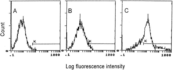
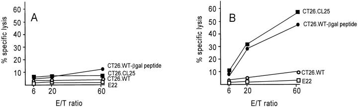
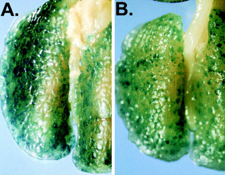
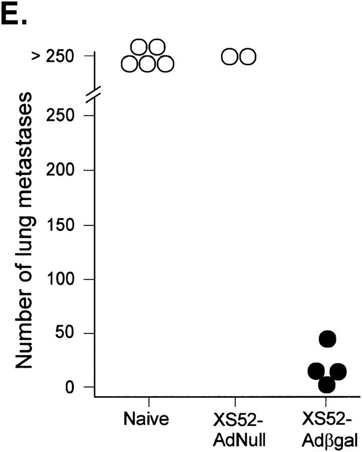
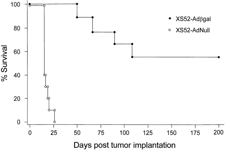
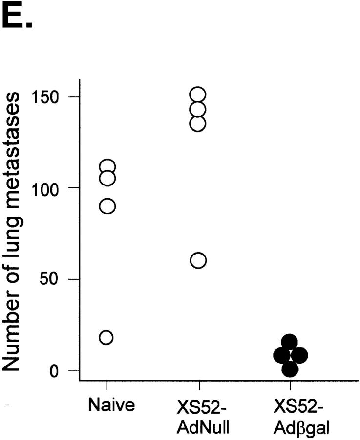
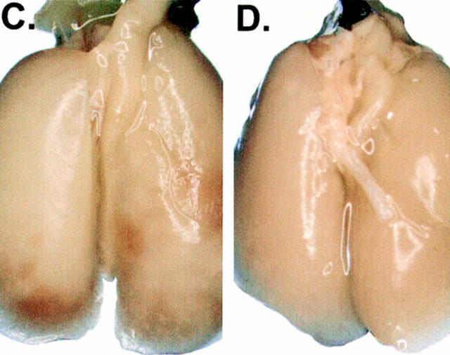
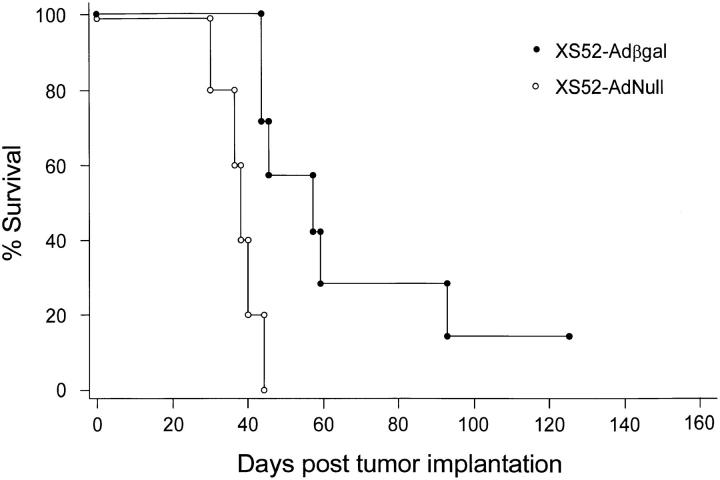
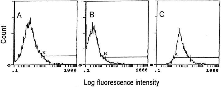
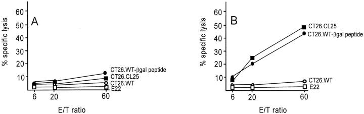
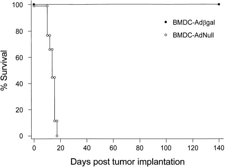
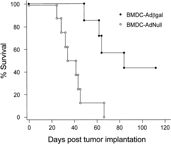
Dendritic cells (DCs) are potent antigen-presenting cells that play a critical role in the initiation of antitumor immune responses. In this study, we show that genetic modifications of a murine epidermis-derived DC line and primary bone marrow-derived DCs to express a model antigen beta-galactosidase (betagal) can be achieved through the use of a replication-deficient, recombinant adenovirus vector, and that the modified DCs are capable of eliciting antigen-specific, MHC-restricted CTL responses. Importantly, using a murine metastatic lung tumor model with syngeneic colon carcinoma cells expressing betagal, we show that immunization of mice with the genetically modified DC line or bone marrow DCs confers potent protection against a lethal tumor challenge, as well as suppression of preestablished tumors, resulting in a significant survival advantage. We conclude that genetic modification of DCs to express antigens that are also expressed in tumors can lead to antigen-specific, antitumor killer cells, with a concomitant resistance to tumor challenge and a decrease in the size of existing tumors.


Figures

Figure 1
Figure 1
Ad vector–mediated gene transfer and expression of βgal to the murine XS52 DC line in vitro. The XS52 cells were infected in vitro with an Ad vector expressing βgal (Adβgal) or the AdNull control vector at moi of 100 for 2 h. 24 h later, βgal expression was quantified by flow cytometry using fluorescein di-β-galactoside. Cells were stained with propidum iodide to facilitate discrimination among live and dead cells. For all panels, the y-axis reflects DC number and the x-axis reflects log fluorescein intensity. The K gate has been set according to the negative control in A. The percentage of βgal-expressing DCs was determined by the right shift of the curve along the K gate. (A) Uninfected XS52 cells. (B) XS52 infected with AdNull. (C) XS52 infected with Adβgal.
Figure 2
Figure 2
Induction of CTL response in BALB/c mice after in vivo administration of Adβgal-infected XS52 DCs. The XS52 DCs were transduced in vitro with AdNull or Adβgal at moi of 100 for 2 h. 24 h later, 3 × 10⁵ cells were administered subcutaneously to syngeneic BALB/ c mice. 14 d later, splenocytes harvested from immunized mice were stimulated for 5 d in vitro with syngeneic fibroblasts (SVBalb) pulsed with a βgal peptide, and then assayed for specific cell lysis against three syngeneic colon carcinoma target cells (parental CT26.WT cells, βgal-expressing CT26.CL25 cells, or CT26.WT pulsed with βgal peptide in vitro [CT26.WT-βgal peptide]) as well as the E22 βgal-expressing allogeneic tumor cell line. (A) CTL response in mice immunized with XS52 DCs infected with AdNull. (B) CTL response in mice immunized with XS52 DCs infected with Adβgal.
Figure 3
Figure 3
Protection against lethal tumor challenge using modified XS52 DCs. Shown are examples and quantitative data of protection against lung metastases after XS52 immunization, using the βgal as a model antigen, BALB/c mice, and syngeneic CT26.CL25 colon carcinoma cells (expressing βgal). XS52 cells were genetically modified by in vitro infection with AdNull or Adβgal (moi 100, 2 h). 24 h after infection, 3 × 10⁵ XS52-AdNull or XS52-Adβgal were injected subcutaneously to syngeneic BALB/c mice. 14 d later, the mice were challenged with intravenous administration of 3 × 10⁵ CT26.CL25 cells. 12–16 d after tumor challenge, the mice were killed, their lungs harvested, fixed, and stained for βgal expression with X-Gal. (A) Example of lungs from a nonimmunized mouse. (B) Example of lungs from a mouse immunized with XS52-AdNull. (C and D) Examples of lungs from mice immunized with XS52-Adβgal. (E) Quantification of the number of lung metastases in nonimmunized, XS52-AdNull–immunized and XS52-Adβgal–immunized mice after tumor challenge. Using a dissecting microscope, surface blue-staining (βgal+) lung metastases were enumerated. Each data point represents an individual animal. Only metastatic deposits ⩽250 could be reliably enumerated; lungs with >250 metastases were assigned an empirical number of 250.
Figure 3
Figure 3
Protection against lethal tumor challenge using modified XS52 DCs. Shown are examples and quantitative data of protection against lung metastases after XS52 immunization, using the βgal as a model antigen, BALB/c mice, and syngeneic CT26.CL25 colon carcinoma cells (expressing βgal). XS52 cells were genetically modified by in vitro infection with AdNull or Adβgal (moi 100, 2 h). 24 h after infection, 3 × 10⁵ XS52-AdNull or XS52-Adβgal were injected subcutaneously to syngeneic BALB/c mice. 14 d later, the mice were challenged with intravenous administration of 3 × 10⁵ CT26.CL25 cells. 12–16 d after tumor challenge, the mice were killed, their lungs harvested, fixed, and stained for βgal expression with X-Gal. (A) Example of lungs from a nonimmunized mouse. (B) Example of lungs from a mouse immunized with XS52-AdNull. (C and D) Examples of lungs from mice immunized with XS52-Adβgal. (E) Quantification of the number of lung metastases in nonimmunized, XS52-AdNull–immunized and XS52-Adβgal–immunized mice after tumor challenge. Using a dissecting microscope, surface blue-staining (βgal+) lung metastases were enumerated. Each data point represents an individual animal. Only metastatic deposits ⩽250 could be reliably enumerated; lungs with >250 metastases were assigned an empirical number of 250.
Figure 4
Figure 4
Survival advantage in mice immunized with modified XS52 cells after a tumor challenge. Animals were immunized with XS52 cells modified with Adβgal or AdNull. The experiment is similar to that depicted in Fig. 3, except that the animals were not killed but were followed for survival. The data is expressed as percent survival as a function of time. Survival for the mice immunized with XS52-Adβgal was significantly prolonged over the XS52-AdNull control, as determined by log-rank analysis of the Kaplan-Meier survival curves (P <0.0001).
Figure 5
Figure 5
Suppression of preestablished lung metastases by administration of modified XS52 DCs. Shown are examples and quantitative data of treatment effect on preestablished lung metastases induced by XS52-Adβgal immunization. 3 d after the establishment of diffuse lung metastases in BALB/c mice with intravenous administration of 3 × 10⁴ CT26.CL25 cells, 3 × 10⁵ XS52-AdNull or XS52-Adβgal were administered subcutaneously to the tumor-bearing mice. 20 d after tumor implantation, the mice were killed, and their lungs were harvested, fixed, and stained for βgal expression with X-Gal. (A) Example of lungs from a nonimmunized mouse. (B) Example of lungs from a mouse receiving XS52-AdNull treatment. (C and D) Examples of lungs from mice receiving XS52-Adβgal treatment. (E) Quantification of the number of lung metastases in untreated, XS52-AdNull– and XS52-Adβgal–treated mice with preestablished lung metastases. Using a dissecting microscope, surface blue-staining (βgal+) lung metastases were enumerated. Each data point represents an individual animal. Only metastatic deposits ⩽250 could be reliably enumerated; lungs with >250 metastases were assigned an empirical number of 250.
Figure 5
Figure 5
Suppression of preestablished lung metastases by administration of modified XS52 DCs. Shown are examples and quantitative data of treatment effect on preestablished lung metastases induced by XS52-Adβgal immunization. 3 d after the establishment of diffuse lung metastases in BALB/c mice with intravenous administration of 3 × 10⁴ CT26.CL25 cells, 3 × 10⁵ XS52-AdNull or XS52-Adβgal were administered subcutaneously to the tumor-bearing mice. 20 d after tumor implantation, the mice were killed, and their lungs were harvested, fixed, and stained for βgal expression with X-Gal. (A) Example of lungs from a nonimmunized mouse. (B) Example of lungs from a mouse receiving XS52-AdNull treatment. (C and D) Examples of lungs from mice receiving XS52-Adβgal treatment. (E) Quantification of the number of lung metastases in untreated, XS52-AdNull– and XS52-Adβgal–treated mice with preestablished lung metastases. Using a dissecting microscope, surface blue-staining (βgal+) lung metastases were enumerated. Each data point represents an individual animal. Only metastatic deposits ⩽250 could be reliably enumerated; lungs with >250 metastases were assigned an empirical number of 250.
Figure 6
Figure 6
Survival advantage in tumor-bearing mice treated with modified XS52 cells. Animals were immunized with XS52 cells modified with Adβgal or AdNull. The experiment is similar to that depicted in Fig. 5, except that the animals were not killed but were followed for survival. The data is expressed as percentage of survival as a function of time. Survival for mice which were treated with XS52-Adβgal was significantly prolonged over the XS52-AdNull control, as determined by log-rank analysis of the Kaplan-Meier survival curves (P <0.002).
Figure 7
Figure 7
Ad vector–mediated gene transfer and expression of βgal in bone marrow DC (BMDC) in vitro. The primary murine DCs were infected in vitro with Adβgal or AdNull control vector at moi of 100 for 2 h. 24 h later, βgal expression was quantified by flow cytometry using fluorescein di-β-galactoside. For all panels, the y-axis reflects DC number and the x-axis reflects log fluorescein intensity. The percentage of βgal-expressing DCs was determined by the right shift of the curve along the K gate. (A) Uninfected bone marrow DCs. (B) Bone marrow DCs infected with AdNull. (C) Bone marrow DCs infected with Adβgal.
Figure 8
Figure 8
Induction of CTL response in BALB/c mice after in vivo administration of bone marrow DCs infected with Adβgal. Bone marrow DCs were transduced in vitro with AdNull or Adβgal at moi of 100 for 2 h. 24 h later, 3 × 10⁵ cells were administered subcutaneously into syngeneic BALB/c mice. 14 d later, splenocytes harvested from immunized mice were stimulated for 5 d in vitro with syngeneic fibroblasts (SVBalb) pulsed with βgal peptide and then assayed for specific cell lysis against three syngeneic colon carcinoma target cells (parental CT26.WT cells, βgal-expressing CT26.CL25 cells, or CT26.WT pulsed with βgal peptide in vitro [CT26.WT-βgal peptide]) as well as the E22 βgal-expressing allogeneic tumor cell line. (A) CTL response in mice immunized with bone marrow DCs infected with AdNull. (B) CTL response in mice immunized with bone marrow DCs infected with Adβgal.
Figure 9
Figure 9
Survival advantage in mice immunized with modified bone marrow DCs after a tumor challenge. Animals were immunized with bone marrow DCs modified with Adβgal or AdNull, followed 14 d later by intravenous challenge of 105 CT26.CL25 tumor cells. The animals were not killed, but were followed for survival. The data is expressed as percentage of survival as a function of time. Survival for the mice immunized with bone marrow DC–Adβgal was significantly prolonged over the bone marrow DC–AdNull control, as determined by log-rank analysis of the Kaplan-Meier survival curves (P <0.0001).
Figure 10
Figure 10
Survival advantage in tumor-bearing mice treated with modified bone marrow DCs. 3 d after the establishment of lung metastases with intravenous administration of 3 × 10⁴ CT26.CL25 tumor cells, BALB/c mice were immunized with bone marrow DCs modified with Adβgal or AdNull. The animals were not killed, but were followed for survival. The data is expressed as percent survival as a function of time. Survival for mice that were treated with bone marrow DC–Adβgal was significantly prolonged over the bone marrow DC–AdNull control, as determined by log-rank analysis of the Kaplan-Meier survival curves (P <0.002).

References

    1. Pardoll DM. Cancer vaccines. Immunol Today. 1993;14:310–316. - PubMed
    1. Houghton AN. Cancer antigens: immune recognition of self and altered self. J Exp Med. 1994;180:1–4. - PMC - PubMed
    1. Boon T, van der Bruggen P. Human tumor antigens recognized by T lymphocytes. J Exp Med. 1996;183:725–729. - PMC - PubMed
    1. Juillard V, Villefroy P, Godfrin D, Pavirani A, Venet A, Guillet JG. Long-term humoral and cellular immunity induced by a single immunization with replication-defective adenovirus recombinant vector. Eur J Immunol. 1995;25:3467–3473. - PubMed
    1. Chen PW, Wang M, Bronte V, Zhai Y, Rosenberg SA, Restifo NP. Therapeutic antitumor response after immunization with a recombinant adenovirus encoding a model tumor-associated antigen. J Immunol. 1996;156:224–231. - PMC - PubMed

Publication types

MeSH terms