. 1997 Oct 20;186(8):1269-75.
doi: 10.1084/jem.186.8.1269.

Peripheral T cell survival requires continual ligation of the T cell receptor to major histocompatibility complex-encoded molecules

Affiliations

Peripheral T cell survival requires continual ligation of the T cell receptor to major histocompatibility complex-encoded molecules

J Kirberg et al. J Exp Med. .

Abstract

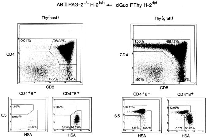
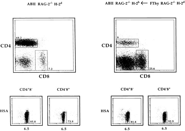
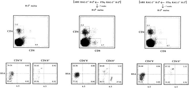
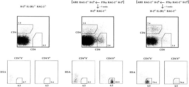
In the thymus, T cells are selected according to their T cell receptor (TCR) specificity. After positive selection, mature cells are exported from primary lymphoid organs to seed the secondary lymphoid tissue. An important question is whether survival of mature T cells is an intrinsic property or requires continuous survival signals, i.e., engagement of the TCR by major histocompatibility complex (MHC) molecules in the periphery, perhaps in a similar way as occurring during thymic positive selection. To address this issue we used recombination-activating gene (Rag)-deficient H-2b mice expressing a transgenic TCR restricted by I-Ed class II MHC molecules. After engraftment with Rag-/- H-2d fetal thymi, CD4+8- peripheral T cells emerged. These cells were isolated and transferred into immunodeficient hosts of H-2b or H-2d haplotype, some of the latter being common cytokine receptor gamma chain deficient to exclude rejection of H-2b donor cells by host natural killer cells. Our results show that in the absence, but not in the presence, of selecting MHC molecules, peripheral mature T cells are short lived and disappear within 7 wk, indicating that continuous contact of the TCR with selecting MHC molecules is required for survival of T cells.


Figures

Figure 1
Figure 1
Thymic development of CD4+8 T cells in H-2d fetal thymus–grafted H-2b ABII TCR Rag−/− mice. H-2b ABII TCR Rag−/− mice were transplanted with BALB/c fetal thymi that had been treated with dGuo. Several weeks after transplantation, thymocytes from grafted and host thymi were recovered and analyzed by four-color flow cytometry including the ABII TCR–specific mAb 6.5. Similar data were obtained when grafting H-2d Rag−/− thymi that, however, grew much more extensively (average of 3.5 × 10⁷ thymocytes).
Figure 2
Figure 2
CD4+8 lymphocytes are present in H-2b ABII TCR Rag−/− mice grafted with an H-2d fetal thymus. Lymphocytes were recovered from H-2d ABII TCR Rag−/− and H-2b ABII TCR Rag−/− mice that had previously been transplanted with fetal thymus lobes from H-2d Rag−/− mice. Cells were analyzed by four-color flow cytometry as shown.
Figure 3
Figure 3
CD4+8 lymphocytes from H-2d thymus-grafted H-2b ABII TCR Rag−/− mice disappear in H-2b adoptive hosts. 7.5 × 10⁵ CD4+8 and 5 × 10⁵ CD48low lymphocytes from H-2d Rag−/− fetal thymus–transplanted H-2b ABII TCR Rag−/− mice were adoptively transferred into H-2b and H-2d nu/nu mice. 7 wk later, cells from lymph nodes and spleen (RBCs lysed) were isolated, depleted of sIg+ cells, and analyzed by four-color flow cytometry. Data from lymph nodes and spleen gave similar results. Calculated numbers of 6.5+HSA cells were 5,600, 6,500, and 1,800 for CD4+8 and 1,800, 463,600, and 400 for CD48low cells in unmanipulated H-2b (one mouse), injected H-2b (average of two mice), and injected H-2d (average of two mice) nu/nu mice, respectively. (Note that numbers also contain the calculated background values of mice not injected.) Another experiment and two experiments using H-2b Rag−/− and H-2d Rag−/− recipients gave similar results.
Figure 4
Figure 4
CD4+8 lymphocytes from H-2d thymus-grafted H-2b ABII TCR Rag−/− mice survive in H-2d NK cell–deficient adoptive hosts. 105 CD4+8 and 105 CD48low lymphocytes from H-2d Rag−/− fetal thymus–transplanted H-2b ABII TCR Rag−/− mice were adoptively transferred into H-2b Rag−/− and H-2d Rag−/− IL-2Rγ−/− mice. 7 wk after transfer, cells from lymph nodes and spleen were analyzed as in Fig. 3. Calculated numbers of 6.5+HSA cells were 900, 13,500, 5,900, and 3 × 10⁶ for CD4+8 and 600, 6,100, 7.6 × 10⁵, and 1.5 × 10⁶ for CD48low cells in unmanipulated H-2d Rag−/− IL-2Rγ−/−, unmanipulated H-2b Rag−/− (not shown), injected H-2b Rag−/−, and injected H-2d Rag−/− IL-2Rγ−/− mice, respectively. Expansion in Rag-deficient H-2d mice was consistent with previous data (41). We also performed this type of experiment with two thymectomized H-2d Rag−/− IL-2Rγ−/− mice and obtained similar results.

References

    1. Kisielow P, Teh HS, Blüthmann H, von Boehmer H. Positive selection of antigen-specific T cells in thymus by restricting MHC molecules. Nature (Lond) 1988;335:730–733. - PubMed
    1. Scott B, Blüthmann H, Teh HS, von Boehmer H. The generation of mature T cells requires interaction of the alpha beta T-cell receptor with major histocompatibility antigens. Nature (Lond) 1989;338:591–593. - PubMed
    1. Anderson G, Owen JJ, Moore NC, Jenkinson EJ. Thymic epithelial cells provide unique signals for positive selection of CD4+CD8+thymocytes in vitro. J Exp Med. 1994;179:2027–2031. - PMC - PubMed
    1. Kisielow P, Miazek A. Positive selection of T cells: rescue from programmed cell death and differentiation require continual engagement of the T cell receptor. J Exp Med. 1995;181:1975–1984. - PMC - PubMed
    1. Pircher H, Ohashi PS, Boyd RL, Hengartner H, Brduscha K. Evidence for a selective and multi-step model of T cell differentiation: CD4+CD8lowthymocytes selected by a transgenic T cell receptor on major histocompatibility complex class I molecules. Eur J Immunol. 1994;24:1982–1987. - PubMed

Publication types

MeSH terms