. 1997 Oct 20;186(8):1277-85.
doi: 10.1084/jem.186.8.1277.

The common cytokine receptor gamma chain controls survival of gamma/delta T cells

Affiliations

The common cytokine receptor gamma chain controls survival of gamma/delta T cells

M Malissen et al. J Exp Med. .

Abstract

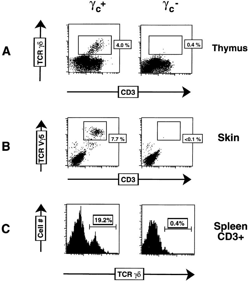
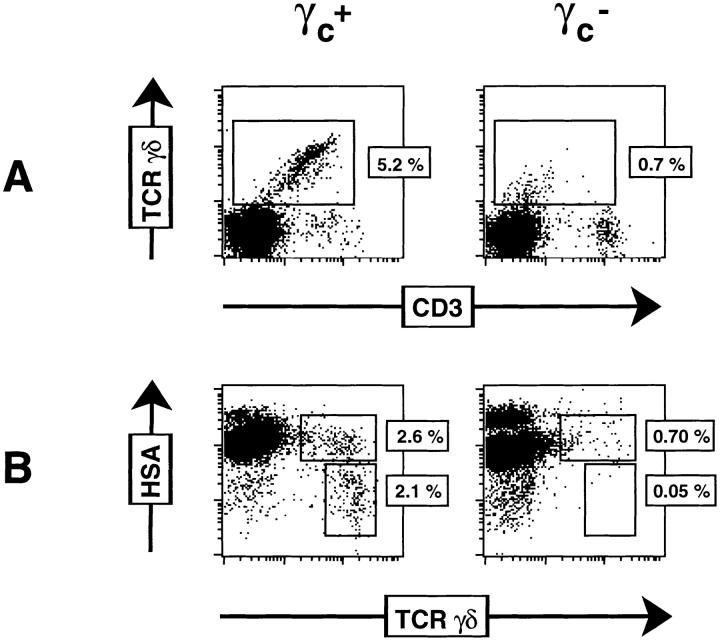
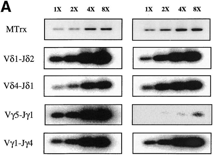
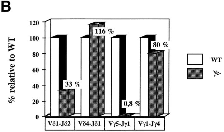
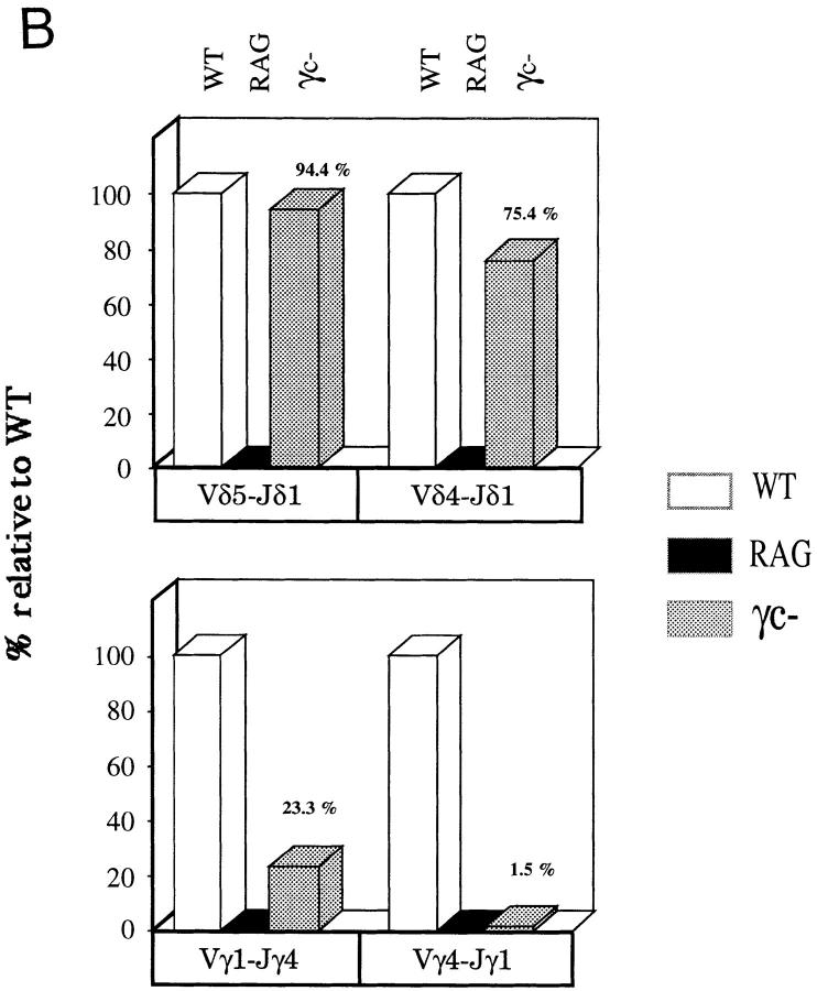
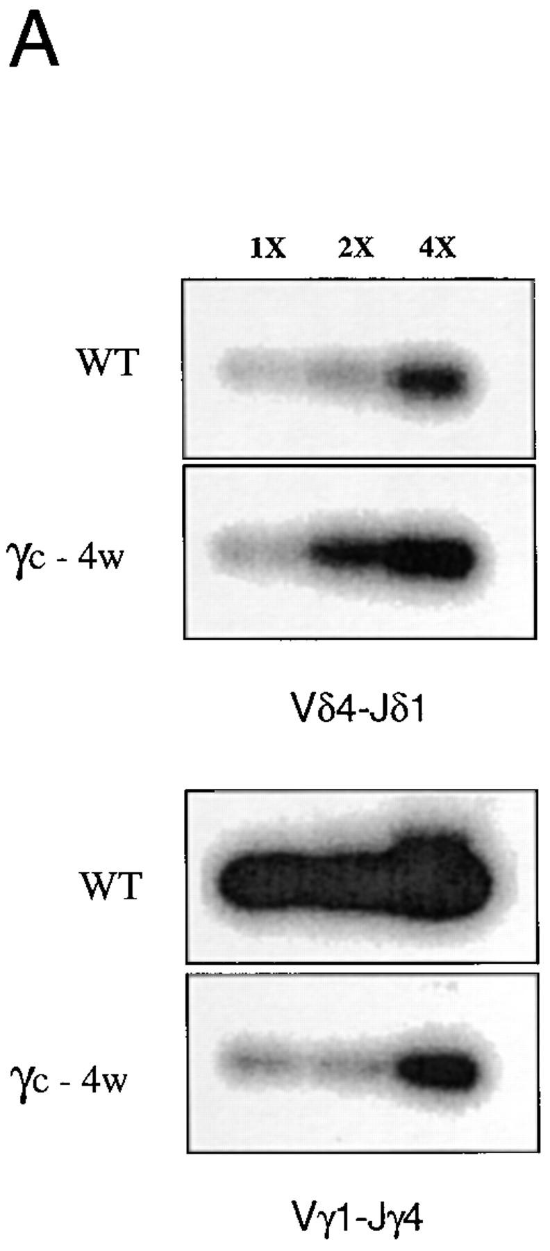
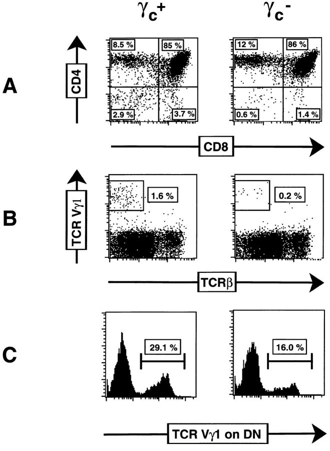
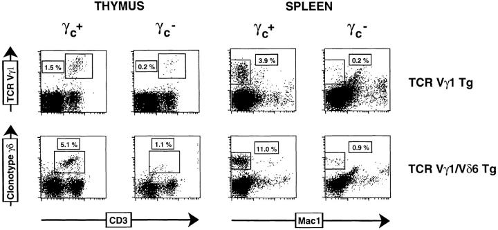
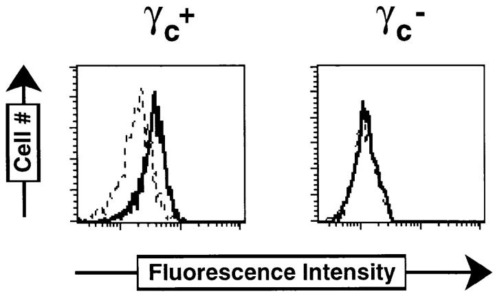
We have investigated the role of common gamma chain (gamma c)-signaling pathways for the development of T cell receptor for antigen (TCR)-gamma/delta T cells. TCR-gamma/delta-bearing cells were absent from the adult thymus, spleen, and skin of gamma c-deficient (gamma c-) mice, whereas small numbers of thymocytes expressing low levels of TCR-gamma/delta were detected during fetal life. Recent reports have suggested that signaling via interleukin (IL)-7 plays a major role in facilitating TCR-gamma/delta development through induction of V-J (variable-joining) rearrangements at the TCR-gamma locus. In contrast, we detected clearly TCR-gamma rearrangements in fetal thymi from gamma c- mice (which fail to signal in response to IL-7) and reduced TCR-gamma rearrangements in adult gamma c thymi. No gross defects in TCR-delta or TCR-beta rearrangements were observed in gamma c- mice of any age. Introduction of productively rearranged TCR V gamma 1 or TCR V gamma 1/V delta 6 transgenes onto mice bearing the gamma c mutation did not restore TCR-gamma/delta development to normal levels suggesting that gamma c-dependent pathways provide additional signals to developing gamma/delta T cells other than for the recombination process. Bcl-2 levels in transgenic thymocytes from gamma c- mice were dramatically reduced compared to gamma c+ transgenic littermates. We favor the concept that gamma c-dependent receptors are required for the maintenance of TCR-gamma/delta cells and contribute to the completion of TCR-gamma rearrangements primarily by promoting survival of cells committed to the TCR-gamma/delta lineage.


Figures

Figure 1
Figure 1
Flow cytometric analysis of γ/δ T cells from adult γc + and γc mice. (A) Thymocytes were stained with FITC–anti–pan-TCR-γ/δ (GL3), PE–anti-CD3, biotin-anti–CD4, and biotin–anti-CD8α. CD4 CD8 (DN) cells were electronically gated. (B) Skin-resident DETCs were isolated as indicated in Materials and Methods, and stained with biotin–anti-TCR Vγ5 and FITC–anti-CD3. (C) Splenocytes were monocyte depleted by adherence and stained with FITC–anti–pan-TCR-γ/δ, PE–anti-CD3, biotin–anti-CD4, and biotin–anti-CD8α. CD3+ DN cells were electronically gated. Biotinylated antibodies were revealed with streptavidin-Tricolor.
Figure 2
Figure 2
Flow cytometric analysis of fetal thymocytes from γc + and γc mice. (A) Thymocytes were stained with FITC–anti–pan-TCR-γ/δ and PE–anti-CD3. (B) Thymocytes were stained with FITC–anti-pan-TCR-γ/δ and PE–anti-HSA. In all experiments, nonspecific binding was blocked by the addition of anti-CD16/32.
Figure 3
Figure 3
Quantitation of TCR-δ and TCR-γ rearrangements in fetal γc + and γc thymocytes. (A) Indicated rearrangements were amplified from DNA isolated from unfractionated D17 fetal γc + or γc thymi. Serial dilutions of DNA template were analyzed (1×, 2×, 4×, and 8×). (B) Quantification of A. Hybridizing bands were scanned using a phosphorimager and the relative levels of rearrangements compared to wild-type (WT) γc + thymi after normalizing for input DNA using MTrx primers. See Materials and Methods for details.
Figure 3
Figure 3
Quantitation of TCR-δ and TCR-γ rearrangements in fetal γc + and γc thymocytes. (A) Indicated rearrangements were amplified from DNA isolated from unfractionated D17 fetal γc + or γc thymi. Serial dilutions of DNA template were analyzed (1×, 2×, 4×, and 8×). (B) Quantification of A. Hybridizing bands were scanned using a phosphorimager and the relative levels of rearrangements compared to wild-type (WT) γc + thymi after normalizing for input DNA using MTrx primers. See Materials and Methods for details.
Figure 4
Figure 4
Quantitation of TCR-δ and TCR-γ rearrangements in adult γc + and γc thymocytes. (A) Rearrangements of Vδ4-Jδ1 and Vγ1-Jγ4 were amplified from DNA isolated from unfractionated γc + or γc thymi. Serial dilutions of DNA template were analyzed (1×, 2×, and 4×). (B) Quantification of A. Relative levels of rearrangements are compared to wild-type (WT) γc + thymi.
Figure 4
Figure 4
Quantitation of TCR-δ and TCR-γ rearrangements in adult γc + and γc thymocytes. (A) Rearrangements of Vδ4-Jδ1 and Vγ1-Jγ4 were amplified from DNA isolated from unfractionated γc + or γc thymi. Serial dilutions of DNA template were analyzed (1×, 2×, and 4×). (B) Quantification of A. Relative levels of rearrangements are compared to wild-type (WT) γc + thymi.
Figure 5
Figure 5
Flow cytometric analysis of thymocytes from γc + or γc mice transgenic for a rearranged TCR Vγ1 receptor. (A) Cells were stained with FITC–anti-CD8α and PE–anti-CD4. (B) Cells were stained with FITC–anti–TCR Vγ1 and biotin–anti–TCR-β. (C) Cells were stained with FITC–anti–TCR Vγ1, PE–anti-CD4, and PE–anti-CD8α. CD4CD8 (DN) cells were electronically gated.
Figure 6
Figure 6
Flow cytometric analysis of thymocytes and splenocytes of γc + and γc mice transgenic for TCR Vγ1 or double transgenic for TCR Vγ1/Vδ6. Cells were stained with combinations of FITC–anti–TCR Vγ1, FITC–anti–TCRγ/δ clonotype, PE–anti-CD3, and PE–anti-Mac1.
Figure 7
Figure 7
Intracellular levels of Bcl-2 are diminished in γc γ/δ T cells. Cells were surface stained with FITC–anti–TCR Vγ1, fixed, and permeabilized. Bcl-2 staining was performed as described in Materials and Methods. Dotted lines indicate staining with hamster Ig and solid lines with hamster anti–mouse Bcl-2–specific Ig.

References

    1. Kisielow P, von Boehmer H. Development and selection of T cells: facts and puzzles. Adv Immunol. 1995;58:87–209. - PubMed
    1. Allison JP. γδ T-cell development. Curr Opin Immunol. 1993;5:241–246. - PubMed
    1. Hass W, Pereira P, Tonegawa S. Gamma/delta cells. Annu Rev Immunol. 1993;11:637–685. - PubMed
    1. Lafaille JJ, DeCloux A, Bonneville M, Takagaki Y, Tonegawa S. Junctional sequences of T cell receptor γδ genes: implications for γδ T cell lineages and for a novel intermediate of V-(D)-J joining. Cell. 1989;59:859–870. - PubMed
    1. Bogue M, Mossman H, Stauffer U, Benoist C, Mathis D. The level of N-region diversity in T cell receptors is not pre-ordained in the stem cell. Eur J Immunol. 1993;23:1185–1188. - PubMed

Publication types

MeSH terms

Substances