. 1997 Oct 20;186(8):1315-22.
doi: 10.1084/jem.186.8.1315.

Heat shock protein-peptide complexes, reconstituted in vitro, elicit peptide-specific cytotoxic T lymphocyte response and tumor immunity

Affiliations

Heat shock protein-peptide complexes, reconstituted in vitro, elicit peptide-specific cytotoxic T lymphocyte response and tumor immunity

N E Blachere et al. J Exp Med. .

Abstract

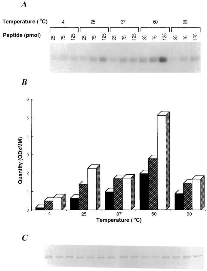
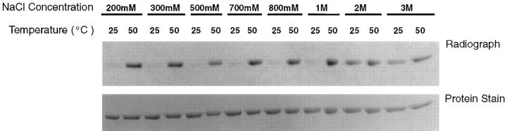
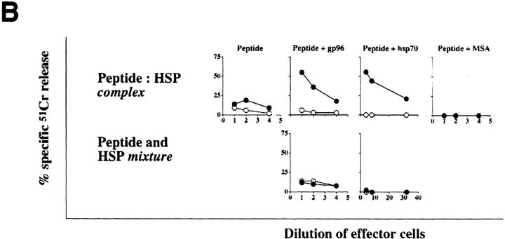
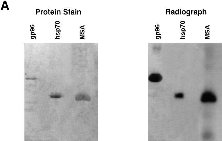
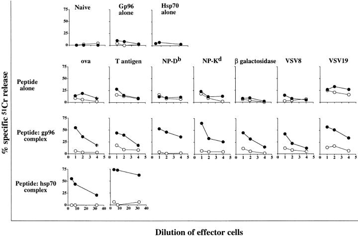
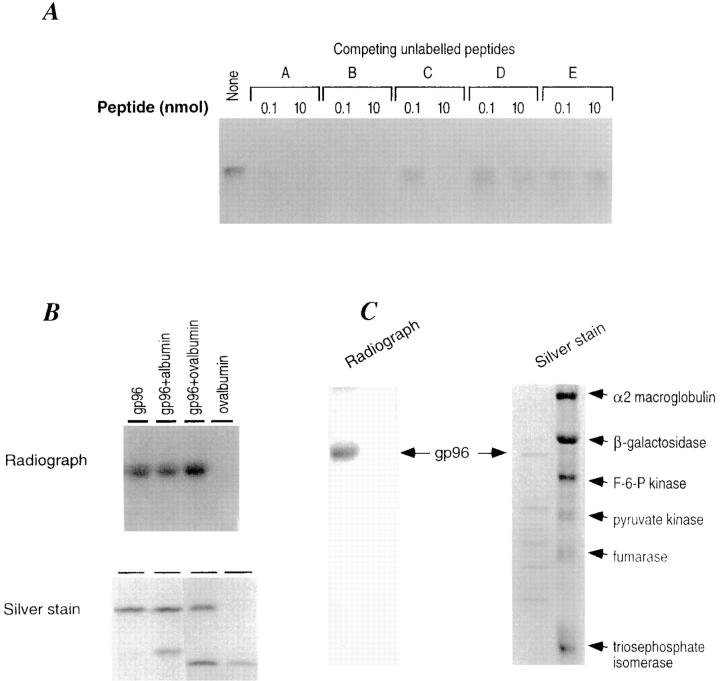
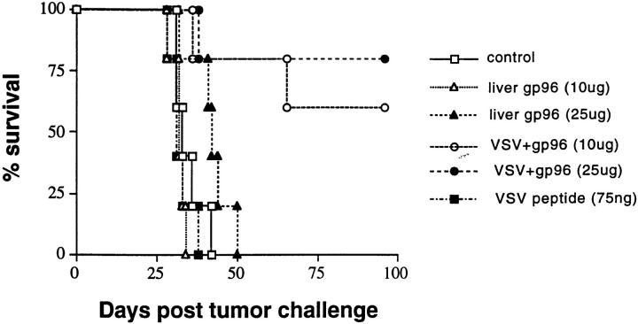
Heat shock protein (HSP) preparations derived from cancer cells and virus-infected cells have been shown previously to elicit cancer-specific or virus-specific immunity. The immunogenicity of HSP preparations has been attributed to peptides associated with the HSPs. The studies reported here demonstrate that immunogenic HSP-peptide complexes can also be reconstituted in vitro. The studies show that (a) complexes of hsp70 or gp96 HSP molecules with a variety of synthetic peptides can be generated in vitro; (b) the binding of HSPs with peptides is specific in that a number of other proteins tested do not bind synthetic peptides under the conditions in which gp96 molecules do; (c) HSP-peptide complexes reconstituted in vitro are immunologically active, as tested by their ability to elicit antitumor immunity and specific CD8+ cytolytic T lymphocyte response; and (d) synthetic peptides reconstituted in vitro with gp96 are capable of being taken up and re-presented by macrophage in the same manner as gp96- peptides complexes generated in vivo. These observations demonstrate that HSPs are CD8+ T cell response-eliciting adjuvants.


Figures

Figure 1
Figure 1
gp96 binds to peptides in vitro. gp96 (10 pmol) was incubated with increasing concentrations of radioiodinated peptide A (25, 75, and 125 pmol) for 10 min at different temperatures in 20 μl reaction buffer, followed by 30 min at room temperature. The reaction was terminated by mixing with sample buffer (0.1% SDS, 20% glycerol, and 5% bromophenol blue) and analyzed by SDS-PAGE. (A) Autoradiogram after 48-h exposure. (B) Densitometric quantification of results in A. An aliquot of each reaction was analyzed in parallel by SDS-PAGE and silver staining (C).
Figure 2
Figure 2
Exchange of exogenous and native-bound peptides at high salt concentrations. gp96 (40 pM) and iodinated synthetic peptide (2 nM, NH2–Ser–Leu–Ser–Asp–Leu–Arg–Gly–Tyr–Val–Tyr–Gln– Gly– Leu– Lys– Ser–Gly–Asn–Val–Ser–COOH) were mixed in phosphate buffer in 20 μl reaction volume and incubated at 25 or 50°C for 10 min. After centrifugation, the mixtures were incubated at 25°C for another 30 min. Samples were analyzed by SDS-PAGE and staining, followed by autoradiography of the stained gel (24-h exposure).
Figure 5
Figure 5
Chaperoning of peptides by HSPs is required for generation of an effective CD8+ T cell response. gp96, hsp70, or mouse serum albumin (MSA) were complexed with radiolabeled VSV9 and analyzed by (A) SDS-PAGE followed by Coomassie blue staining and autoradiography. In addition, mice were immunized (B) with peptides complexed or simply mixed with each of the proteins. Splenocytes of these mice were tested for induction of CD8+ T lymphocytes, as described in legend to Fig. 4. N1 (closed circle) and EL4 (open circle) were used as targets.
Figure 5
Figure 5
Chaperoning of peptides by HSPs is required for generation of an effective CD8+ T cell response. gp96, hsp70, or mouse serum albumin (MSA) were complexed with radiolabeled VSV9 and analyzed by (A) SDS-PAGE followed by Coomassie blue staining and autoradiography. In addition, mice were immunized (B) with peptides complexed or simply mixed with each of the proteins. Splenocytes of these mice were tested for induction of CD8+ T lymphocytes, as described in legend to Fig. 4. N1 (closed circle) and EL4 (open circle) were used as targets.
Figure 4
Figure 4
Hsp–peptide complexes reconstituted in vitro prime mice for CD8+ T cell response. Mice were immunized with HSPs alone (20–50 μg), peptides alone (10 μg), or HSP–peptide complexes (20–50 μg), as indicated. 1 wk after the last immunization, spleens were removed and stimulated with the cognate peptide or with cells transfected with the gene encoding the relevant antigen. The lymphocyte cultures were tested for their ability to lyse cells transfected with the antigen of interest (closed circle) and the nontransfected parental line (open circle). For the top panel, HSPs alone were tested for their immunizing ability in each antigenic system and were found to be consistently negative. The CTL responses were tested in many but not all systems and where tested, were found to be MHC class I and CD8 restricted.
Figure 3
Figure 3
Specificity of peptide binding by gp96. (A) Unlabeled peptides A, B, C, D, and E (0.1 and 10 nM) were used to compete with labeled peptide A (25 pmol) for the binding to gp96 (10 pmol). The sequences of these peptides are described in the text. The binding assay described in the legend to Fig. 1 was used, except that binding was carried out at 50°C. (B) Albumin and ovalbumin do not compete with gp96 for binding to peptide A. gp96 (10 pmol) was incubated with 25-pmol radiolabeled peptide A and analyzed as in the legend to Fig. 1. Albumin and ovalbumin (10 pmol each) were included in the binding assay. The autoradiogram and the silver stained gel are shown. (C) A partially degraded preparation of gp96 and a mixture of six purified proteins (i.e., α–2 macroglobulin, β-galactosidase, fructose-6-phosphokinase, pyruvate kinase, fumarase, and triosephosphate isomerase) were tested for binding to iodinated peptide A. Only the intact gp96 molecule is able to form a stable complex with radioactive peptide A. None of the other six proteins tested are able to bind peptide A.
Figure 6
Figure 6
gp96–VSV8 complexes reconstituted in vitro elicit peptide-specific, protective immunity. Mice were immunized twice at weekly intervals with gp96–VSV8 complexes (10 μg or 25 μg liver gp96 complexed with 1–2 ng VSV8 peptide), liver gp96 alone (10 or 25 μg), VSV8 peptide alone (75 ng), or RPMI medium control. gp96–VSV8 complexes were washed extensively using a minicon 50 to remove unbound peptides. All mice were challenged intraperitoneally with 5,000 live N1 cells 1 wk after the second immunization; survival was monitored.
Figure 7
Figure 7
VSV8 peptide chaperoned by gp96 is re-presented by macrophage. Pristane-induced peritoneal exudate cells (104) and VSV peptide-specific CTL (5 × 10⁴) were cocultured with gp96– VSV8 complexes (3–20 μg/ml) reconstituted at 37, 60, 85, or 98°C in a 96-well U-bottomed plate at 37°C. After 24 h, supernatants were collected and assayed for TNF-α production in a cytotoxicity assay as described (15).

References

    1. Srivastava PK, Das MR. The serologically unique cell surface antigen of Zajdela ascitic hepatoma is also its tumor associated transplantation antigen. Int J Cancer. 1984;33:417–422. - PubMed
    1. Srivastava PK, DeLeo AB, Old LJ. Tumor rejection antigens of chemically induced tumors of inbred mice. Proc Natl Acad Sci USA. 1986;83:3407–3411. - PMC - PubMed
    1. Ullrich SJ, Robinson EA, Law LW, Willingham M, Appella E. A mouse tumor-specific transplantation antigen is a heat shock–related protein. Proc Natl Acad Sci USA. 1986;83:3121–3125. - PMC - PubMed
    1. Palladino MA, Srivastava PK, Oettgen HF, DeLeo AB. Expression of a shared tumor-specific antigen by two chemically induced BALB/c sarcomas. Cancer Res. 1987;47:5074–5079. - PubMed
    1. Udono H, Srivastava PK. Comparison of tumor-specific immunogenicities of stress-induced proteins gp96, hsp90, and hsp70. J Immunol. 1994;152:5398–5403. - PubMed

Publication types

MeSH terms

Substances