Comparative Study
. 1997 Nov 1;17(21):8083-92.
doi: 10.1523/JNEUROSCI.17-21-08083.1997.

Parapinopsin, a novel catfish opsin localized to the parapineal organ, defines a new gene family

Affiliations
Comparative Study

Parapinopsin, a novel catfish opsin localized to the parapineal organ, defines a new gene family

S Blackshaw et al. J Neurosci. .

Abstract

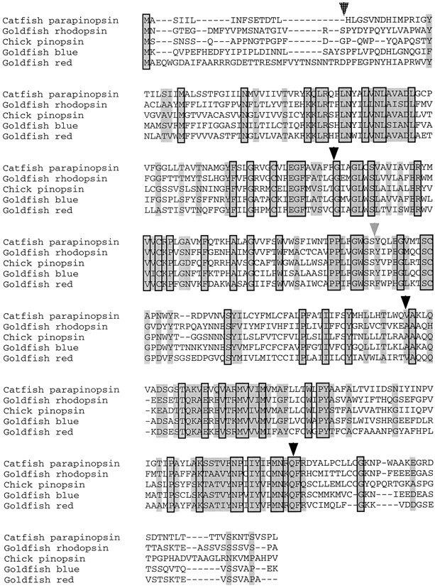
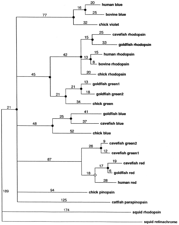
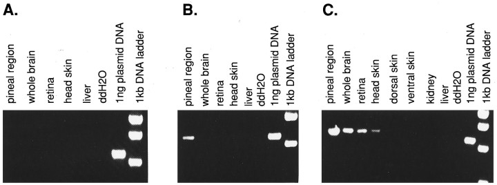
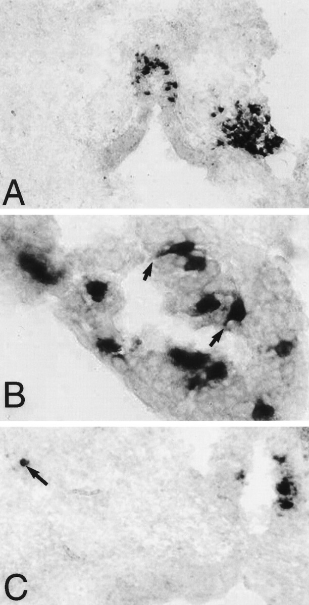
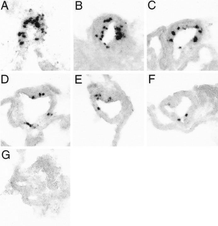
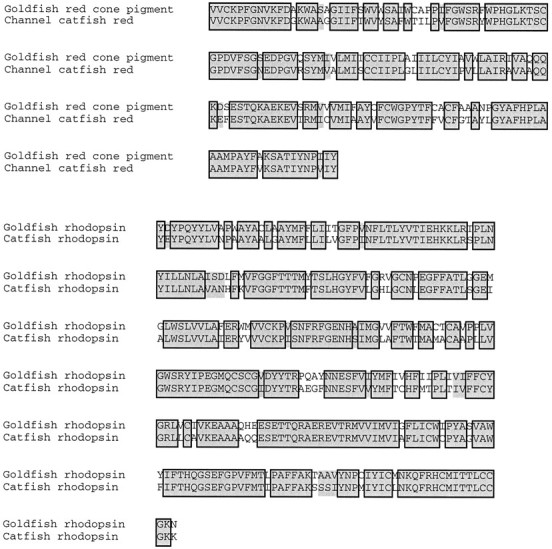
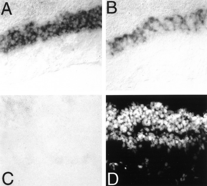
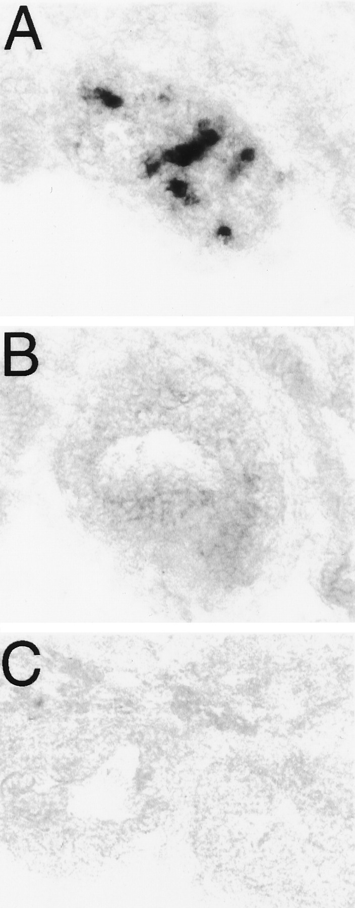
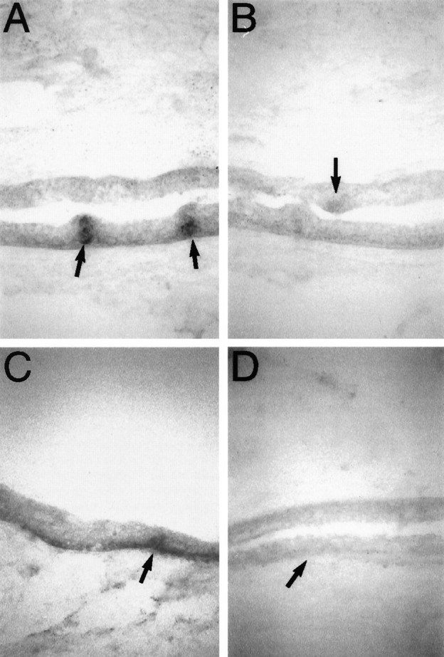
Multiple sites of extraretinal photoreception are present in vertebrates, but the molecular basis of extraretinal phototransduction is poorly understood. This study reports the cloning of the first opsin specifically expressed in the directly photosensitive pineal and parapineal of cold-blooded vertebrates. This opsin, identified in channel catfish and termed parapinopsin, defines a new gene family of vertebrate photopigments and is expressed in a majority of parapinealocytes and a subset of pineal photoreceptor cells. Parapinopsin shows a caudal-rostral gradient of expression within the pineal organ. This study also reports the cloning of partial cDNAs encoding the channel catfish orthologues of rhodopsin and the red cone pigment-the full complement of retinal opsins in the species. In situ hybridization studies using probes derived from these retinal opsins, together with parapinopsin, reveal no expression of retinal opsins in pineal and parapineal organ and no expression of any opsin tested in the "deep brain," iris, or dermal melanophores. These data imply that phototransduction in these sites of extraretinal photoreception must be mediated by novel opsins.


Figures

Fig. 1.
Fig. 1.
Primary sequence of catfish parapinopsin aligned to chick pinopsin and selected goldfish retinal opsins. Residues conserved in all sequences are boxed. Residues similar in all sequences are shaded. Black arrowheads indicate introns present in all genes. Thehatched arrow indicates the position of the N-terminal intron found in the red/green-sensitive opsins. The gray arrowhead indicates the position of the intron found at the end of transmembrane domain 4, which is absent in parapinopsin but found in all other vertebrate opsins studied to date.
Fig. 2.
Fig. 2.
A space-filling dendrogram plot of catfish parapinopsin aligned to other vertebrate and invertebrate opsins. The PAUP sequence alignment program is used. Statistically significant groupings (p < 0.05) are indicated by afilled circle at the node, whereas those that are not statistically significant (p > 0.05) are indicated by an open circle.
Fig. 3.
Fig. 3.
RT-PCR analysis of parapinopsin expression. Primers and conditions are described in Materials and Methods.A, PCR conducted for 30 cycles. B, PCR conducted for 35 cycles. C, PCR conducted for 40 cycles.
Fig. 4.
Fig. 4.
Parapinopsin expression, as determined byin situ hybridization. A, Expression in pineal stalk and right-sided parapineal organ (magnification, 200×).B, Expression in pineal photoreceptor cells.Black arrows indicate selected cells with prominent inner segments that show parapinopsin expression (magnification, 1000×). C, Expression in pineal organ and a displaced cell of the lateral habenula, which is indicated by a black arrow (magnification, 200×). Sections are counterstained with Fast Green.
Fig. 5.
Fig. 5.
A caudal–rostral gradient exists in the number of cells expressing parapinopsin in the catfish pineal.A–G, Shown are consecutive 10 μm sections, starting from the most caudal region of the pineal stalk and taken 20 μm apart, in a typical pineal organ (magnification, 400×). Sections are counterstained with Fast Green.
Fig. 6.
Fig. 6.
Alignment of catfish rhodopsin and red cone pigment partial-length cDNAs to their goldfish orthologues. Residues identical in both genes are boxed; similar residues areshaded. The catfish rhodopsin cDNA runs from the middle of the N-terminal domain to the middle of the C-terminal domain of the gene and shows 79% identity and 84% similarity to its goldfish orthologue. The catfish red cone pigment cDNA runs from the middle of the second cytoplasmic domain to the middle of transmembrane domain 7 and shows 78% identity and 82% similarity to its goldfish orthologue.
Fig. 7.
Fig. 7.
Parapinopsin is not expressed in the retina.A, Rhodopsin expression in catfish retina.B, Red cone pigment expression in catfish retina.C, Parapinopsin expression in catfish retina.D, DAPI stain of the section used in C. Color reaction in these sections was allowed to run for 20 min. A faint level of parapinopsin expression was observed in the photoreceptor cell layer of the retina after 3 d of exposure (data not shown). All photographs were taken at 200× magnification.
Fig. 8.
Fig. 8.
Retinal opsins are not expressed in the pineal complex. A, Parapinopsin expression in a catfish pineal organ. B, Rhodopsin expression in caudally adjacent section of catfish pineal organ. C, Red cone pigment in a rostrally adjacent section of catfish pineal organ. Three different pineal complexes were analyzed in this manner, and no rhodopsin or red cone pigment expression was observed in either the pineal or parapineal organs.
Fig. 9.
Fig. 9.
Rhodopsin and parapinopsin are expressed in taste buds and the basal layer of epidermis. A, Expression of rhodopsin in catfish skin. Expression is visible in the basal layer of epidermis and taste buds. Taste buds are indicated by black arrows. B, Expression of red cone pigment in catfish skin. A taste bud is indicated by the black arrow. C, Expression of parapinopsin in catfish skin. Expression is visible in the basal layer of epidermis and taste buds. A taste bud is indicated by the black arrow.D, A sense control probe for parapinopsin hybridized to a section adjacent to C and exposed for an identical period of time. A taste bud is indicated by the black arrow.

Similar articles

Cited by

References

    1. Barr L. Photomechanical coupling in the vertebrate sphincter pupillae. CRC Crit Rev Neurosci. 1989;4:325–366. - PubMed
    1. Boni SG, Foster RG. A novel and ancient vertebrate opsin. FEBS Lett. 1997;406:279–283. - PubMed
    1. Cepko CL. The patterning and onset of opsin expression in vertebrate retina. Curr Opin Neurol. 1996;6:542–546. - PubMed
    1. Chang BSW, Crandall KA, Carulli JP, Hartl DL. Opsin phylogeny and evolution: a model for blue shifts in wavelength regulation. Mol Phylogenet Evol. 1995;4:31–43. - PubMed
    1. Chou W-H, Hall KJ, Wilson DB, Wideman CL, Townson SM, Chadwell LV, Britt SG. Identification of a novel Drosophila opsin reveals specific patterning of the R7 and R8 photoreceptor cells. Neuron. 1996;17:1101–1115. - PubMed

Publication types

MeSH terms

Associated data