. 1997 Nov 1;17(21):8270-82.
doi: 10.1523/JNEUROSCI.17-21-08270.1997.

Analysis of rat vestibular hair cell development and regeneration using calretinin as an early marker

Affiliations

Analysis of rat vestibular hair cell development and regeneration using calretinin as an early marker

J L Zheng et al. J Neurosci. .

Abstract

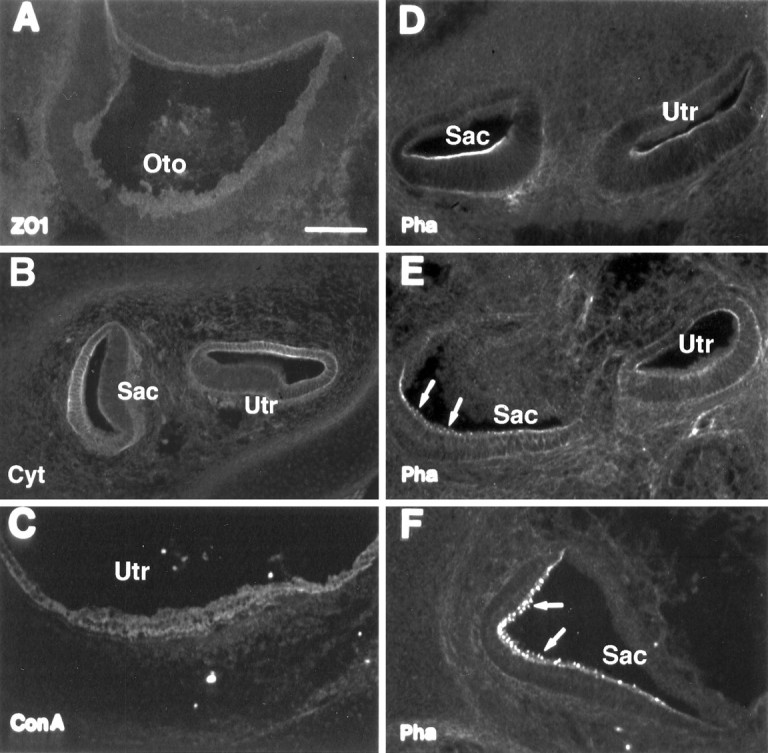
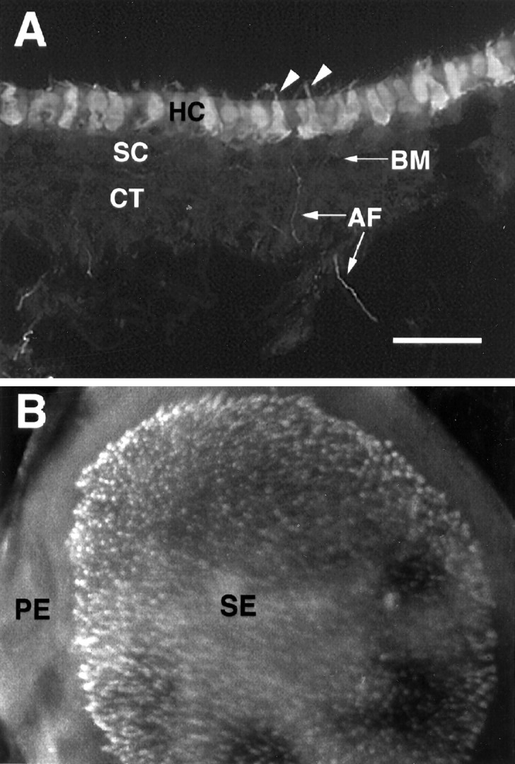
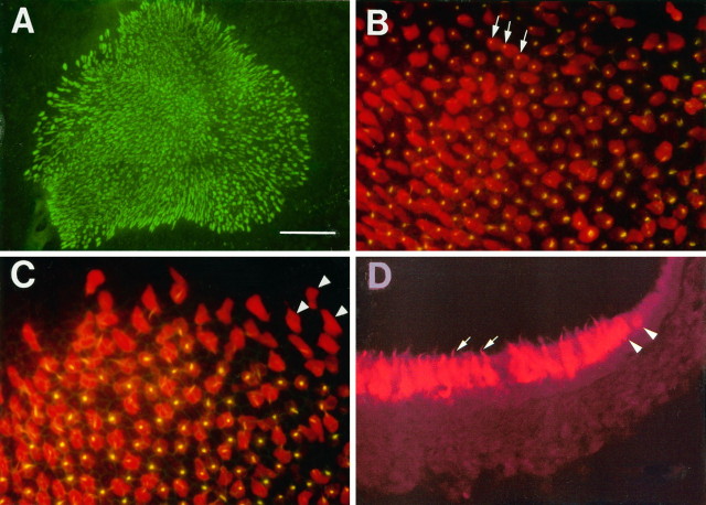
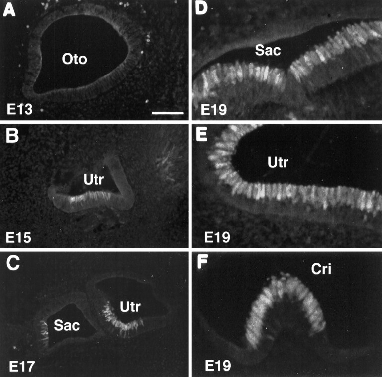
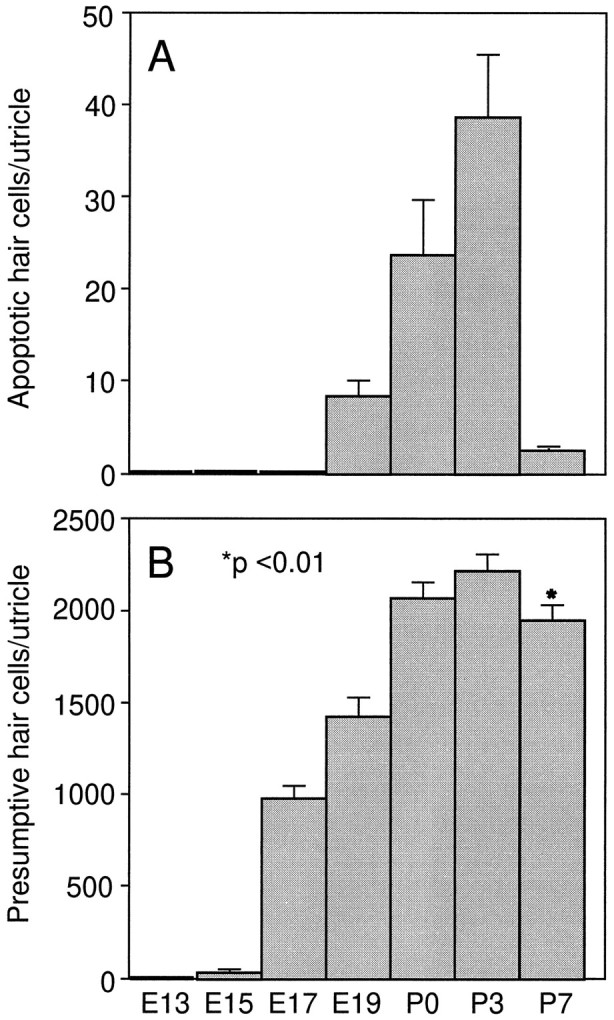
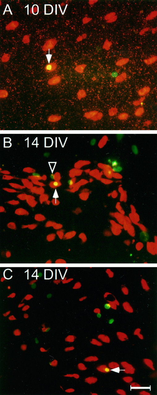
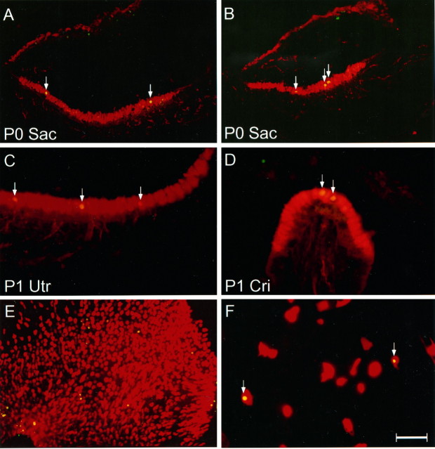
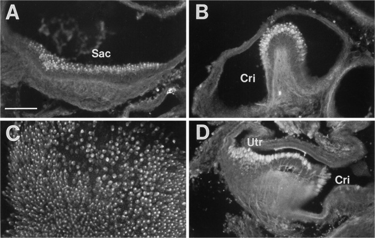
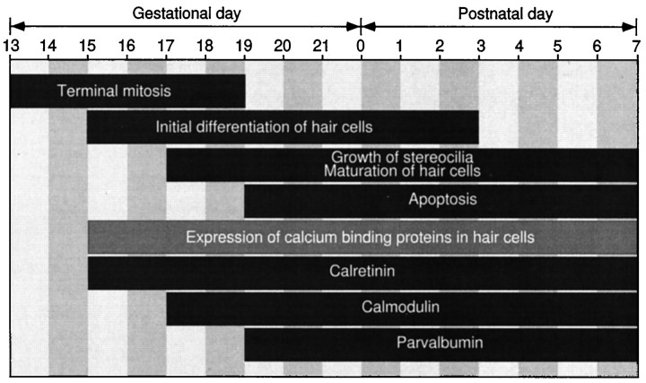
Despite increased interest in inner ear hair cell regeneration, it is still unclear what exact mechanisms underlie hair cell regeneration in mammals because of our limited understanding of hair cell development and the lack of specific hair cell markers. In this report, we studied hair cell development using immunohistochemistry on sections prepared from embryonic day (E) 13 to postnatal day 7 rat inner ear tissues. Of many epithelial, neuronal, and glial markers, we found that calcium-binding protein antibodies recognizing calretinin, calmodulin, or parvalbumin labeled immature hair cells in rat vestibular end organs. In particular, calretinin antiserum labeled the initial differentiating hair cells at E15, a stage immediately after the terminal mitosis of hair cell progenitors. The selective immunoreactivity of postmitotic presumptive hair cells, but not supporting cells or peripheral epithelial cells, was confirmed in utricular epithelial sheet cultures. Double labeling with calretinin and bromodeoxyuridine antibodies in long-term cultures showed that only a few mitotic utricular supporting cells became calretinin positive. Thus, although proliferation-mediated regeneration of new hair cells might directly contribute to hair cell regeneration in rat utricles after injury, it is very limited. In addition, double labeling with calretinin and terminal deoxynucleotidyl transferase-mediated dUTP nick end labeling (TUNEL) revealed that differentiated hair cells underwent apoptosis during normal development at late embryonic and early postnatal stages in vivo and in vitro. Therefore, these experiments lay the groundwork for the time course of differentiation, regeneration, and apoptosis of mammalian vestibular hair cells. This work also suggests that calcium-binding proteins are useful markers for studies on inner ear hair cell differentiation and regeneration.


Figures

Fig. 2.
Fig. 2.
Immunolabeling of developing inner ear tissues with general epithelial markers. A, ZO1 immunostaining at E13. B, Cytokeratin antibody labeling at E17.C, ConA labeling at P0. D–F, Phalloidin labeling at E15, E17, and E19, respectively. Arrowsindicate the appearance of hair cell stereociliary bundles.Oto, Otocyst; Utr, utricle;Sac, saccule. Scale bar (shown in A):A, 50 μm; B–F, 100 μm.
Fig. 1.
Fig. 1.
Calretinin immunolabeling of early postnatal utricular section and whole mount. A, High magnification micrographs of calretinin-labeled hair cells in the lumenal layer of P4 rat utricular epithelium. B, P4 utricular whole mount.Arrowheads indicate hair cell stereociliary bundles.HC, Hair cells; BM, basement membrane;SC, supporting cells; CT, connective tissue; AF, afferent nerve fibers from spiral ganglion neurons; SE, sensory epithelium; PE, peripheral epithelium. Scale bar (shown in A):A, 50 μm; B, 100 μm.
Fig. 4.
Fig. 4.
Calretinin antibody and phalloidin labeling in utricular sheets maintained in cultures for 2 d and in P3 utricular sections. A, Calretinin antibody labeling (FITC-mediated) of a cultured P3 utricular epithelial sheet. B, C, High magnification of cultured P3 utricular epithelial sheets double-labeled with calretinin antibody (red) and phalloidin (green). The yellow color of the stereocilia (arrows) indicates the overlapping satining. D, Calretinin antibody labeling (Texas Red-mediated) of a P3 utricular section showing that two presumptive hair cells (arrowheads) have not acquired stereociliary bundles. Note that a few calretinin-positive cells at the border between sensory epithelium and nonsensory epithelium were not double-labeled by phalloidin (arrowheads) inC. Scale bar (shown in A):A, 200 μm; B, C, D, 50 μm.
Fig. 3.
Fig. 3.
Calretinin immunolabeling of embryonic inner ear tissues. A, B, C, E13, E15, and E17, respectively.D, E, F, The saccule, utricle, and crista at E19, respectively. Note the earliest appearance of calretinin in the lumenal layer of the inner ear epithelium at E15. Oto, Otocyst;Sac, saccule; Utr, utricle;Cri, crista. Scale bar (shown in A):A–C, 100 μm; D–F, 50 μm.
Fig. 7.
Fig. 7.
Cell counts of apoptotic hair cells and total presumptive hair cells in the rat utricle at different developmental time points. A, Number of calretinin and TUNEL double-labeled cells in the utricular epithelium. B, Total number of calretinin-positive presumptive hair cells in the utricle. In A, data were collected from serial cryostat utricular sections prepared from five utricles for each of the various time points. In B, data were collected from serial sections prepared from 5 E13, 5 E15, 5 E19, 5 P0, 10 P3, and 10 P7 rat utricles. Data are expressed as mean ± SEM. There is a statistically significant reduction in hair cell number from P3 to P7 (p < 0.01).
Fig. 5.
Fig. 5.
Double calretinin and BrdU immunocytochemistry in three undissociated utricular epithelial sheet cultures maintained 10–14 DIV. The cultures were exposed to 3 mm gentamicin on the second day for 2 d, and BrdU was included in the medium after gentamicin was added. Texas Red- and FITC-conjugated secondary antibodies were used to reveal the staining patterns of calretinin and BrdU, respectively. Note that the arrows point to the double-labeled presumptive hair cells, and the arrowheadindicates that a supporting cell (BrdU positive but calretinin negative) is localized close to a presumptive newly generated hair cell, which may arise from the same mitotic precursor cell. Scale bar, 40 μm.
Fig. 6.
Fig. 6.
Double labeling of calretinin antibody (red) and TUNEL (green) in neonatal inner ear sections and in cell cultures. A, B, P0 saccules; C, P1 utricle; D, P1 crista;E, F, cultured utricular epithelial sheet and partially dissociated utricular epithelial cells prepared from P3 rat at 2 DIV, respectively. Arrows indicate the apoptotic hair cells. Overlapping label appears yellow. Sac, Saccule; Utr, utricle; Cri, crista. Scale bar (shown in F): A, B, E, 100 μm; C, D, F, 50 μm.
Fig. 8.
Fig. 8.
Immunolabeling of developing inner ear tissues with calmodulin and parvalbumin antibodies. A, B, Calmodulin immunostaining at E20. C, Calmodulin immunostaining of a cultured P4 utricular sheet at 2 DIV.D, Parvalbumin immunostaining at E20.Sac, Saccule; Cri, crista;Utr, utricle. Scale bar, 100 μm.
Fig. 9.
Fig. 9.
Developmental time course of rat vestibular hair cell differentiation and apoptosis. Note that there is a developmental gradient from the central to the peripheral region of the sensory epithelium.

Similar articles

Cited by

References

    1. Adler HJ, Raphael Y. New hair cells arise from supporting cell conversion in the acoustically damaged chick inner ear. Neurosci Lett. 1996;205:17–20. - PubMed
    1. Baimbridge KG, Celio MR, Roger JH. Calcium-binding proteins in the nervous system. Trends Neurosci. 1992;15:303–309. - PubMed
    1. Baird RA, Torres MA, Schuff NR. Hair cell regeneration in the bullfrog vestibular otolith organs following aminoglycoside toxicity. Hear Res. 1993;65:164–174. - PubMed
    1. Baird RA, Steyger PS, Schuff NR. Mitotic and nonmitotic hair cell regeneration in the bullfrog vestibular otolith organs. Ann NY Acad Sci. 1996;781:59–70. - PubMed
    1. Balak KJ, Corwin JT, Jones JE. Regenerated hair cells can originate from supporting cell progeny: evidence from phototoxicity and laser ablation experiments in the lateral line system. J Neurosci. 1990;10:2505–2512. - PMC - PubMed

MeSH terms

LinkOut - more resources