Comparative Study
. 1997 Nov 1;17(21):8283-92.
doi: 10.1523/JNEUROSCI.17-21-08283.1997.

Angiotensin II AT1A receptor mRNA expression is induced by estrogen-progesterone in dopaminergic neurons of the female rat arcuate nucleus

Affiliations
Comparative Study

Angiotensin II AT1A receptor mRNA expression is induced by estrogen-progesterone in dopaminergic neurons of the female rat arcuate nucleus

O Jöhren et al. J Neurosci. .

Abstract

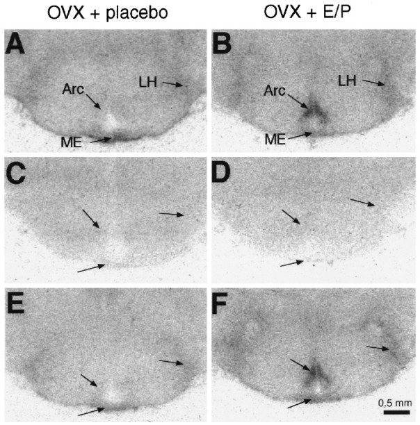
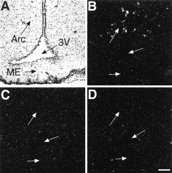
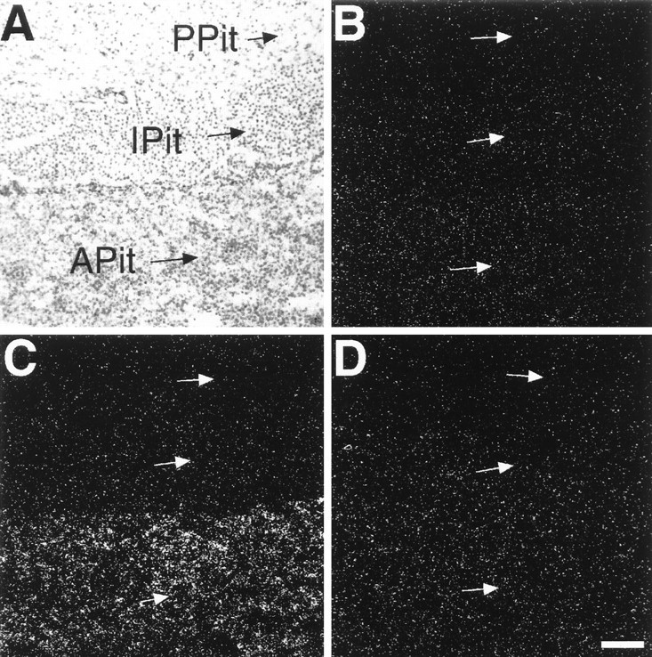
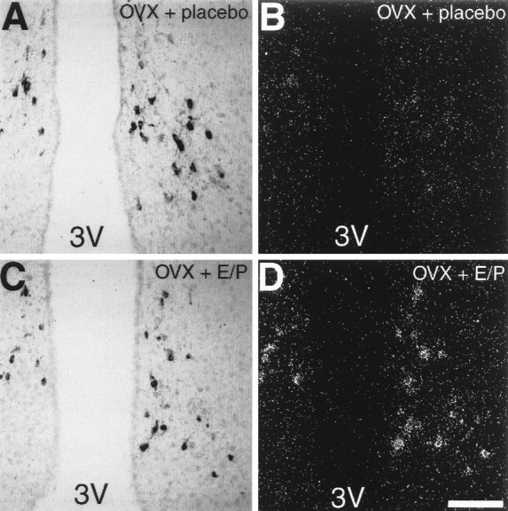
Brain angiotensin II (Ang II) inhibits pituitary prolactin release by an indirect mechanism requiring stimulation of dopamine formation and release. We report that [125I]Sar1-Ang II binding to AT1 receptors and AT1A receptor mRNA expression increase selectively in the dorsomedial arcuate nucleus of 17beta-estradiol-primed ovariectomized rats after treatment with progesterone. In hormone-treated rats, arcuate nucleus AT1A receptor mRNA expression is associated with tyrosine hydroxylase-positive neurons. No AT1A receptor mRNA was detected in tyrosine hydroxylase-positive cells of the arcuate nucleus of intact male rats. Conversely, in the anterior pituitary, where local or circulating Ang II stimulates prolactin release, [125I]Sar1-Ang II binding to AT1 receptors and AT1B receptor mRNA expression are decreased in 17beta-estradiol/progesterone-treated ovariectomized rats. Thus, AT1A receptors in the dorsal arcuate nucleus and AT1B receptors in the anterior pituitary are regulated inversely by estrogen/progesterone treatment, supporting the hypothesis of a dual role for brain and pituitary Ang II on prolactin release. The colocalization of AT1A receptor mRNA and tyrosine hydroxylase in neurons of the arcuate nucleus furthermore indicates that within this area central Ang II acts directly on dopaminergic neurons. These results support the hypothesis that central Ang II inhibits pituitary prolactin release indirectly via modulation of dopaminergic activity in the arcuate nucleus.


Figures

Fig. 10.
Fig. 10.
Bright-field (A) and dark-field (B) photomicrographs showing tyrosine hydroxylase-like immunoreactivity (arrows) and the absence of AT1A receptor mRNA (B) in neurons of the arcuate nucleus of male rats. Sections were double-labeled for AT1A receptor mRNA and tyrosine hydroxylase-like immunoreactivity. No immunoreactivity was observed in the arcuate nucleus after immunostaining of an adjacent section without the first antibody against tyrosine hydroxylase (C). B is a dark-field view ofA. 3V, Third ventricle. Scale bar, 100 μm (applies to all panels).
Fig. 9.
Fig. 9.
Photomicrographs showing tyrosine hydroxylase-like immunoreactivity and AT1A receptor mRNA in the A13 cell group (A, B) and the lateral hypothalamus (C, D) of estrogen/progesterone-treated rats. Sections were double-labeled for AT1A receptor mRNA and tyrosine hydroxylase-like immunoreactivity. Note the presence of scattered AT1A receptor mRNA-expressing neurons in the lateral hypothalamus (arrows). B andD are dark-field views of A andC, respectively. ZI, Zona incerta;LH, lateral hypothalamus. Scale bar, 100 μm (applies to all panels).
Fig. 1.
Fig. 1.
Film autoradiographs of coronal brain sections showing [125I]Sar1–Ang II binding in the arcuate nucleus (Arc), the lateral hypothalamus (LH), and the median eminence (ME) of placebo-treated OVX rats (A, C, E) and estrogen/progesterone-treated OVX rats (B, D, F). Brain sections were incubated with 5 × 10⁻¹⁰m [125I]Sar1–Ang II alone (A, B; total binding), or they were incubated in the presence of 10−5m the AT1 receptor antagonist losartan (C, D) or 10−7m the AT2 receptor ligand CGP 42112 (E, F).
Fig. 2.
Fig. 2.
Film autoradiographs of sections from pituitary showing [125I]Sar1–Ang II binding in the anterior lobe of the pituitary (APit) of placebo-treated OVX rats (A, C, E) and estrogen/progesterone-treated OVX rats (B, D, F). Sections were incubated with 5 × 10⁻¹⁰m [125I]Sar1–Ang II alone (A, B; total binding), or they were incubated in the presence of 10−5m the AT1 receptor antagonist losartan (C, D) or 10−7m the AT2 receptor ligand CGP 42112 (E, F). PPit, Posterior lobe of the pituitary.
Fig. 3.
Fig. 3.
Quantitative analysis of Ang II receptor subtypes in the arcuate nucleus, the median eminence, the lateral hypothalamus, and the anterior pituitary of placebo-treated OVX rats and estrogen/progesterone-treated OVX rats. Shown is the mean ± SEM obtained by quantitative autoradiography from six rats per group.specific, Amount of total [125I]Sar1–Ang II binding displaced by 5 × 10⁻⁶m unlabeled Ang II (specific binding); AT1, amount of total binding displaced by 10−5m losartan; *p ≤ 0.001 (arcuate nucleus) or p ≤ 0.01 (anterior pituitary).
Fig. 4.
Fig. 4.
Bright-field (A) and dark-field (B–D) photomicrographs taken from adjacent coronal brain sections through the arcuate nucleus of estrogen/progesterone-treated OVX rats after in situhybridization with AT1A receptor-specific (A, B) and AT1Breceptor-specific (C) antisense probes or with control AT1A receptor sense probes (D). B is a dark-field view ofA. Arc, Arcuate nucleus;ME, median eminence; 3V, third ventricle. Scale bar, 100 μm (applies to all panels).
Fig. 6.
Fig. 6.
Bright-field (A) and dark-field (B–D) photomicrographs taken from adjacent pituitary sections of placebo-treated OVX rats after in situ hybridization with AT1A(B) and AT1B (A,C) receptor-specific antisense probes or with control AT1A receptor sense probes (D).C is a dark-field view of A.APit, Anterior lobe of the pituitary;IPit, intermediate lobe of the pituitary;PPit, posterior lobe of the pituitary. Scale bar, 100 μm (applies to all panels).
Fig. 5.
Fig. 5.
Semiquantitative analysis of AT1A and AT1B receptor mRNA levels in the arcuate nucleus, the median eminence, the lateral hypothalamus, and the anterior pituitary of OVX rats and estrogen/progesterone-treated OVX rats. Silver grains were counted over single cells after in situhybridization, using AT1A and AT1Breceptor-specific antisense or control sense probes. Shown is the mean ± SEM of silver grains per cell from six rats per group; *p ≤ 0.001.
Fig. 7.
Fig. 7.
Photomicrographs showing tyrosine hydroxylase-like immunoreactivity (A, C) and AT1A receptor mRNA (B, D) in neurons of the arcuate nucleus of placebo-treated OVX rats (A, B) and estrogen/progesterone-treated OVX rats (C, D). Sections were double-labeled for AT1A receptor mRNA and tyrosine hydroxylase-like immunoreactivity. B andD are dark-field views of A andC, respectively. 3V, Third ventricle. Scale bar, 100 μm (applies to all panels).
Fig. 8.
Fig. 8.
High-power photomicrographs illustrate the colocalization of AT1A receptor mRNA expression with tyrosine hydroxylase immunoreactivity in neurons of the arcuate nucleus of estrogen/progesterone-treated OVX rats (arrows). B shows a polarized epifluorescence illumination of A to visualize silver grains. Note the absence of silver grains over one tyrosine hydroxylase-positive neuron (arrowhead). Scale bar, 20 μm.

Similar articles

Cited by

References

    1. Aguilera G, Hyde C, Catt KJ. Angiotensin II receptors and prolactin release in pituitary lactotrophs. Endocrinology. 1982;111:1045–1050. - PubMed
    1. Arbogast LA, Voogt JL. Progesterone reverses the estradiol-induced decrease in tyrosine hydroxylase mRNA levels in the arcuate nucleus. Neuroendocrinology. 1993;58:501–510. - PubMed
    1. Barron WM, Schreiber J, Lindheimer MD. Effect of ovarian sex steroids on osmoregulation and vasopressin secretion in the rat. Am J Physiol. 1986;250:E352–E361. - PubMed
    1. Becú-Villalobos D, Lacau-Mengido IM, Thyssen SM, Díaz-Torga GS, Libertun C. Effects of LHRH and Ang II on prolactin stimulation are mediated by hypophysial AT1 receptor subtype. Am J Physiol. 1994;266:E274–E278. - PubMed
    1. Ben-Jonathan N, Oliver C, Weiner HJ, Mical RS, Porter JC. Dopamine in hypophysial portal plasma of the rat during the estrous cycle and throughout pregnancy. Endocrinology. 1977;100:452–458. - PubMed

Publication types

MeSH terms