Comparative Study
. 1997 Nov 1;17(21):8300-12.
doi: 10.1523/JNEUROSCI.17-21-08300.1997.

Expression of neural RNA-binding proteins in the postnatal CNS: implications of their roles in neuronal and glial cell development

Affiliations
Comparative Study

Expression of neural RNA-binding proteins in the postnatal CNS: implications of their roles in neuronal and glial cell development

S Sakakibara et al. J Neurosci. .

Abstract

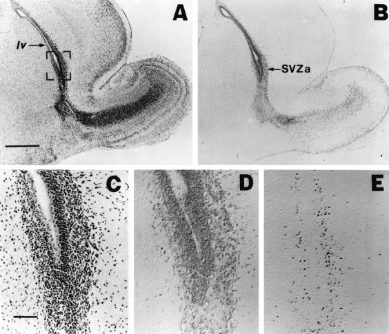
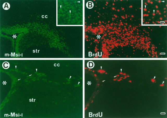
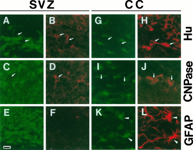
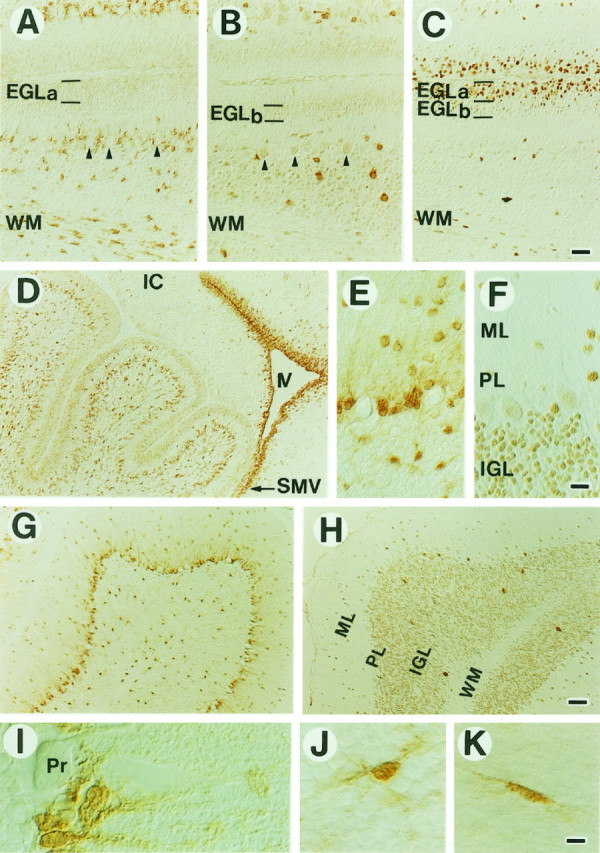
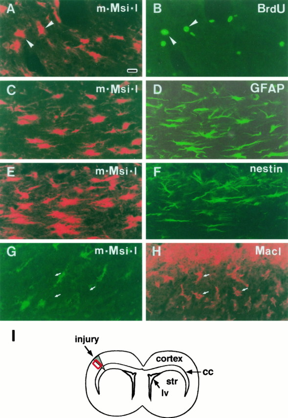
There is an increasing interest in the role of RNA-binding proteins during neural development. Mouse-Musashi-1 (m-Msi-1) is a mouse neural RNA-binding protein with sequence similarity to Drosophila musashi (d-msi), which is essential for neural development. m-Msi-1 is highly enriched in neural precursor cells that are capable of generating both neurons and glia during embryonic CNS development. The present study characterized m-Msi-1-expressing cells in the postnatal and adult CNS. Postnatally, m-Msi-1 was expressed in proliferative neuronal precursors in the external granule cell layer of the cerebellum and in the anterior corner of the subventricular zone of the lateral ventricles. In gliogenesis, the persistent expression of m-Msi-1 was observed in cells of the astrocyte lineage ranging from proliferative glial precursors in the subventricular zone (SVZ) to differentiated astrocytes in the parenchyma. In addition, we showed that m-Msi-1 was still expressed in proliferating cells in the adult SVZ, which may contain neural precursor or stem cells. Another neural RNA-binding protein Hu (the mammalian homolog of a Drosophila neuronal RNA-binding protein Elav) was present in postmitotic neurons throughout the development of the CNS, and its pattern of expression was compared with that of m-Msi-1. These observations imply that these two RNA-binding proteins may be involved in the development of neurons and glia by regulating gene expression at the post-transcriptional level.


Figures

Fig. 1.
Fig. 1.
m-Msi-1 expression in P3 forebrain. Serial sagittal sections of the forebrain at P3 with hematoxylin–eosin staining (A, C), immunolocalizations of m-Msi-1 (B, D), and PCNA (E). Anterior is to the right and dorsal is up. C–E, Higher magnifications of the anterior corner of the lateral ventricle (SVZa) shown in the bracketed portion ofA. m-Msi-1 immunostaining was observed in PCNA-positive proliferating cells in SVZa. Note that m-Msi-1 expression was also detected in the posterior SVZ in addition to the migratory route of neuronal precursor cells from SVZa into the olfactory bulb described previously (Luskin, 1993). Scale bars: A, B, 500 μm;C–E, 50 μm. SVZa, Anterior region of the SVZ; lv, lateral ventricle.
Fig. 6.
Fig. 6.
Immunolocalization of m-Msi-1 and Hu in the developing and adult cerebrum. A, B, Serial sagittal sections showing the expression of m-Msi-1 (A) and PCNA (B) in the SVZ and the adjacent intermediate zone at P3. Dorsal is to the right. The lateral ventricle is marked with asterisks. At P3, m-Msi-1 is strongly expressed in tightly packed, small round cells in the SVZ, which seem to correspond to PCNA-positive proliferating cells. It is also noted that there are some m-Msi-1-positive cells with a few processes extending into the dorsal parenchyma as if they were migrating out of the SVZ. C–F, Serial sagittal sections of the P7 (C, D) and adult (E, F) cerebral cortex showing the distribution of m-Msi-1 (C, E) and Hu antigens (D, F). The pial surface is at the top. G–J, High-power photomicrographs of individual cells expressing m-Msi-1 in the gray matter (layers III–IV, H and I) and white matter (layer I, G; subcortical white matter,J) in the adult cerebral cortex. By P7, in addition to the high level of expression of Msi-1 in the SVZ, small scattered m-Msi-1-positive cells become localized throughout the cortex. At the same time, Hu expression is almost confined to neurons residing in the cortical gray matter. In the adult cerebral cortex, a larger number of m-Msi-1-positive cells are distributed uniformly in both gray and white matter, and these have small oval cell bodies with multiple short processes (G, I) or bipolar processes (H, J). Hu antigens continue to be expressed in large round neurons in the gray matter.MZ, Marginal zone; CP, cortical plate;CC, corpus callosum; SVZ, subventricular zone. I, II, III, V, and VI represent the cortical layers with the same designation. K–N, Double-immunofluorescent labeling of sections through the adult cerebral cortex: m-Msi-1 (K), GFAP (L), m-Msi-1 (M), and CNPase (N). m-Msi-1-positive cells, which have multiple branched processes, are seen throughout the cortex. Astrocytes in the superficial molecular layer and near the pial surface show colocalization of m-Msi-1 and GFAP (arrowheads inK and L), whereas CNPase-positive oligodendroglial cell bodies predominantly present in the molecular layer are m-Msi-1-negative (arrows in Mand N). Scale bars: A, B, 18 μm; C, D, 71 μm; E, F, 36 μm; G–J, 8 μm; J–N, 10 μm.
Fig. 2.
Fig. 2.
m-Msi-1 expression in proliferating cells residing in the postnatal and adult SVZ. To label the entire constitutively proliferating population surrounding the lateral ventricles, P7 and adult mice received 3 and 12.5 hr of BrdU injections, respectively. Double-immunofluorescent labeling of coronal sections through the SVZ surrounding the lateral ventricle in P7 forebrain (A, B) and adult forebrain (C, D) with antibodies to m-Msi-1 (A, C; FITC) and BrdU (B, D; Cy3). Lateral is to the right and dorsal is up. Insets inA and B, Higher magnifications of the dorsolateral corner of the P7 SVZ. Many dividing, small, densely packed cells that are brightly immunostained with both m-Msi-1 and BrdU are observed in the postnatal developing SVZ (arrowheads ininsets in A and B). Similarly, a considerable number of m-Msi-1- and BrdU-positive dividing cells are also present in the adult subependyma (arrowheads in C and D), although it is obvious that there is a subpopulation of m-Msi-1-positive but BrdU-negative cells (arrows inC and D). Scale bars: A, B, 36 μm; insets, C and D, 18 μm. Asterisks, Lateral ventricle; cc, corpus callosum; str, striatum.
Fig. 3.
Fig. 3.
Cell type of m-Msi-1-positive cells in the developing SVZ and the corpus callosum. Double-labeling of coronal sections just dorsolateral to the region of the P7 SVZ (A–F) and the P7 corpus callosum (G–L) with anti-m-Msi-1 antibody (A, C, E, G, I, and K; FITC), anti-Hu (B, H; Cy3), anti-CNPase (D, J; Cy3), and anti-GFAP (F, L; Cy3). The bar in Erepresents 18 μm. Antibodies to CNPase and Hu label populations that are distinct from m-Msi-1-positive cells in both the SVZ and the corpus callosum. The arrows point to the Hu-positive but m-Msi-1-negative cells (A, B, G, H), and CNPase-positive but m-Msi-1-negative cells (C, D, I, J). Although GFAP immunoreactivity is not detected in the SVZ, a few m-Msi-1- and GFAP-positive cells that have multiple processes or short branched processes are now present in the developing corpus callosum (arrowheads in K andL) among the more numerous m-Msi-1-positive cells.
Fig. 4.
Fig. 4.
Top. Absence of m-Msi-1 expression in PDGFα-R-positive, early O-2A cells lying in the SVZ. Scanning confocal images representing the expression of PDGFα-R (red) and m-Msi-1 (green).A, Coronal section through the SVZ surrounding the dorsolateral corner of the lateral ventricle and the adjacent corpus callosum of the P2 mouse forebrain. B, Higher magnification of the SVZ surrounding the lateral ventricle. Many PDGFα-R-positive cells, which seem to have small cell bodies with a few processes, are observed not only in the corpus callosum and the striatum but also in the SVZ, where m-Msi-1-positive small cells are condensed. Note that these two populations of cells distribute intermingled but never overlap with each other. Scale bars: A, 20 μm;B, 10 μm. lv, Lateral ventricle.
Fig. 7.
Fig. 7.
Distribution of m-Msi-1 and Hu in the developing (A–D) and adult (E–K) cerebellum. D, m-Msi-1 expression in the P3 cerebellum. m-Msi-1 is expressed in neuronal precursor cells in the external granule cell layer (EGL) and in the vast number of small cells that have a few processes in the deep cerebellar regions. Intense m-Msi-1 staining is also noted in densely packed small cells in the SVZ lining the fourth ventricle (IV) and in the superior medullary velum (SMV) located at the base of the cerebellum.A–C, Serial sagittal sections showing the immunolocalization of m-Msi-1 (A), Hu (B), and PCNA (C) in the EGL, the deep cerebellar regions, and folia white matter (WM) at P7. The EGL is toward thetop of the panels. At P7, weak expression of m-Msi-1 is seen mainly in the EGLa, which contains the PCNA-positive outer proliferating zone of the EGL, whereas Hu expression is observed in the EGLb, which contains the PCNA-negative inner early differentiating neurons. Granule neurons within the internal granule layer (IGL) and Purkinje cells (arrowheads) forming a single row show immunoreactivity for Hu but not for m-Msi-1. m-Msi-1 expression is retained by small oval cells that have a few processes and reside in the deep cerebellar regions and WM. G, H, Sagittal sections showing the expression of m-Msi-1 (G) and Hu (H) in the adult cerebellum.E, F, Higher magnifications of the molecular layer (ML), Purkinje cell layer (PL), and IGL shown in G and H, respectively.I–K, High-power photomicrographs of individual cells expressing m-Msi-1 in the PL (I), IGL (J), and WM (K) of the adult cerebellum. In the PL, m-Msi-1 is expressed in Bergmann glia, the cell bodies of which are located adjacent to the large cell bodies of Purkinje cells (Pr) and extend their tangential processes into the ML. In addition, sparsely distributed putative astrocytes in the IGL and WM, which exhibit oval or elongated cell bodies with multiple short processes (J) or bipolar processes (K), are labeled with the m-Msi-1 antibody. IC, Inferior colliculus. Scale bars:A–C, 36 μm; D, G, H, 71 μm;E, F, 25 μm; I–K, 8 μm.
Fig. 8.
Fig. 8.
m-Msi-1 expression in reactive astrocytes in the injured region of adult cerebral cortex. Double-label fluorescent localization of m-Msi-1 and BrdU, GFAP, nestin, or MacI at 4 d postlesion. I, Schematic illustration of a coronal section through the injured forebrain. All photomicrographs correspond to the boxed region in I. The lesioned sites are toward the top in all panels. A, m-Msi-1 (Cy3). B, Same field, BrdU (FITC). To label a proliferating population after brain injury, mice received BrdU injections for 3 hr before they were killed. m-Msi-1 is expressed in a population of BrdU-positive proliferating cells (arrowheads) that lie close to the injury site and have enlarged cell bodies with multiple processes. C, m-Msi-1 (Cy3). D, Same field, GFAP (FITC). Intense m-Msi-1 staining is observed in an increased number of GFAP-positive reactive astrocytes surrounding the lesioned site; these cells exhibit enlarged, elongated cell bodies with multiple processes. E, m-Msi-1 (Cy3). F, Same field, nestin (FITC). The simultaneous expression of nestin is induced in a subpopulation of m-Msi-1-positive reactive cells near the lesioned site.G, m-Msi-1 (FITC). H, Same field, MacI (Cy3). Large numbers of macrophages or ameboid microglia, which are MacI-positive but m-Msi-1-negative, form a dense plaque surrounding the lesion site. Arrows indicate the MacI-positive ramified microglia that are immunonegative for m-Msi-1. Scale bar (shown inA): 18 μm. lv, Lateral ventricle;cc, corpus callosum; str, striatum.
Fig. 9.
Fig. 9.
Schematic representation of cells expressing m-Msi-1 during embryonic and postnatal CNS development. Solid patterns represent m-Msi-1-positive cells. In the embryonic CNS, m-Msi-1 is expressed in proliferating neural precursors in the ventricular zone (VZ); these cells are capable of self-renewal and generate postmitotic neurons, as described previously (Sakakibara et al., 1996). In the perinatal stage, the VZ shrinks and a second proliferative SVZ appears. Postnatally, m-Msi-1 expression is seen in proliferating glial precursors in the SVZ and cells of astrocyte lineage, including ependymal cells and Bergmann glia. At present, it is not known whether neural precursors in the embryonic VZ generate glial precursors in the postnatal SVZ, or whether bipotent glial precursors in the postnatal SVZ generate the astrocyte and O-2A-oligodendrocyte lineages through an “asymmetric cell division.” However, this asymmetric division at least may occur when neuronal precursors give rise to neurons in the embryonic VZ (Chenn and McConnell, 1995). The expression of m-Msi-1 in some populations of neurons is not represented for reasons of simplicity.

Similar articles

Cited by

References

    1. Altman J. Postnatal development of the cerebellar cortex in the rat. J Comp Neurol. 1972;145:353–398. - PubMed
    1. Bignami A, Dahl D. Astrocyte-specific protein and neuroglial differentiation. An immunofluorescence study with antibodies to the glial fibrillary acidic protein. J Comp Neurol. 1974;153:27–38. - PubMed
    1. Bignami A, Dahl D. The astroglial response to stabbing. Immunofluorescence studies with antibodies to astrocyte-specific protein (GFA) in mammalian and submammalian vertebrates. Neuropathol Appl Neurobiol. 1976;2:99–110.
    1. Chenn A, McConnell SK. Cleavage orientation and the asymmetric inheritance of Notch1 immunoreactivity in mammalian neurogenesis. Cell. 1995;82:631–641. - PubMed
    1. Craig CG, Tropepe V, Morshead CM, Reynolds BA, Weiss S, van der Kooy D. In vivo growth factor expansion of endogenous subependymal neural precursor cell populations in the adult mouse brain. J Neurosci. 1996;16:2649–2658. - PMC - PubMed

Publication types

MeSH terms

LinkOut - more resources