

Origin and route of tangentially migrating neurons in the developing neocortical intermediate zone
- PMID: 9334406
- PMCID: PMC6573720
- DOI: 10.1523/JNEUROSCI.17-21-08313.1997
Origin and route of tangentially migrating neurons in the developing neocortical intermediate zone
Abstract
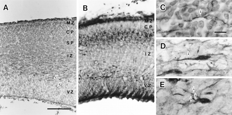
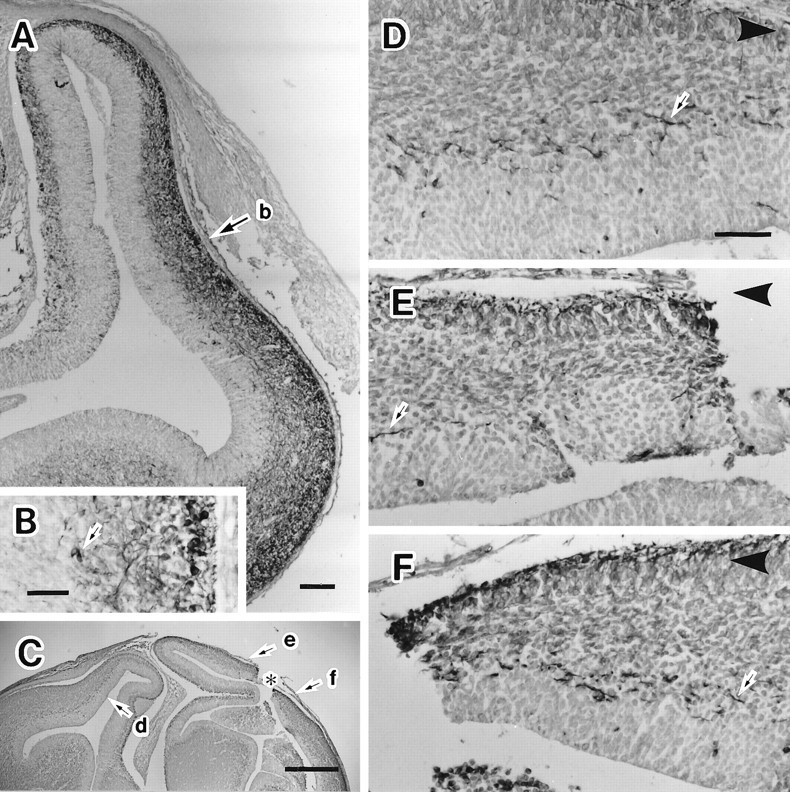
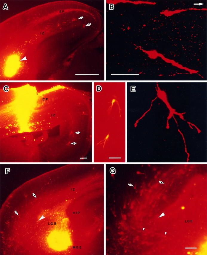
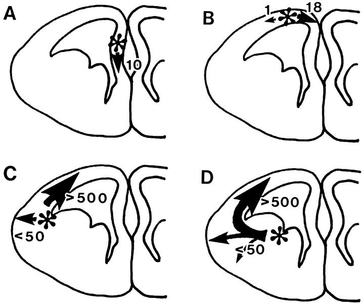
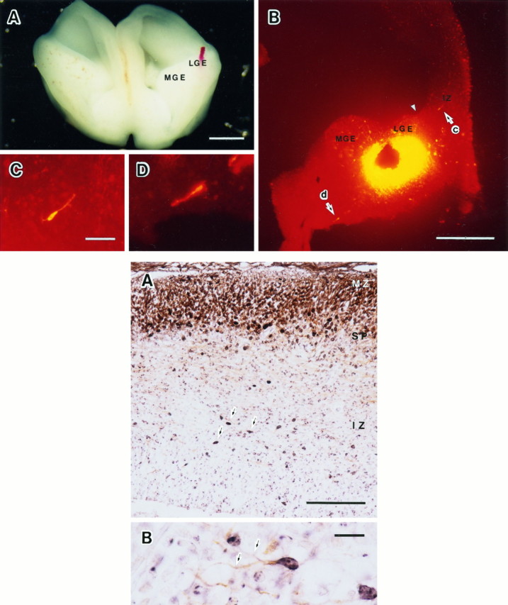
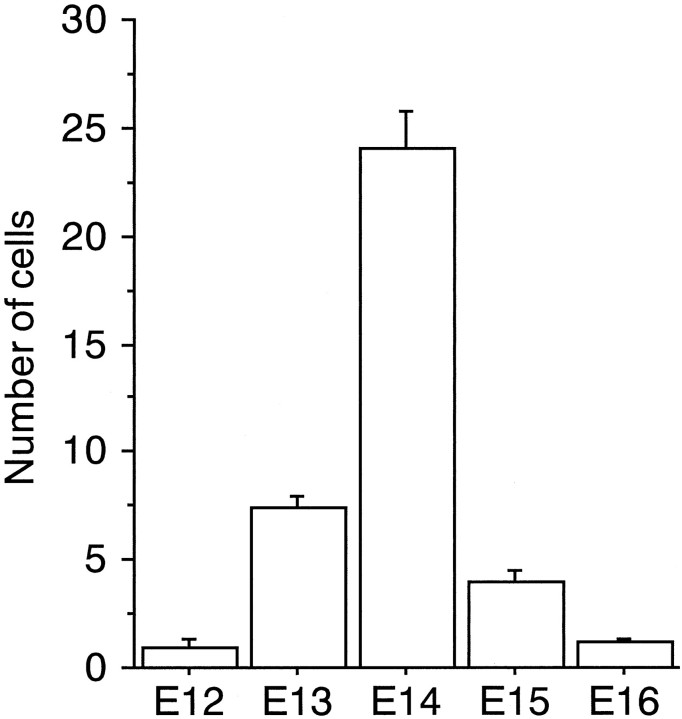
Neuroblasts produced in the ventricular zone of the neocortex migrate radially and form the cortical plate, settling in an inside-out order. It is also well known that the tangential cell migration is not negligible in the embryonic neocortex. To have a better understanding of the tangential cell migration in the cortex, we disturbed the migration by making a cut in the neocortex, and we labeled the migrating cells with 1,1'-dioctodecyl-3,3,3',3'-tetramethylindocarbocyanine perchlorate (DiI) in vivo and in vitro. We also determined the birth dates of the cells. Disturbance of tangential cell migration caused an accumulation and disappearance of microtubule-associated protein 2 immunoreactive (MAP2-IR) cells on the ventral and dorsal side of the cut, respectively, which indicated that most of the MAP2-IR cells in the intermediate zone (IZ) were migrating toward the dorsal cortex. The DiI injection study in vivo confirmed the tendency of the direction of cell migration and suggested the origin of the cells to be in the lateral ganglionic eminence (LGE). DiI injection into the LGE in vitro confirmed that the LGE cells cross the corticostriatal boundary and enter the IZ of the neocortex. The migrating cells acquired multipolar shape in the IZ of the dorsal cortex and seemed to reside there. A 5-bromo-deoxyuridine incorporation study revealed that the migrating MAP2-IR cells in the IZ were early-generated neurons. We concluded that the majority of tangentially migrating cells were generated in the LGE and identified as a distinct population that was assumed not to have joined the cortical plate.
Figures
Similar articles
-
Differential localization of high- and low-molecular-weight variants of microtubule-associated protein 2 in the developing rat telencephalon.J Comp Neurol. 2002 Aug 5;449(4):330-42. doi: 10.1002/cne.10286. J Comp Neurol. 2002. PMID: 12115669
-
Evidence that Sema3A and Sema3F regulate the migration of GABAergic neurons in the developing neocortex.J Comp Neurol. 2003 Jan 6;455(2):238-48. doi: 10.1002/cne.10476. J Comp Neurol. 2003. PMID: 12454988
-
Tangential migration in neocortical development.Dev Biol. 2002 Apr 1;244(1):155-69. doi: 10.1006/dbio.2002.0586. Dev Biol. 2002. PMID: 11900465
-
Involvement of filamin A and filamin A-interacting protein (FILIP) in controlling the start and cell shape of radially migrating cortical neurons.Anat Sci Int. 2005 Mar;80(1):19-29. doi: 10.1111/j.1447-073x.2005.00101.x. Anat Sci Int. 2005. PMID: 15794127 Review.
-
Telencephalic cells take a tangent: non-radial migration in the mammalian forebrain.Nat Neurosci. 2001 Nov;4 Suppl:1177-82. doi: 10.1038/nn749. Nat Neurosci. 2001. PMID: 11687827 Review.
Cited by
-
Accumulation of GABAergic neurons, causing a focal ambient GABA gradient, and downregulation of KCC2 are induced during microgyrus formation in a mouse model of polymicrogyria.Cereb Cortex. 2014 Apr;24(4):1088-101. doi: 10.1093/cercor/bhs375. Epub 2012 Dec 16. Cereb Cortex. 2014. PMID: 23246779 Free PMC article.
-
Sonic hedgehog and bone morphogenetic protein regulate interneuron development from dorsal telencephalic progenitors in vitro.J Neurosci. 2003 Oct 29;23(30):9862-72. doi: 10.1523/JNEUROSCI.23-30-09862.2003. J Neurosci. 2003. PMID: 14586015 Free PMC article.
-
Effects of Developmental Nicotine Exposure on Frontal Cortical GABA-to-Non-GABA Neuron Ratio and Novelty-Seeking Behavior.Cereb Cortex. 2020 Mar 14;30(3):1830-1842. doi: 10.1093/cercor/bhz207. Cereb Cortex. 2020. PMID: 31599922 Free PMC article.
-
DISC1: a key lead in studying cortical development and associated brain disorders.Neuroscientist. 2013 Oct;19(5):451-64. doi: 10.1177/1073858412470168. Epub 2013 Jan 8. Neuroscientist. 2013. PMID: 23300216 Free PMC article. Review.
-
The external granule layer of the developing chick cerebellum generates granule cells and cells of the isthmus and rostral hindbrain.J Neurosci. 2001 Jan 1;21(1):159-68. doi: 10.1523/JNEUROSCI.21-01-00159.2001. J Neurosci. 2001. PMID: 11150332 Free PMC article.
References
-
- Altman J, Das GD. Autoradiographic and histological studies of postnatal neurogenesis. 1. A longitudinal investigation of the kinetics, migration and transformation of cells incorporating tritiated thymidine in neonate rats, with special reference to postnatal neurogenesis in some brain regions. J Comp Neurol. 1966;127:337–390. - PubMed
-
- Anderson SA, Eisenstat D, Shi L, Rubenstein JLR (1997) Interneuron migration from basal forebrain to neocortex: dependence on Dlx genes. Science, in press. - PubMed
-
- Angevine JB, Jr, Sidman RL. Autoradiographic study of cell migration during histogenesis of cerebral cortex in the mouse. Nature. 1961;192:766–768. - PubMed
-
- Austin CP, Cepko CL. Cellular migration patterns in the developing mouse cerebral cortex. Development. 1990;110:713–732. - PubMed
-
- Bayer SA. 3H-thymidine-radiographic studies of neurogenesis in the rat olfactory bulb. Exp Brain Res. 1983;50:329–340. - PubMed
Publication types
MeSH terms
Substances
LinkOut - more resources
Full Text Sources
Other Literature Sources