Comparative Study
. 1997 Nov 1;17(21):8402-7.
doi: 10.1523/JNEUROSCI.17-21-08402.1997.

Developmental synaptic depression underlying reorganization of visceral reflex pathways in the spinal cord

Affiliations
Comparative Study

Developmental synaptic depression underlying reorganization of visceral reflex pathways in the spinal cord

I Araki et al. J Neurosci. .

Abstract

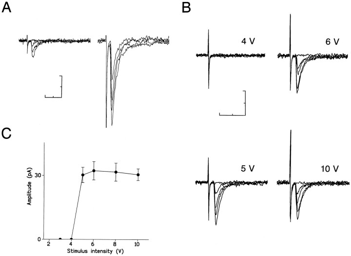
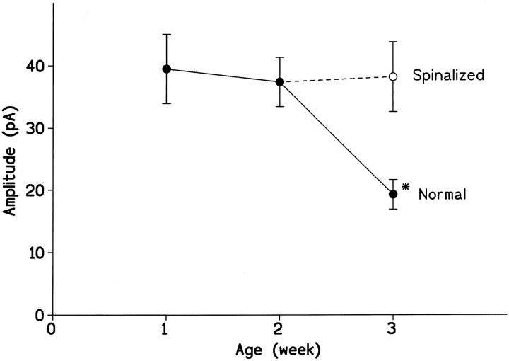
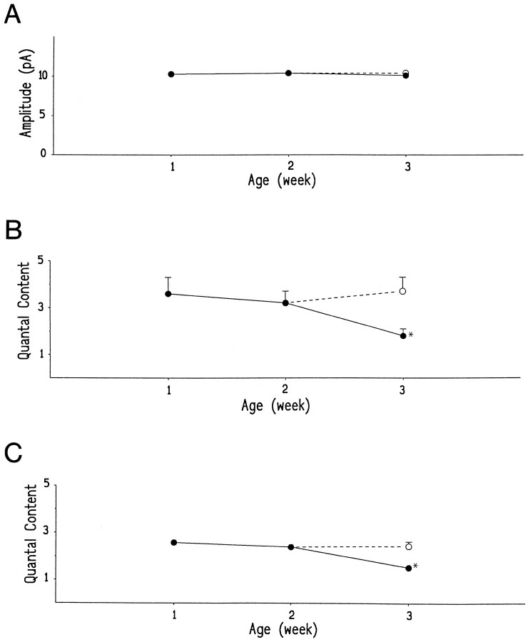
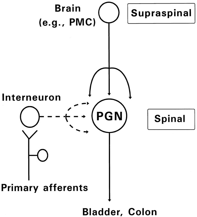
During development, neuronal connectivity has a remarkable plasticity. Synaptic refinement in the spinal autonomic nucleus might be involved in the elimination of primitive segmental reflexes and the emergence of mature spinobulbospinal reflexes, which occurs a few weeks after birth. To address this possibility, we examined the postnatal changes of segmental excitatory synaptic transmission by applying the whole-cell recording technique to parasympathetic preganglionic neurons in slice preparations of the rat lumbosacral spinal cord. The mean magnitude of unitary excitatory synaptic currents evoked in preganglionic neurons by stimulation of single interneurons remained unchanged during the first two postnatal weeks but was reduced by 50% during the third postnatal week. This reduction in synaptic efficacy was associated with a decrease in the amount of transmitter release from interneurons. Moreover, this developmental depression of segmental synaptic transmission was prevented by spinal cord transection at the thoracic level on postnatal day 14. Thus, developmental modification of excitatory synapses on preganglionic neurons appears to be attributable to competition between segmental interneuronal and descending bulbospinal inputs, which results in the developmental reorganization of parasympathetic excretory reflex pathways.


Figures

Fig. 1.
Fig. 1.
Schematic diagram for the neural circuit of parasympathetic spinal segmental reflex and the neurons studied in slice preparations of the lumbosacral cord. PGN, Parasympathetic preganglionic neurons; INT, dorsal interneurons; LCP, lateral collateral pathway of primary afferents; MPG, major pelvic ganglia.
Fig. 2.
Fig. 2.
EPSCs in segmental parasympathetic reflex pathways. A, Composite EPSCs evoked in a dorsal interneuron by stimulation of primary afferent fibers. Four responses evoked by threshold (left) or supramaximal (right) intensities of stimulus are superimposed.B, Unitary EPSCs evoked in a PGN by stimulation of a dorsal interneuron. Five responses at each stimulus intensity indicated above tracing are superimposed. C, Relationship between the stimulus intensity and the mean peak amplitude of 30 consecutive EPSCs (including failures) in the same cell as in B.Vertical bars represent SEM. The holding potential was −60 mV. Scale bars, 10 msec and 40 pA.
Fig. 3.
Fig. 3.
Developmental change in the efficacy of excitatory transmission at interneuronal–PGN synapses and its modification by chronic spinal transection. Mean peak amplitudes of unitary EPSCs recorded from PGNs in intact (•) or spinalized (○) rats are plotted against ages. Mean peak amplitudes were measured from averaged responses of >30 consecutive EPSCs (including failures). The holding potential was −60 mV. Each point represents the mean ± SEM (vertical bar) from 14 or 15 cells. Theasterisk represents significant difference from the values observed in other preparations (p < 0.006).
Fig. 4.
Fig. 4.
Quantal analysis indicated a presynaptic mechanism for the developmental synaptic depression. Peak amplitude distributions of unitary EPSCs recorded from PGNs at a holding potential of −60 mV.A, From an 8-d-old rat. Bin width was 1.6 pA.a, In the standard external solution (2 mmCa2+ and 1 mm Mg2+), 23 failures in 339 trials; b, in an external solution containing 1 mm Ca2+ and 5 mm Mg2+ to reduce the release probability, 124 failures in 363 trials. B, From a 20-d-old rat. Bin width was 1.2 pA, 48 failures in 363 trials.C, From a 21-d-old rat spinalized at T10level 2 weeks after birth. Bin width was 1.6 pA, 27 failures in 581 trials. Insets, Frequency distributions of background noise. The smooth lines superimposed on the histograms represent the sum of Gaussian distributions that best fit the data. In all recordings, the SD of the noise (1.5 ± 0.3 pA;n = 31) was less than that of any Gaussian curve fitted to the EPSC amplitude histogram. The failures of response are not shown.
Fig. 5.
Fig. 5.
Developmental changes of quantal size (quantal amplitude) and quantal content. A, Mean quantal amplitudes measured at a holding potential of −60 mV.B, Mean quantal contents calculated by dividing the mean amplitude of unitary EPSCs by the quantal size in each cell.C, Mean quantal contents calculated from the following equation, based on Poisson’s law: m = loge(N/n0) (m, quantal content; N, total number of trials; and n0, number of failures of response). Mean values obtained from PGNs in intact (•) or spinalized (○) rats are plotted against ages. Each mean value was measured from 7 or 11 cells in which >300 responses were evoked. Eachpoint represents the mean ± SEM (vertical bar). SEMs <0.15 are omitted. The asteriskrepresents significant difference from the values observed in other preparations (p < 0.03).
Fig. 6.
Fig. 6.
Diagram indicating two sources of excitatory inputs to PGNs during development. In neonates (broken lines), interneurons synaptically activated by primary afferent inputs mediate segmental somatovisceral reflexes. In adults (solid lines), descending projections from higher center in the brain (e.g., PMC) control PGN activity. The interneuronal inputs and the bulbospinal inputs could compete for synaptic sites on PGNs during postnatal development.

Similar articles

Cited by

References

    1. Anderson CR, Edwards SL. Intraperitoneal injections of fluorogold reliably labels all sympathetic preganglionic neurons in the rat. J Neurosci Methods. 1994;53:137–141. - PubMed
    1. Araki I. Inhibitory postsynaptic currents and the effects of GABA on visually identified sacral parasympathetic preganglionic neurons in neonatal rats. J Neurophysiol. 1994;72:2903–2910. - PubMed
    1. Araki I, de Groat WC. Unitary excitatory synaptic currents in preganglionic neurons mediated by two distinct groups of interneurons in neonatal rat sacral parasympathetic nucleus. J Neurophysiol. 1996;76:215–226. - PubMed
    1. Balice-Gorden RJ, Lichtman JW. Long-term synapse loss induced by focal blockade of postsynaptic receptors. Nature. 1994;372:519–524. - PubMed
    1. Beach FA. Ontogeny of “coitus-related” reflexes in the female guinea pig. Proc Natl Acad Sci USA. 1966;56:526–533. - PMC - PubMed

Publication types

MeSH terms