. 1997 Nov 1;17(21):8408-26.
doi: 10.1523/JNEUROSCI.17-21-08408.1997.

Transition from growth cone to functional motor nerve terminal in Drosophila embryos

Affiliations

Transition from growth cone to functional motor nerve terminal in Drosophila embryos

M Yoshihara et al. J Neurosci. .

Abstract

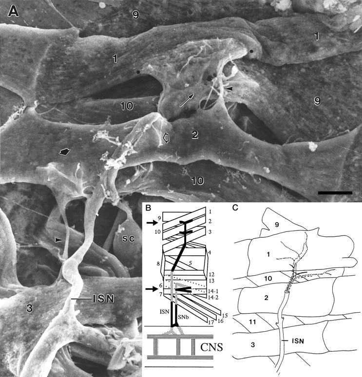
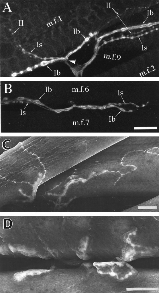
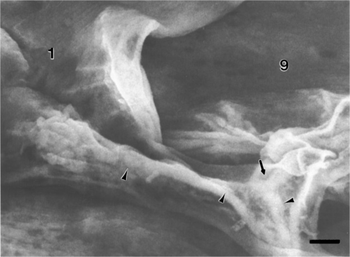
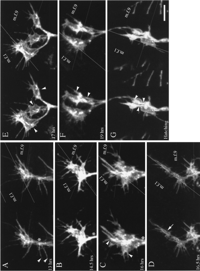
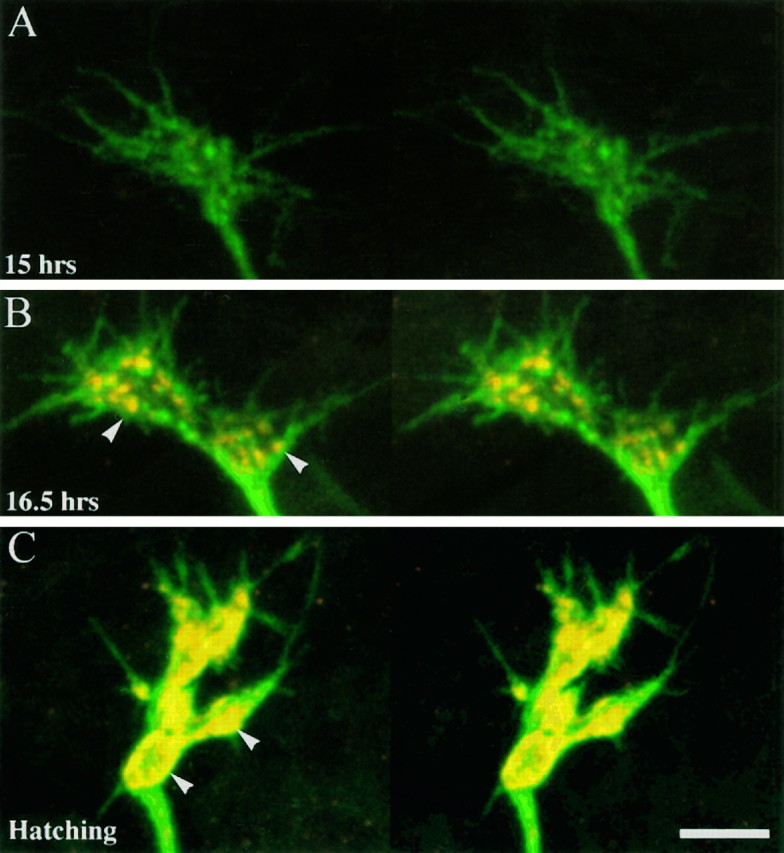
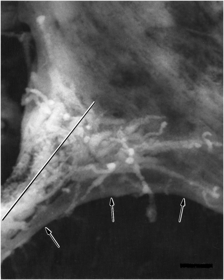
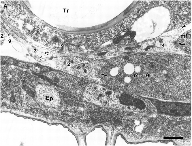
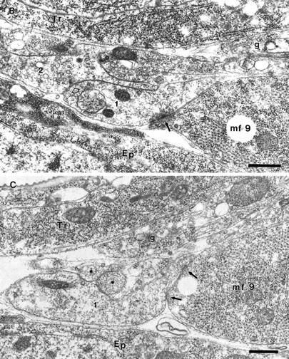
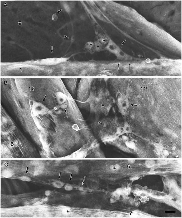
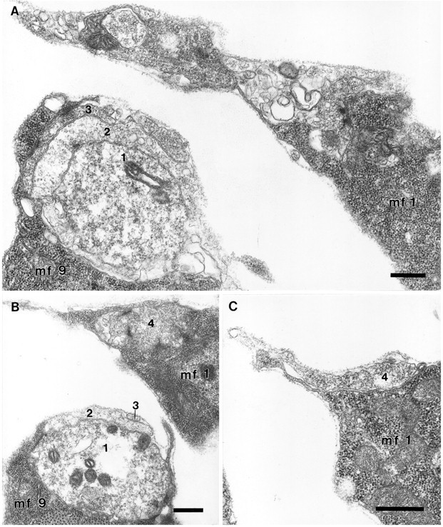
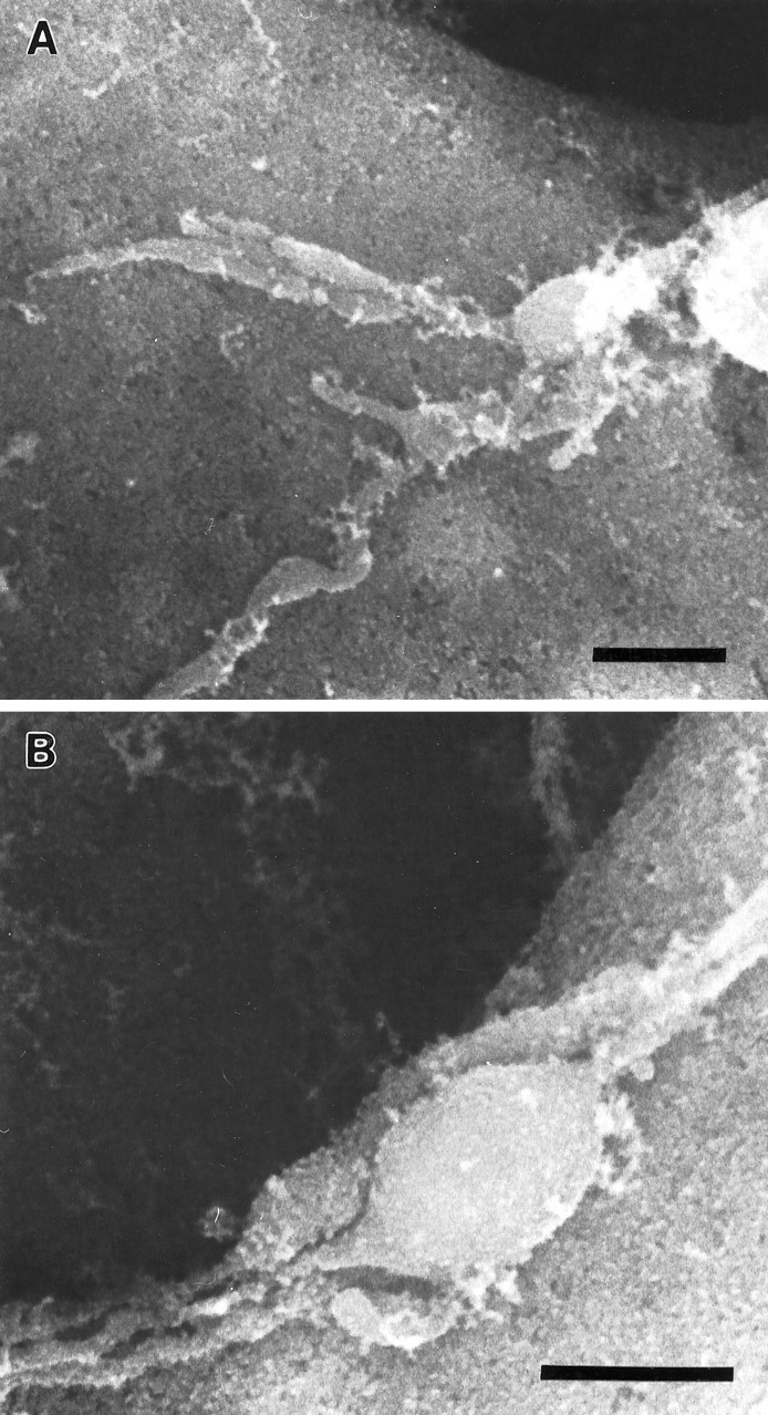
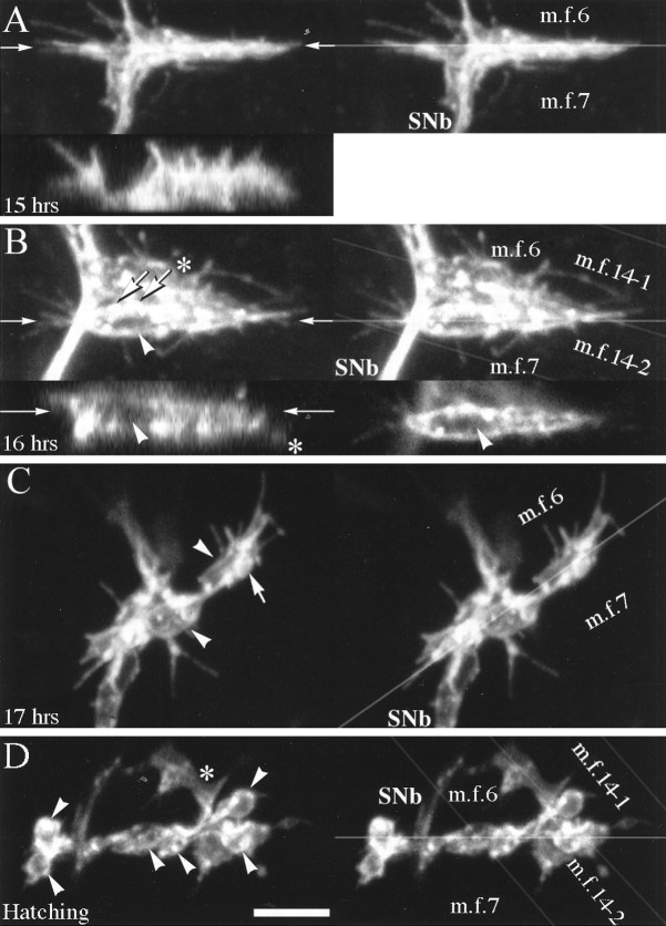
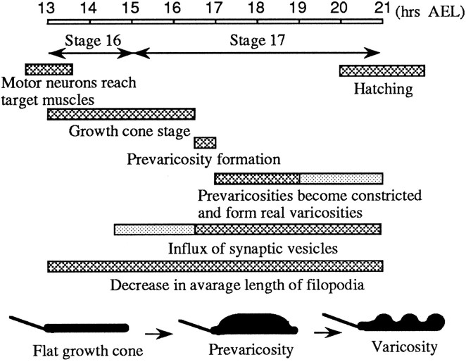
As a motor axon grows from the CNS to its target muscle, the terminal has the form of a flattened growth cone with a planar central region, lamellipodia, and filopodia. A mature terminal usually has a stereotyped shape that may be elongated with varicosities, as in several invertebrate species, or have short branches with boutons, as in mammals. We examined in Drosophila the developmental changes between growth cone and mature terminal using ultrastructural and immunocytochemical methods. The transition period, which occurs 2-3 hr after the first growth cone reaches its target muscle, is marked by the formation of "prevaricosities," smoothly contoured enlargements of the axons at the point where the nerve trunk first contacts the muscle fiber (MF). There is a 15-30 min ventral-to-dorsal gradient in the formation of prevaricosities on the individual abdominal MFs. Multineuronal innervation of each MF has occurred by this time, and two or more different axons undergo prevaricosity formation while they are intimately intertwined at the nerve entry point (NEP). Presynaptic active zones, both nerve-nerve and nerve-muscle, occur within the prevaricosities along broad contact regions. Synaptotagmin immunoreactive clusters form concurrently. The first varicosities then develop as a result of constrictions of the larger prevaricosities rather than as enlargement of discrete portions of the filopodia or neurites. The prevaricosity stage therefore may include the key steps that lead to the differentiation of functional differences in terminal subtypes as well as those leading to the formation of a stable neuromuscular junction.


Figures

Fig. 1.
Fig. 1.
A, Scanning electron micrograph of the dorsal abdominal muscles, right side, from an embryo 16.75 hr AEL. Anterior is to the left. Compare with Band C. The ISN crosses over MFs 3 and 2, innervating them in passing, to form a final bifurcation (thin arrow) to innervate MFs 1 and 9. The junctional aggregates (the one on MF 1 is bracketed betweenasterisks) typify those seen late in the growth cone period or early in the prevaricosity period. On MF 1 the terminals/growth cones are spread out and partially overlapping. They are still relatively flat. An isolated terminal on MF 2 (short arrow) illustrates the thickening and swelling of branch near the NEP that is characteristic of the prevaricosity period. Residual MF to MF connections are often seen (arrowheads). Filopodia are found both above and below the basal lamina of the MF, and a few are exploring “inappropriate” areas away from the typical junctional sites (open arrow), as is also seen in Figure4A. The large lateral sensory cells (SC) are exposed between MFs 10 and 3. Prepared with anti-HRP labeling and OTO method. See Figure 2 for interpretation of the method. Photographed at accelerating voltage of 10 kV. Scale bar, 5 μm. B, Schematic diagram of a single abdominal segment of a Drosophila embryo or larva showing the nomenclature (after Crossley, 1978) and the innervation of the muscles examined in this study. The general innervation pattern is segmentally repeated in abdominal segments A2–A7. Dorsal is to the top, and anterior is to the left. All subsequent figures illustrating part of an abdominal segment, except Figure 9, are oriented in this manner. The arrows indicate the locations of the junctions that were explored. The intersegmental nerve (ISN) innervates MFs 1 and 9 primarily on their medial surfaces after its final bifurcation, and segmental nerve b (SNb) innervates MFs 6 and 7 along the space between them. C, Diagram of the dorsal portion of a typical larval abdominal segment comparable to the region of the embryo shown above. The data available are consistent with an axonal branch pattern such that MFs 1 and 9 share innervation from one large motor axon (solid line) and from one Type II axon (longer dotted line), but each is innervated by two different motor axons (dashed lines), making a total of four axons supplying the two muscles.
Fig. 2.
Fig. 2.
Nerve terminals in mature third instar larvae. InA and B, nerve cell membranes were stained with fluorescein-tagged anti-HRP, and the projections of confocal optical sections were photographed. For comparison, inC and D the location of anti-HRP was visualized with DAB and the peroxidase reaction; the precipitate is highly osmiophilic, so labeled structures have enhanced secondary electron emission compared with surrounding tissue in the scanning electron microscope. This method allows clearer visualization of adjacent structures and relationships and is particularly useful in interpreting embryonic structures illustrated subsequently. A, C, Three morphological types of nerve terminals innervate MFs 1 (m.f.1) and 9 (m.f.9). They resemble the Types Ib, Is, and II described on other abdominal muscles (after the nomenclature of Johansen et al., 1989a; Atwood et al., 1993). Each terminal branch consists of a chain of varicosities (boutons) connected by thinner neurites. The terminal types may be distinguished on the basis of the relative sizes of their varicosities, with Ib having the largest varicosities, Is having intermediate-sized varicosities, and II having the smallest varicosities. The terminal branches on MF 9 typically spread posteriorly from the ISN across the medial surface of the fiber. The terminal branches on MF 1 extend both anteriorly and posteriorly; most branches lie on the internal surface of the fiber or along the ventral edge of the fiber, but occasionally some occur on the external surface. The point at which the nerve contacts the edge of MF 1 is indicated by the arrowhead in A.B, D, MFs 6 (m.f.6) and 7 (m.f.7) are innervated by Type Is and Ib terminals and usually lack Type II terminals (Atwood et al., 1993). The parts of the junctions extending onto the internal surfaces are easily seen in SEM, but a significant part of the innervation of these two muscles lies within the cleft between them and is extending in a plane perpendicular to the surface. The latter regions can be visualized only if the MFs are rendered transparent, as in fluorescence imaging. Scale bars, 20 μm.
Fig. 3.
Fig. 3.
Distal-most bifurcation of the ISN and innervation of MFs 1 and 9 from an embryo 18.5 hr AEL, late in the prevaricosity period. A bifurcation of one of the large motor axons (thin arrow) occurs at the point where the ISN divides to innervate MFs 1 and 9. Small varicosities of the sort typically associated with Type II axons within the nerve trunk are indicated by the arrowheads. The junctional aggregate of MF 1 is divided into two parts in this animal, with the right-hand portion forming an enlarged prevaricosity with six shortened filopodia. Both parts appear to be composed of two or more layers of overlapping or intertwined axonal branches. Labeled with anti-HRP, followed by osmium-thiocarbohydrazide treatment. Accelerating voltage, 10 kV. Scale bar, 1 μm.
Fig. 4.
Fig. 4.
Embryonic development of the innervation of MFs 1 and 9. In each panel, the two confocal images form stereo pairs in thex-y plane. The edges of the MFs are indicated bygray lines in the right image, and anasterisk is placed on the nerve trunk at the point above which all axons would be destined for MFs 1 and 9. Labeling: fluorescein-conjugated secondary antibody to anti-HRP. Scale bar (shown in G): 5 μm for all panels. A, After the embryonic nerve trunk has reached the dorsal-most target muscles (13 hr AEL). The junctional aggregates of MFs 1 (m.f.1) and 9 (m.f.9) have a common origin at this developmental stage and consist of one or more overlapping veil-like growth cones that are not distinguishable from one another. It is not possible to determine from these images how many separate axons contribute to the dorsal-most growth cones. Note the long filopodia, including some (arrowheads) that originate below the point at which the junctions typically form on MFs 1 and 9. The long filopodium at the lower right of the ISN belongs to a growth cone on MF 2, the remainder of which is out of view.B, The junctional aggregates appear structurally more complex (14.5 hr AEL). Portions of the growth cones innervating MFs 1 and 9 are spreading to both the internal and external surfaces of MF 1, as indicated by the two very different planes that can be seen in the stereo pair. Regional thickenings or portions that are more heavily labeled can be distinguished, seeming to condense from the more evenly labeled growth cone of earlier stages. C, Branches or divisions of the growth cones (16 hr AEL) are extending both over the internal surface of MF 1 and along its external surface in the cleft between MF 1 and MF 9 (which lies more lateral to MF 1). The divisions of the junctions that typically extend from the NEP both anteriorly and posteriorly on MF 1 appear to be forming (arrowheads). The junctional aggregate of MF 9 typically spreads posteriorly from the ISN. D, The distinction between the junctional aggregates of MFs 1 and 9 is now discernible because of the increase in length of the connecting portion of the ISN (16.5 hr AEL). A small cylindrical thickening is visible in a posterior branch on MF 1 (arrow). Overall, the impression at this stage is that the planar growth cone is condensing to form thicker, branch-like structures, but numerous filopodia are still present. E, There are three separate regions (arrowheads) in which terminals have formed noticeably thicker, three-dimensional structures (17 hr AEL). Only the surface membrane is labeled, so the impression of hollow structures is given. On the anterior (left) growth cone on MF 1, the stereo pair shows what could either be a large flattened structure or two separate thinner structures lying on top of each other. Electron microscopic observations of this stage indicate that both interpretations are possible. We refer to the single enlarged regions of a terminal as prevaricosities. F, G, By 19 hr AEL (F), and continuing through hatching (G), the enlarged structures along terminal branches began to have more clear-cut constrictions on either side of them. Their shapes ranged from tubular to spherical. We view this process as giving rise to the first real varicosities of the developing terminal. The dimensions of the individual spherical structures were typically less than those of the prevaricosities. Multiple terminal branches appear to be present. The arrowheads in each panel point to much thinner terminal branches, which are superimposed on the larger varicosities; these could be either Type Is or II, if, as might be expected, the largest varicosities are from Ib terminals. Filopodia are still present but tend to be somewhat fewer in number and shorter relative to the dimensions of the enlarged thicker regions. This transition is diagrammed in Figure 7C,D.
Fig. 12.
Fig. 12.
Development of anti-synaptotagmin immunoreactivity at synaptic terminals innervating MFs 1 and 9. The surface membranes of presynaptic nerve terminals were labeled with fluorescein-conjugated secondary antibodies to anti-HRP (green), and the synaptic vesicles were labeled with Cy-3-tagged antibodies to synaptotagmin (red). Each set is a stereo pair. A, There is little immunoreactivity to synaptotagmin (15 hr AEL), and the growth cones of the junctional aggregates are planar. B, Prevaricosity stage (16.5 hr AEL). Synaptotagmin immunoreactivity is clustered into discrete patches (arrowheads) within the prevaricosities. C, Hatching (21 hr AEL). Synaptotagmin immunoreactivity is denser, it is accumulated into larger patches, and the distribution of patches is restricted primarily to the outer margins of the varicosities (arrowheads).
Fig. 5.
Fig. 5.
Scanning electron micrograph of an anti-HRP-labeled junctional aggregate at the NEP on MF 1 from an embryo 17 hr AEL, during the prevaricosity period. Processes from two or three of the innervating axons overlie one another. The filopodia are shorter than those seen during the growth cone period, and some axonal branches have begun to enlarge at the NEP. Because the animal was dissected and fixed flat as a “fillet” for SEM, as were the animals examined confocally in Figure 4, we find that the ISN is pulled ventrally and part of the MF membrane (arrows) is pulled with it. The plane of section for the junctional aggregate seen in TEM in Figure 6is indicated by a line. Scale bar, 1 μm.
Fig. 6.
Fig. 6.
Junctional aggregates of MFs 1 and 9 at the prevaricosity stage (17 hr AEL). The animal was not filleted but fixed by perfusion through the tail end, so the nerves and muscles are lying in their natural positions. A, B, and Care sections 12, 32, and 52, respectively, from a series.Ep, Epidermal cell; Tr, main dorsal tracheole; g, parts of glial process on the distal part of ISN. A, This section passes through the center of the NEP for MF 9 (mf 9) and just off center for the NEP on MF 1 (mf 1). Notice the swellings in the axonal profiles at the NEPs compared with their diameter at the dorsal edge of MF 2, at the left of the micrograph. These enlarged profiles can be compared with the prevaricosities shown in Figures 1, 3, and 4. Three overlapping axonal branches are found in the NEP of MF 9 in this plane of section. Axon 3 (numbers applied solely for identification in this animal) is bifurcating at this point (open arrows), with one branch following the axons that are headed toward MF 1 and one branch continuing eventually to the surface of MF 9. Elementary synapses are seen at the contact points of axons 1 and 4 with their respective muscles (arrows). A profile of a filopodium from a growth cone lying more dorsally (out of the field of view) next to MF 1 is indicated with an arrowhead. One edge of the glial covering of the ISN is indicated (g), but the axons and terminals in this region are largely naked. At higher magnification it can be determined that the nerve passing over MF 9 en route to MF 1 actually consists of several axonal branches cut very obliquely. Scale bar, 1 μm. B, NEP on MF 9, anterior to 6A. Axon 1 has formed a synapse with MF 9 (arrow), and axon 2 has formed a dense body in apposition to axon 1 (open arrow). Axon 2 branches on both sides of another axonal profile. Note the difference in synaptic vesicle size between axon 1 and axon 2. Scale bar, 0.5 μm. C, Anterior edge of NEP on MF 9. Axon 1 remains in contact with MF 9; the other axonal branches have turned laterally and are now seen in cross section (asterisks). Compare with Figure 5 to visualize. Focal contacts and elementary synapses are formed by axon 1 with MF 9 (arrows). Scale bar, 0.5 μm. Figure continues.
Fig. 6.
Fig. 6.
Junctional aggregates of MFs 1 and 9 at the prevaricosity stage (17 hr AEL). The animal was not filleted but fixed by perfusion through the tail end, so the nerves and muscles are lying in their natural positions. A, B, and Care sections 12, 32, and 52, respectively, from a series.Ep, Epidermal cell; Tr, main dorsal tracheole; g, parts of glial process on the distal part of ISN. A, This section passes through the center of the NEP for MF 9 (mf 9) and just off center for the NEP on MF 1 (mf 1). Notice the swellings in the axonal profiles at the NEPs compared with their diameter at the dorsal edge of MF 2, at the left of the micrograph. These enlarged profiles can be compared with the prevaricosities shown in Figures 1, 3, and 4. Three overlapping axonal branches are found in the NEP of MF 9 in this plane of section. Axon 3 (numbers applied solely for identification in this animal) is bifurcating at this point (open arrows), with one branch following the axons that are headed toward MF 1 and one branch continuing eventually to the surface of MF 9. Elementary synapses are seen at the contact points of axons 1 and 4 with their respective muscles (arrows). A profile of a filopodium from a growth cone lying more dorsally (out of the field of view) next to MF 1 is indicated with an arrowhead. One edge of the glial covering of the ISN is indicated (g), but the axons and terminals in this region are largely naked. At higher magnification it can be determined that the nerve passing over MF 9 en route to MF 1 actually consists of several axonal branches cut very obliquely. Scale bar, 1 μm. B, NEP on MF 9, anterior to 6A. Axon 1 has formed a synapse with MF 9 (arrow), and axon 2 has formed a dense body in apposition to axon 1 (open arrow). Axon 2 branches on both sides of another axonal profile. Note the difference in synaptic vesicle size between axon 1 and axon 2. Scale bar, 0.5 μm. C, Anterior edge of NEP on MF 9. Axon 1 remains in contact with MF 9; the other axonal branches have turned laterally and are now seen in cross section (asterisks). Compare with Figure 5 to visualize. Focal contacts and elementary synapses are formed by axon 1 with MF 9 (arrows). Scale bar, 0.5 μm. Figure continues.
Fig. 7.
Fig. 7.
A–D, Diagrammatic representation of steps in development of the junctional aggregate on a single MF.A, Growth cone stage. During the growth cone stage, the developing terminal is thin and flat, with long filopodia. The growth cone from a single axon is illustrated. However, it often appears that toward the end of the growth cone stage two or more growth cones are overlapping in the vicinity of the NEP. B, Prevaricosity stage. The prevaricosities formed by a single axon are shown for simplicity. The growth cone condenses into several recognizable branches that have distinct thickness and rounded contours. Filopodia are shorter. Simple presynaptic specializations form along broad contact regions with the MF. C, Junctional aggregate at the prevaricosity stage, ∼16.5–18.5 hr AEL. During the prevaricosity stage, several terminals of differing degrees of development are usually found overlapping at the NEP. In this example, a growth cone is shown slightly diverging to the left, and two overlapping terminals with prevaricosities are to theright. Many additional configurations have been observed. When terminals have entered at the same point, they often remain spatially close, with membrane-to-membrane contact for some distance away from the NEP. This leads to a very complicated appearance when seen at either the light microscope or SEM levels. Thebracket indicates the regions that were measured to quantify prevaricosity formation in E. D, At 18–19 hr AEL, distinct varicosities, swellings with constrictions on either side, resolve from the enlarged branches of the prevaricosity. A single swelling may divide itself into two or three discrete varicosities. Filopodia are shorter, the elements of the SSR begin to separate the broad nerve–muscle contact regions, and individual bouton types can begin to be recognized. Subsequent development between this stage and first instar is a matter of degree of development of individual varicosities. E, Time course of prevaricosity formation. The percentage of junctions having prevaricosities and/or layered structures with a total thickness >2 μm was determined for each stage for MFs 1 and 9 (m.f. 1 & 9) and for MFs 6 and 7 (m.f. 6 & 7). The dimension of 2 μm was an arbitrary criterion to mark prevaricosity formation, with some individual prevaricosities being larger and some being smaller than this size. By 17 hr AEL nearly all junctions on MFs 1 and 9 included prevaricosities by subjective criteria, but not all reached the 2 μm thickness criterion. Subjectively, in a given animal or at a given age, the swelling of the prevaricosity seemed to be greater in the more ventral muscles than in the more dorsal ones (Figs.1, 4, 11). To quantify this difference, the 2 μm criterion was applied to MFs 6 and 7 in the same animals. There appears to be a delay of ∼15–30 min in the initiation of the process from dorsal to ventral muscles. For MFs 1 and 9, the total number of junctions measured at 15.5 hr AFL was n = 23 (4 animals); 16.0 hr,n = 42 (8 animals); 16.5 hr, n= 56 (10 animals); 17.0 hr, n = 46 (6 animals). MFs 6 and 7, at 15.5 hr AEL, n = 22; 16 hr,n = 46; 16.5 hr, n = 56; 17 hr,n = 36, respectively, for the same animals. The fraction of terminals reaching prevaricosity stage was tabulated for each animal; the points plotted represent the mean of these values ± SEM. F, Decrease in filopodial length with increasing age. The lengths of filopodia of the terminals innervating MFs 1 and 9 were measured from projected images along the z axis for three terminals at each age. At 13–14.5 hr AEL, some very long filopodia, >10 μm, are present; by hatching, all the filopodia are <6 μm. The arrows indicate the average filopodial length at each stage.
Fig. 8.
Fig. 8.
Junctional aggregates at the varicosity forming stage, 19 hr AEL, from three sets of muscles of the same abdominal segment. The varicosities are less well defined in MFs 1and 9 (A) than in the ventral muscles 12 and 13(B) or 6 and 7(C). These SEM specimens were prepared only with OTO. Accelerating voltage, 10 kV. Scale bar, 1 μm for all.A, The transition from the prevaricosity to constricted varicosities is evident in two terminal branches on MF 1. Two angular elongated shapes (thick arrows) appear to be in the process of each being subdivided into two varicosities. The developing varicosities contain “hollow” regions similar to those seen in larval junctions. (Dark appearance implies absence of osmiophilic structures such as organelles or synaptic vesicles.) In addition to major divisions, each terminal branch of an apparent Type Ib axon continues to extend thin filopodial or sprout-like processes to the MF. The strong adhesive nature of the nerve–muscle contacts (thin arrows) is evident because of the tension placed on the ISN. Type II axons form varicosities within the nerve by this time (arrowheads). The third axon, presumably Is, forms branches on the lateral side of the MF that are not seen here. Spherical, granular-appearing cells, which may be the persistent twist cells (asterisks), are often seen at the NEP of MF 1. B, On MFs12 and 13 it is possible to recognize several different terminal types by 19 hr AEL. Two or three large Type Ib varicosities are found on each fiber (thick arrows) as well as varicosities of other types (thin arrow andarrowheads). C, On MFs 6and 7, large and small varicosities (thick and thin arrows) have formed in the region of adherence between the two fibers. Other approximately spherical structures (stars) within the MF can be distinguished from varicosities on the basis of their focal plane and absence of connecting neurites.
Fig. 9.
Fig. 9.
Junctional aggregates on MFs 1 and 9 at 19 hr AEL.A, B, C, Serial sections 231, 242, and 254, respectively. The embryo was dissected flat for fixation, so the tension on the ISN has pulled the NEP on MF 1 toward MF 2. Scale bars, 0.5 μm. A, On MF 9 (mf 9), axon 1 has formed a spherical varicosity that is almost completely wrapped by two other terminals (2, 3). In this plane of section, axon 2 has formed a synapse in apposition to axon 1, and axon 3 has formed electron-dense specializations in apposition to a thin arm of the MF. The vesicle sizes in axon 1 were more uniform and smaller than those seen in axon 2. Axon 2 had numerous dense-cored vesicles elsewhere in the series. A growth cone was part of the aggregate in another plane (not shown). At this level, numerous small tangled profiles were seen on MF 1; they included branches from axon 2 just seen on MF 9, and from a growth cone-like structure as well as an axon that formed Type I varicosities in apposition to the MF. B, Axon 1 on MF 9 (mf 9) formed a total of eight presynaptic active zones of the multi-branched type in apposition with the MF in this and subsequent sections through this varicosity (0.7 × 1.0 × 1.7 μm), and three more in a second smaller one. The varicosity (axon 4) shown on MF 1 (mf 1) was 1.7 × 1.0 × 1.1 μm and housed six active zones; in this grazing section their multipronged nature can be seen. C, At the edge of the NEP, a single relatively unspecialized profile of axon 4 continues across the surface of MF 1.
Fig. 10.
Fig. 10.
Varicosity formation, MF 9. At 19 hr AEL, the more distal regions (A) of nerve terminals have irregular sprout-like shapes; varicosities are formed in the neurites closest to the NEP (B). In both regions the processes from one or more different axons are typically intimately intertwined, with membrane-to-membrane contact (verified by TEM views; see Fig. 9A,B), which is maintained over long distances as seen here. This is different from the mature larval form in which Types Ib and Is are at least separated from each other by layers of subsynaptic reticulum. The basal lamina was removed by treatment with 25% KOH for 2 min at 60°C after fixation, followed by osmium-thiocarbohydrazide. Scale bars, 0.5 μm.
Fig. 11.
Fig. 11.
Prevaricosity formation was compared in the terminals innervating MFs 6 and 7. A, At 15 hr AEL, the stereo pair illustrates growth cone lamellae and filopodia, which are spreading both upward over the surface of MF 6 (m.f.6) as well as along the cleft between MFs 6 and 7 (m.f.7). In the cleft, part of the junction appears as a vertical plate-like structure, with subdivisions that extend directly into and out of the plane of optical section. In thex-z image (bottom left inA), the upwardly directed filopodia and the vertical extent of the growth cone are illustrated. The location of thex-z section, in the center of the cleft between MFs 6 and 7, is indicated by the pair ofarrows. It is not possible in the confocal images to determine whether apparent subdivisions of the growth cone are derived from different axons. B, Beginning of prevaricosity formation (16 hr AEL). In this stereo pair the image of the innervation of MFs 6 and 7 is superimposed on that of subsequent branches of SNb (asterisks) that innervate MFs 14–1 (m.f.14–1) and 14–2 (m.f.14–2) (Bate, 1990). A tubular prevaricosity belonging to MFs 6 and 7 is indicated by an arrowhead. The cylindrical nature of the prevaricosity is shown more clearly in a single x-zsection, bottom left (B), and anx-y section, bottom right(B), (arrowheads). Thinner terminals with small varicose regions (thick arrows) may lie on top of this prevaricosity; in addition, however, small “spots” of increased fluorescence are seen where small processes emerge from the prevaricosity perpendicular to the plane of optical section. The pairs of thin arrowsindicate the planes of section of the x-z andx-y images. C, In this junctional aggregate on MFs 6 and 7 (17 hr AEL), two large prevaricosities are seen (arrowheads). Additional thinner terminal branches with small swellings (arrow) also appear to be present and are closely intertwined with the prevaricosities. It is possible that these are terminal branches from the second axon, which is known to be present at this time from physiological findings (Broadie and Bate, 1993). Note, however, that the bases of filopodia also give rise to small dots of increased fluorescence, which at first glance may appear to be small varicosities (above lower arrowhead).D, Hatching, 21 hr AEL. Six large varicosities are present in the cleft between MFs 6 and 7 (arrowheads), as well as some smaller ones. Note that the varicosities are smaller than the enlarged swellings shown inC. Asterisk indicates the 14–1, 14–2 junctions. The stereo pairs are helpful to exclude their varicosities from any counts of the varicosities of the 6–7 junction. Scale bar (shown in D): 5 μm for all panels.
Fig. 13.
Fig. 13.
The approximate time course of the stages of development of the presynaptic terminals of MFs 1 and 9 at 25°C. Several aspects of development were explored. Barsrepresent the duration of each stage. The schematic drawings illustrate the changes in shape undergone by the terminals during this period.Hatched regions of bars indicate time when characteristic was the most obvious; dotted regionsindicate time period when it was observed only occasionally, or less clearly.

Similar articles

Cited by

References

    1. Anderson MD, Halpern ME, Keshishian H. Identification of the neuropeptide transmitter proctolin in Drosophila larvae: characterization of muscle fiber-specific neuromuscular endings. J Neurosci. 1988;8:242–255. - PMC - PubMed
    1. Ashburner M. Drosophila: a laboratory manual. Cold Spring Harbor Laboratory; Cold Spring Harbor, NY: 1989.
    1. Atwood HL, Wojtowicz JM. Short-term and long-term plasticity and physiological differentiation of crustacean motor synapses. Int Rev Neurobiol. 1986;28:275–362. - PubMed
    1. Atwood HL, Govind CK, Wu C-F. Differential ultrastructure of synaptic terminals on ventral longitudinal abdominal muscles in Drosophila larvae. J Neurobiol. 1993;24:1008–1024. - PubMed
    1. Bate M. The embryonic development of larval muscles in Drosophila. Development. 1990;110:791–804. - PubMed

Publication types

MeSH terms

LinkOut - more resources