Comparative Study
. 1997 Nov 1;17(21):8476-90.
doi: 10.1523/JNEUROSCI.17-21-08476.1997.

Nerve growth factor treatment increases brain-derived neurotrophic factor selectively in TrkA-expressing dorsal root ganglion cells and in their central terminations within the spinal cord

Affiliations
Comparative Study

Nerve growth factor treatment increases brain-derived neurotrophic factor selectively in TrkA-expressing dorsal root ganglion cells and in their central terminations within the spinal cord

G J Michael et al. J Neurosci. .

Abstract

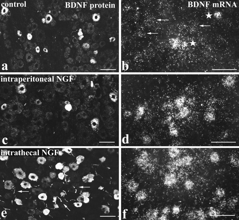
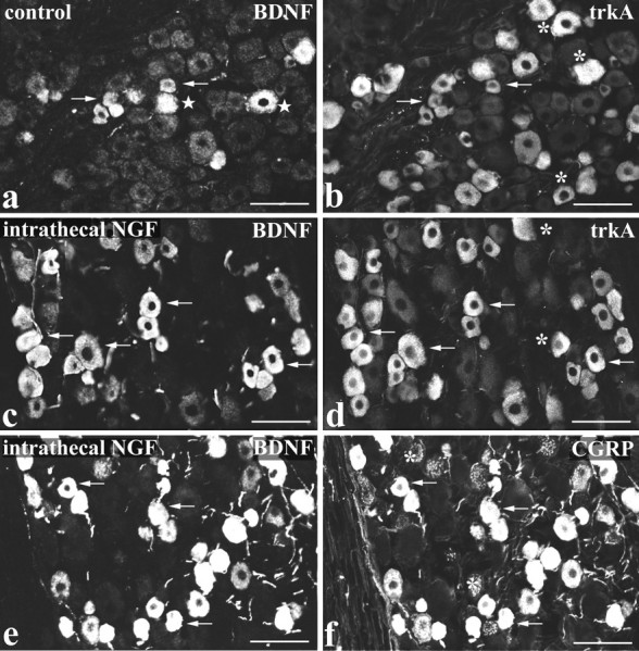
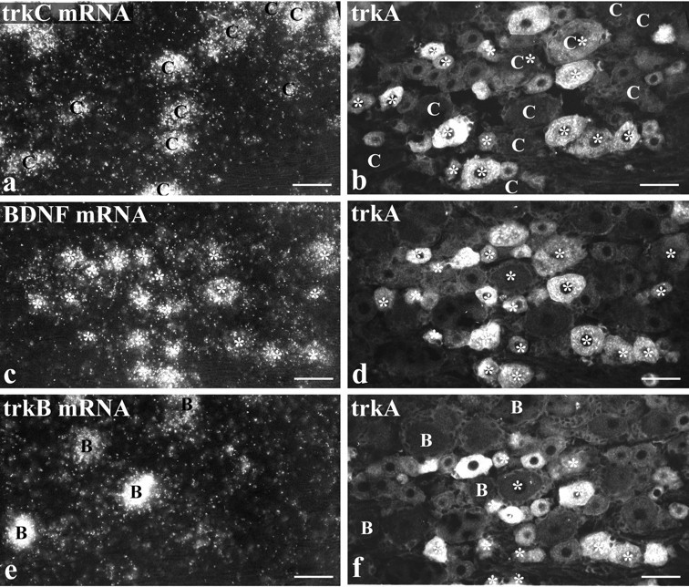
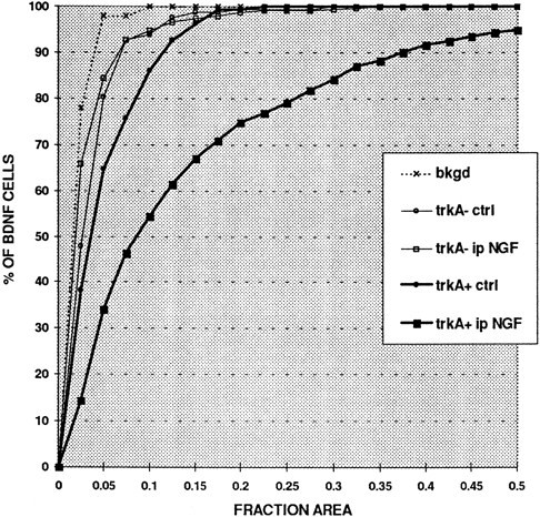
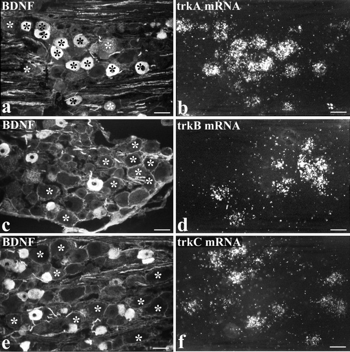
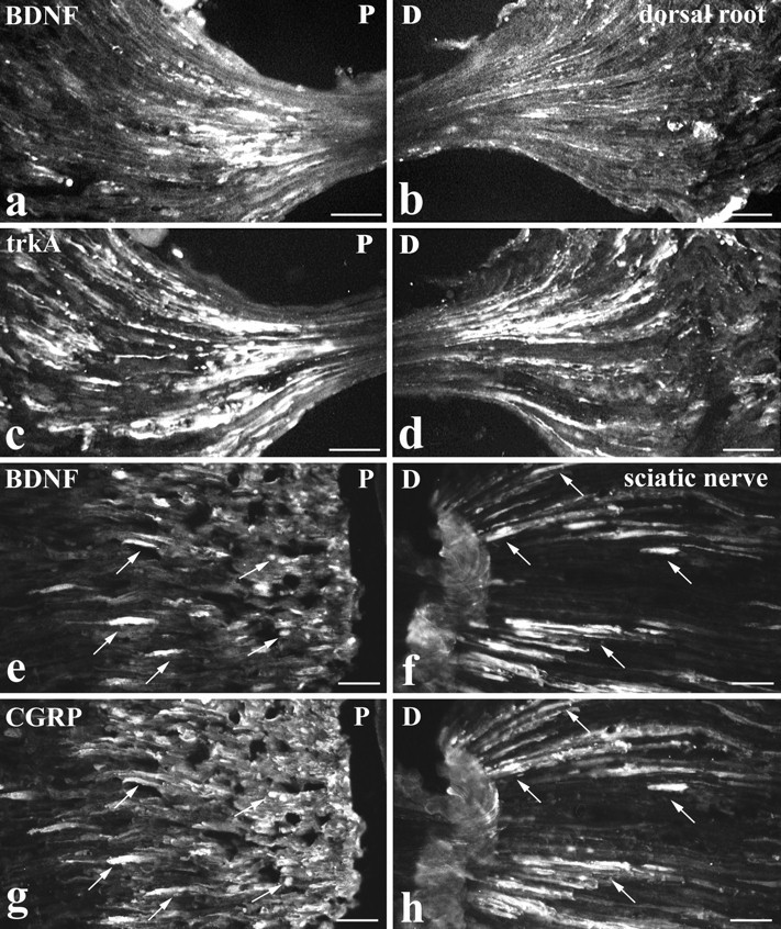
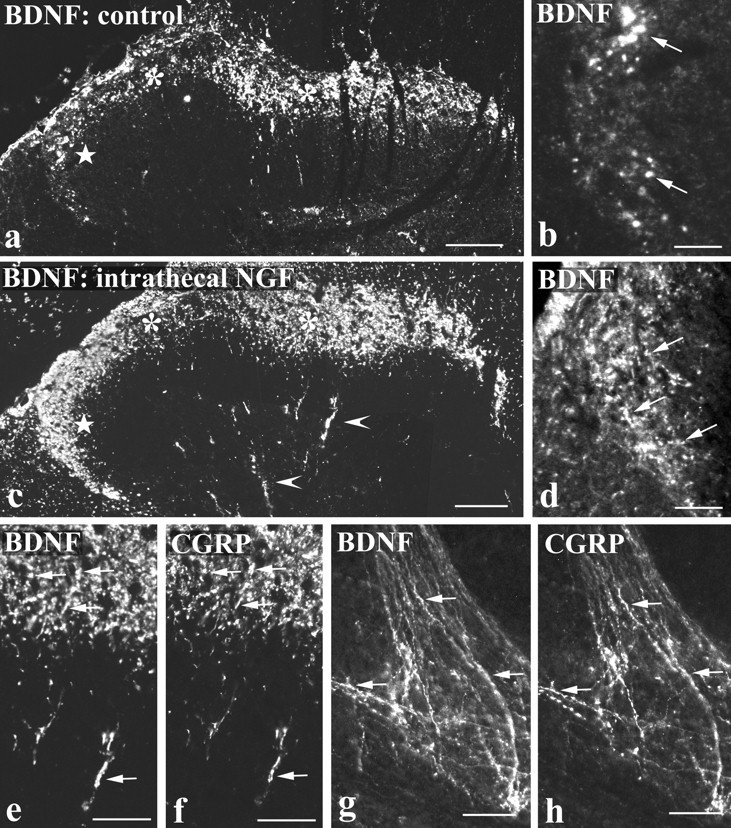
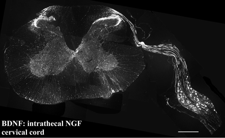
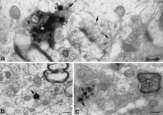
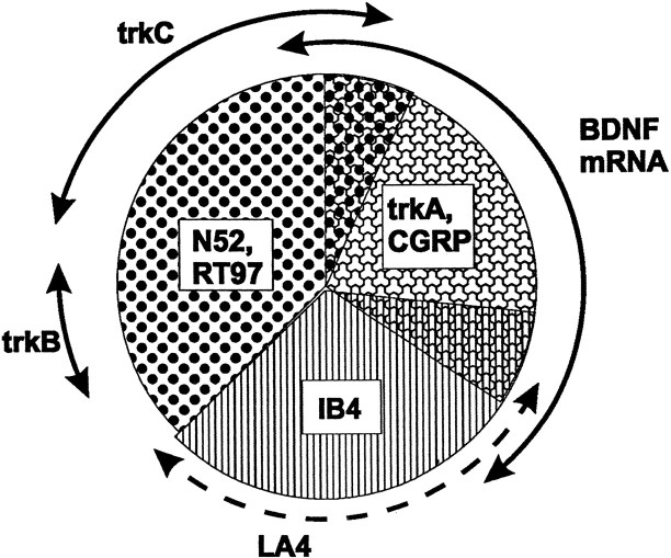
Using immunocytochemistry and in situ hybridization, we have examined the expression of brain-derived neurotrophic factor (BDNF) and of neurotrophin receptors in dorsal root ganglion cells. In the adult rat, BDNF mRNA and protein were found mainly in the subpopulation of cells that express the nerve growth factor (NGF) receptor trkA and the neuropeptide calcitonin gene-related peptide (CGRP). NGF increased BDNF within the trkA/CGRP cells to the extent that almost 90% of trkA cells contained BDNF mRNA after intrathecal NGF treatment, and 80-90% of BDNF-expressing cells contained trkA. Non-trkA cells that expressed BDNF included some trkC cells and some small cells that labeled with the lectin Griffonia simplicifolia IB4, a marker for cells that do not express trks. However, very few trkB cells expressed either BDNF mRNA or protein, and NGF did not increase BDNF expression in non-trkA cells. BDNF protein was anterogradely transported both peripherally and centrally. The central transport resulted in BDNF immunoreactivity in CGRP containing terminal arbors in the dorsal horn of the spinal cord, and this immunoreactivity was increased by NGF treatment. Electron microscopic analysis revealed that the BDNF immunoreactivity was present in finely myelinated and unmyelinated axons and in axon terminals, where it was most concentrated over dense-cored vesicles. Our data do not support an autocrine or paracrine role for BDNF within normal dorsal root ganglia, but indicate that BDNF may act as an anterograde trophic messenger. NGF levels in the periphery could influence dorsal horn neurons via release of BDNF from primary afferents.


Figures

Fig. 1.
Fig. 1.
BDNF mRNA and protein are increased by NGF treatment. BDNF immunofluorescence (a, c, e) andin situ hybridization (b, d, f) in lumbar ganglia of control (a, b), intraperitoneal NGF-treated (c, d), and intrathecal NGF-treated (e, f) rats. BDNF immunoreactivity is present in small to medium sized DRG cells and is increased after NGF treatment. The increase is most evident after intrathecal NGF (e), where immunoreactivity is seen not only in a larger number of DRG cells but also in neighboring axons (arrows). NGF treatment also increases expression of BDNF mRNA. In control tissue (b), a few heavily labeled cells are seen (stars) together with scattered light labeling (arrows). The number of heavily labeled cells is increased after intraperitoneal NGF (d) and increased even more by intrathecal NGF (f). Scale bars, 100 μm.
Fig. 2.
Fig. 2.
BDNF immunoreactivity is present in trkA/CGRP cells and is increased by NGF treatment. Double-labeling for BDNF (a, c, e) and either trkA (b, d) or CGRP (f) immunofluorescence in lumbar ganglia of control (a, b) and intrathecal NGF-treated (c–f) rats is shown. In both control and intrathecally treated animals, the majority of BDNF-immunoreactive DRG cells are also trkA immunoreactive (a–d). However, because intrathecal NGF increases the level of BDNF immunoreactivity, the number of trkA-immunoreactive cells that double-label for BDNF is increased in c and d compared witha and b. A similar situation occurs for BDNF and CGRP double-labeling, with extensive coexistence of BDNF and CGRP evident after intrathecal NGF treatment (e, f). Arrows indicate BDNF/trkA or BDNF/CGRP double-labeled cells; asterisks indicate cells single-labeled for trkA or CGRP; stars indicate cells single-labeled for BDNF. Scale bars, 100 μm.
Fig. 3.
Fig. 3.
BDNF mRNA is expressed by numerous trkA cells but by few trkB or trkC cells. Intraperitoneal NGF treatment is shown.a, c, and e show three serial sections processed for trkC (a), BDNF (c), and trkB (e) mRNAs using in situ hybridization. Each section was also immunostained for trkA (b, d, f). Comparison ofc and d shows that the majority of cells labeled for BDNF mRNA (asterisks) are also trkA immunoreactive. Those BDNF-labeled cells that are visible in the serial sections (a, b, e, f) are marked by asterisks in b and f. Comparison of a and b shows that the majority of trkC-labeled cells (marked C) do not express BDNF mRNA. However, two BDNF/trkC double-labeled cells are visible inb, one of which is trkA immunoreactive. Comparison ofe and f allows the distribution of DRG cells that express trkB (marked B) and BDNF (asterisks) to be compared. They seem to form two discrete populations, and no double-labeled cells are visible. Scale bars, 50 μm.
Fig. 4.
Fig. 4.
NGF increases BDNF mRNA expression only in trkA-immunoreactive cells as shown in Q sum plot of BDNF mRNA expression in trkA-immunoreactive (trkA+) and trkA-nonimmunoreactive (trkA−) lumbar DRG cells in control (ctrl) and intraperitoneal NGF-treated (ip NGF) animals. BDNF expression was quantified by measuring the fraction of the cell body (FRACTION AREA) that was covered by silver grains. Note that intraperitoneal NGF does not change the level of BDNF expression in BDNF-labeled but trkA-nonimmunoreactive cells. In contrast intraperitoneal NGF greatly increases BDNF expression in trkA immunoreactive cells, and even in control animals these cells show higher labeling than the trkA nonimmunoreactive population.bkgd indicates the level of background labeling in control and NGF-treated animals.
Fig. 5.
Fig. 5.
Intrathecal NGF treatment. BDNF immunoreactivity is present in numerous trkA cells but in few trkB or trkC cells.a–f show preparations double-labeled for BDNF immunoreactivity (a, c, e) and either trkA (b), trkB (d), or trkC (f) in situhybridization. The asterisks indicate the location of trk-expressing cells, revealed by the in situhybridization autoradiograms. Many BDNF-immunoreactive cells show trkA labeling (a, b), but the trkB- and trkC-expressing cells are distinct from the BDNF-immunoreactive ones. Scale bars, 50 μm.
Fig. 6.
Fig. 6.
Intraperitoneal NGF treatment. BDNF is anterogradely transported. BDNF (a, b, e, f), trkA (c, d), and CGRP (g, h) immunoreactivity proximal (P: a, c, e, g) and distal (D: b, d, f, h) to lumbar dorsal root (a–d) or sciatic nerve (e–h) ligations are shown. At a dorsal root ligation, both BDNF (a) and trkA (c) accumulate proximal to the ligation (i.e., the DRG side). Distal to the ligation, trkA immunoreactivity accumulates (d) but only occasional BDNF-immunoreactive axons are visible (b). At a sciatic nerve ligation, BDNF accumulates both proximal (e) and distal (f) to the ligation. CGRP shows a similar accumulation (g, h), and double-staining reveals that the BDNF-immunoreactive axons are also CGRP immunoreactive (arrows indicate double-labeled axons). Scale bars, 50 μm.
Fig. 7.
Fig. 7.
BDNF immunoreactivity in the spinal cord is increased by NGF treatment and is present in CGRP-immunoreactive axons.a–d show BDNF immunofluorescence in the dorsal horn of control tissue (a, b) and after intrathecal NGF treatment (c, d). BDNF immunoreactivity in laminae I and II (asterisks in a and c) and in deep dorsal horn (arrowheads in c) is increased by NGF. The increase is particularly striking in lateral lamina II (stars in a andc), and this region is shown at high magnification inb and d. Immunoreactive axons (arrows in b and d) are more abundant after NGF treatment. e–h, Double-labeling showing extensive coexistence of BDNF (e, g) and CGRP (f, h) after intrathecal NGF treatment. In the dorsal horn (e, f) and in lamina X dorsolateral to the aqueduct (g, h), numerous double-labeled axons and varicosities (arrows) are visible. Scale bars:a, c, 100 μm; b, d, 25 μm;e–h, 50 μm.
Fig. 8.
Fig. 8.
Photomontage of a transverse section showing the cervical spinal cord and attached DRG of an animal treated intrathecally with NGF via an upper cervical cannula. BDNF protein is transported by DRG cells along their central processes and into the spinal cord. BDNF levels have increased to such an extent that immunoreactive axons can be traced from DRG cells, along a dorsal root and into the dorsal horn of the spinal cord. Scale bar, 250 μm.
Fig. 9.
Fig. 9.
Preembedding ultrastructural immunocytochemistry showing BDNF immunoreactivity in axons (b, c) and an axon terminal (a) in lamina II of the lumbar spinal cord. a, An immunoreactive terminal (asterisk) shows heavy staining over a region packed with small agranular vesicles and over individual large dense-cored vesicles (open arrows). Arrows indicate dense-cored vesicles in an adjoining unstained terminal.b, A heavily stained unmyelinated axon (arrow) is visible among a group of similar, but unstained, axons. c, An immunostained preterminal axon and an immunostained finely myelinated axon (asterisk) are visible. The preterminal axon shows staining exclusively over large dense-cored vesicles (arrows). Scale bars, 0.25 μm.
Fig. 10.
Fig. 10.
Pie chart summarizing the relationship between BDNF and trk expression in neurochemically defined DRG subclasses. DRG neurons can be divided broadly into large-diameter cells, which innervate low-threshold mechanoreceptors, and small-diameter cells, which innervate mainly nociceptors. BDNF mRNA is expressed by the subpopulation of small cells that contain the neuropeptide CGRP and the NGF receptor trkA. Small cells that do not express any trk receptor, and that can be labeled using the lectin Griffonia simplicifolia IB4 or the monoclonal antibody LA4, mainly do not express BDNF. With the exception of those trkC cells that coexpress trkA, BDNF mRNA is also largely not expressed by trkB or trkC cells. TrkC and trkB are present mainly in large-diameter cells, which can be identified using the neurofilament antisera N52 or RT97. BDNF is constitutively expressed in the cell types illustrated, but expression in trkA cells is increased further by NGF treatment.

Similar articles

Cited by

References

    1. Acheson A, Conover JC, Fandl JP, DeChlara TM, Russell M, Thadani A, Squinto SP, Yancopoulos GD, Lindsay RM. A BDNF autocrine loop in adult sensory neurons prevents cell death. Nature. 1995;374:450–453. - PubMed
    1. Apfel SC, Wright DE, Wiideman AM, Dormia C, Snider WD, Kessler JA. Nerve growth-factor regulates the expression of brain-derived neurotrophic factor messenger RNA in the peripheral nervous system. Mol Cell Neurosci. 1996;7:134–142. - PubMed
    1. Averill S, McMahon SB, Clary DO, Reichardt LF, Priestley JV. Immunocytochemical localization of trkA receptors in chemically identified subgroups of adult rat sensory neurons. Eur J Neurosci. 1995;7:1484–1494. - PMC - PubMed
    1. Averill S, Michael GJ, Shortland PJ, Priestley JV. BDNF increases in large diameter dorsal root ganglion cells and in their central projections following peripheral axotomy. Soc Neurosci Abstr. 1997;23:134.8.
    1. Bennett DLH, French J, Priestley JV, McMahon SB. NGF but not NT-3 or BDNF prevents the A fibre sprouting into lamina II of the spinal cord that occurs following axotomy. Mol Cell Neurosci. 1996a;8:211–220. - PubMed

Publication types

MeSH terms