Comparative Study
. 1997 Nov 1;17(21):8506-19.
doi: 10.1523/JNEUROSCI.17-21-08506.1997.

Neurturin and glial cell line-derived neurotrophic factor receptor-beta (GDNFR-beta), novel proteins related to GDNF and GDNFR-alpha with specific cellular patterns of expression suggesting roles in the developing and adult nervous system and in peripheral organs

Affiliations
Comparative Study

Neurturin and glial cell line-derived neurotrophic factor receptor-beta (GDNFR-beta), novel proteins related to GDNF and GDNFR-alpha with specific cellular patterns of expression suggesting roles in the developing and adult nervous system and in peripheral organs

J Widenfalk et al. J Neurosci. .

Abstract

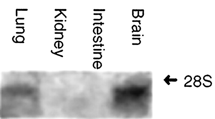
Cloning strategies were used to identify a gene termed glial cell line-derived neurotrophic factor receptor-beta (GDNFR-beta) related to GDNFR-alpha. In situ hybridization was then used to map cellular expression of the GDNF-related trophic factor neurturin (NTN) and GDNFR-beta mRNA in developing and adult mice, and comparisons with GDNFR-alpha and RET were made. Neurturin is expressed in postnatal cerebral cortex, striatum, several brainstem areas, and the pineal gland. GDNFR-beta mRNA was more widely expressed in the developing and adult CNS, including cerebral cortex, cerebellum, thalamus, zona incerta, hypothalamus, brainstem, and spinal cord, and in subpopulations of sensory neurons and developing peripheral nerves. NTN colocalized with RET and GDNFR-alpha in ureteric buds of the developing kidney. The circular muscle layer of the developing intestines, smooth muscle of the urether, and developing bronchiolae also expressed NTN. GDNFR-beta was found in myenteric but not submucosal intestinal plexuses. In developing salivary glands NTN had an epithelial expression, whereas GDNFR-beta was expressed in surrounding tissue. Neurturin and GDNFR-beta were present in developing sensory organs. In the gonads, NTN appeared to be expressed in Sertoli cells and in the epithelium of the oviduct, whereas GDNFR-beta was expressed by the germ cell line. Our findings suggest multiple roles for NTN and GDNFR-beta in the developing and adult organism. Although NTN and GDNFR-beta expression patterns are sometimes complementary, this is not always the case, suggesting multiple modi operandi of GDNF and NTN in relation to RET and the two binding proteins, GDNFR-alpha and GDNFR-beta.


Figures

Fig. 1.
Fig. 1.
NTN mRNA hybridization signals in the mouse brain during P7 and P14. Expression of NTN mRNA 1 week after birth is seen, e.g., in the central gray (A), corpus pineale (P) (A) and parts of the entorhinal cortex (B, arrowhead). Two weeks after birth there is labeling of the caudate putamen (CP) and cingulum (Cg) (C). The caudate putamen appears somewhat diffusely labeled with the NTN probe, and one observes an increasing gradient of labeling laterally. Scale bars:A, B (shown in A), 1 mm;C, 1 mm.
Fig. 2.
Fig. 2.
GDNFR-β mRNA signal in adult cortex and lateral septum (A, dark-field view). Higher magnification (B, bright-field view) reveals signal in parts of the frontal cortex in layers 3 and 4. Scale bars:A, 1 mm; B, 50 μm.
Fig. 3.
Fig. 3.
Comparison between prominent RET, GDNFR-α, and GDNFR-β mRNA expression in 1-week-old mouse brain, dark-field view. All three genes show strong expression in zona incerta (ZI) and the reticular thalamic nucleus (Rt) (A, C, E). In the dentate gyrus, a positive signal is seen for GDNFR-α and GDNFR-β (C, E), but not for RET. Dopamine neurons of substantia nigra (SN) and the ventral tegmental area have a distinct expression pattern of RET and GDNFR-α (B, D), whereas GDNFR-β is absent or very weakly expressed (F). Cortex cerebri represents another area where the signal distribution differs markedly: GDNFR-β labels two layers in cingulum and frontal and parietal cortices (E), GDNFR-α labels only medial cortex (C), and RET labels almost the same areas as GDNFR-β, but slightly weaker and in two different cortical layers (A). GDNFR-β appears diffusely in many areas in the brainstem, such as the superior colliculus (F). Distinct signals for GDNFR-α, but not for RET and GDNFR-β, are seen in the medial habenula (MHb), corpus pineale (P), and cornu ammonis caudally. The trigeminal ganglion shows signal for GDNFR-α, GDNFR-β, and RET, although strongest for the latter. Scale bars: A, C, E (shown in E), 100 μm;B, D, F (shown in F), 100 μm.
Fig. 4.
Fig. 4.
Dark-field photomicrographs depicting distribution of the different GDNF receptor mRNA species in adjacent sections of adult substantia nigra. Both RET and GDNFR-α mRNA give rise to robust hybridization signals in the dopamine neurons of substantia nigra and the ventral tegmental area (AD), whereas GDNFR-β is only weakly expressed in the ventral tegmental area (F). All three probes are positive in the supramammillary nucleus (A–F). The dentate gyrus is positive for GDNFR-α and GDNFR-β but not for RET mRNA (A, C, E). Other areas of GDNFR-β mRNA signal include the peripeduncular nucleus, superior colliculus, and two layers in cortex. Note that in contrast to GDNFR-β, RET and GDNFR-α are not detectable in cortex. Scale bars: B, D, F (shown inF), 1 mm; A, C, E (shown inE), 2 mm.
Fig. 5.
Fig. 5.
High-magnification bright-field photomicrographs of some of the most prominent neuronal GDNFR-β hybridization signals in the brain. A, The lateral dentate gyrus in a 1-week-old mouse shows distinct hybridization signal. B,In cingulum at the same stage several layers are positively labeled.C, A distinct signal for GDNFR-β mRNA is found in the adult zona incerta. D, The lateral septum in the 2-week-old mouse also shows strong labeling. Scale bars: A, B, C (shown in A), 50 μm; D, 50 μm.
Fig. 6.
Fig. 6.
Dark-field images depicting GDNFR-α and GDNFR-β mRNA expression in neonatal mouse CNS. Prominent hybridization of the GDNFR-β probe is seen in zona incerta, dentate gyrus, the trigeminal ganglia, and the superior and inferior colliculus (B). No robust GDNFR-β signal is observable in the substantia nigra area (SN) in contrast to GDNFR-α (A). Cerebellum is positive for GDNFR-β, especially in the Purkinje cell layer (C). Cb, Cerebellum;DG, dentate gyrus; IC, inferior colliculus; fn, facial nucleus; m, mesencephalic flexure; Mo5, trigeminal motor nucleus;SC, superior colliculus; TG, trigeminal ganglion; ZI, zona incerta. Scale bar (shown inC): A, B, 250 μm; C, 125 μm.
Fig. 7.
Fig. 7.
GDNFR-β mRNA hybridization in developing spinal cord. Hybridization is observed in most neurons of the spinal cord during development (bright-field, A; and dark-field,B, D) and in a subset of neurons in the dorsal root ganglia (dark-field image, D). The positive labeling is continuous from medulla oblongata and downward at birth as shown in sagittal section (B). One week later (P7) GDNFR-β mRNA signals are still present throughout gray matter, labeling most neurons, including α-motor neurons (A,D). Compared with GDNFR-α (C), which has a similar expression pattern, the most striking difference is the relatively weak expression of GDNFR-β mRNA in α-motor neurons (compare arrows in C, D).CC, Canalis centralis. C1, First cervical vertebrae. Scale bars: A, 50 μm; B, 125 μm; C, D (shown in C), 250 μm.
Fig. 8.
Fig. 8.
Examples of NTN and GDNFR-β mRNA hybridization in the P0 trigeminal system (A--E) and of GDNFR-β mRNA in the E20 lung. In the olfactory epithelium NTN and GDNFR-β mRNA appear in complementary patterns (dark-field, A, C). NTN mRNA is found in the olfactory epithelium, especially in Bowman’s glands (C, arrows), and GDNFR-β is found in lamina propria of the olfactory mucosa, a tissue containing abundant nerves and vessels. Vibrissae are positive for GDNFR-β (bright-field image, B; dark-field image, E), as are peripheral nerves exemplified by nervus maxillaris (E, arrowheads). D, A subpopulation of neurons in the trigeminal ganglion are positively labeled for GDNFR-β mRNA. In the developing lung GDNFR-β is found in the stromal tissue (dark-field, F). Scale bars: A, 500 μm; B, 50 μm; C, 500 μm;D, 50 μm; E, 250 μm;F, 200 μm.
Fig. 9.
Fig. 9.
E20 salivary glands and adult testis. Neurturin and GDNFR-β mRNA show a complementary expression pattern in both organs. The developing tubules in the salivary glands are labeled by NTN hybridization (B), whereas GDNFR-β mRNA is represented in the stroma surrounding the epithelial structures (A). In testis a very strong neurturin mRNA signal is seen in the periphery of some of the seminiferous tubules, suggesting localization to the Sertoli cells (C, dark-field view; D, bright-field view). GDNFR-β is also expressed in some of the tubules, but at levels indicating localization to the germ cells (E, F). Scale bars: A, B, 200 μm; C–F, 100 μm.
Fig. 10.
Fig. 10.
Dark-field photomicrographs showing the mRNA distribution of all known GDNF family members in the developing kidney at embryonal day 19 (A–E). NTN mRNA is expressed in the developing epithelial buds. GDNF mRNA, in contrast, is localized in peripheral mesenchyme. GDNFR-α and RET show similar patterns of probe labeling in the peripheral epithelial buds, whereas GDNFR-β could not be detected in the periphery. Weak GDNFR-β as well as weak NTN hybridization signals were found in pelvic regions of the developing kidney. Scale bar (shown in A), 500 μm.
Fig. 11.
Fig. 11.
Northern blot analysis of GDNFR-β levels in postnatal week 6 mouse tissues. Total RNA from indicated tissues (30 gm/slot) is shown. A 1 kb 32P-labeled GDNFR-β cDNA probe was used for hybridization. Note single bands.

Similar articles

Cited by

References

    1. Angrist M, Bolk S, Halushka M, Lapchak P, Chakravarti A. Germline mutations in glial cell line-derived neurotrophic factor (GDNF) and RET in a Hirschsprung disease patient. Nat Genet. 1996;14:341–344. - PubMed
    1. Baloh R, Tansey M, Golden J, Creedon D, Heuckeroth R, Keck C, Zimonjic D, Popescu N, Johnson E, Jr, Milbrandt J. TrnR2, a novel receptor that mediates neurturin and GDNF signaling through Ret. Neuron. 1997;18:793–802. - PubMed
    1. Buj-Bello A, Adu J, Piñón L, Horton A, Thompson J, Rosenthal A, Chinchetru M, Buchman V, Davies A. Neurturin responsiveness requires a GPI-linked receptor and the Ret receptor tyrosine kinase. Nature. 1997;387:721–724. - PubMed
    1. Dagerlind Å, Friberg K, Bean A, Hökfelt T. Sensitive mRNA detection using unfixed tissue: combined radioactive and non-radioactive in situ hybridization histochemistry. Histochemistry. 1992;98:39–49. - PubMed
    1. Durbec P, Marcos-Gutierrez C, Kilkenny C, Grigoriou M, Suvanto P, Wartiovaara K, Smith D, Ponder B, Constantini F, Saarma M, Sariola H, Pachnis V. Glial cell line-derived neurotrophic factor signalling through the Ret receptor tyrosine kinase. Nature. 1996;381:789–792. - PubMed

Publication types

MeSH terms

Substances

LinkOut - more resources