. 1997 Nov 1;17(21):8566-79.
doi: 10.1523/JNEUROSCI.17-21-08566.1997.

Neuronal activity in monkey superior colliculus related to the initiation of saccadic eye movements

Affiliations

Neuronal activity in monkey superior colliculus related to the initiation of saccadic eye movements

M C Dorris et al. J Neurosci. .

Abstract

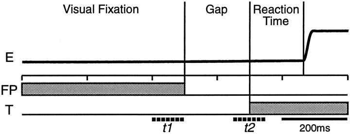
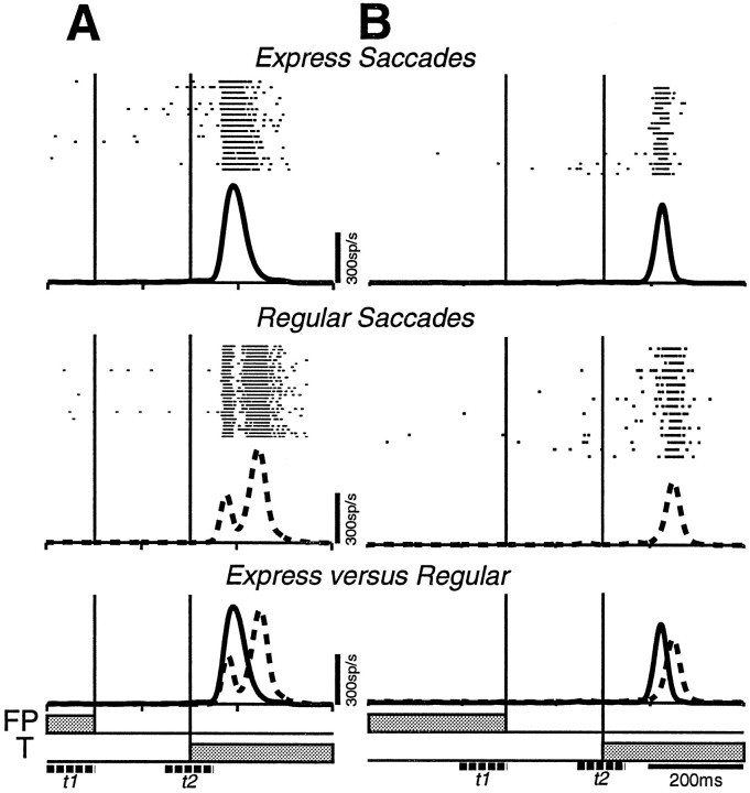
The introduction of a temporal gap between the disappearance of an initially fixated target and the appearance of an eccentric saccadic target results in a general reduction of saccadic reaction times (SRTs)-the gap effect-and often in the production of express saccades, the latencies of which approach the conduction time of the shortest neural pathways from the retina to the eye muscles. We investigated saccade initiation by recording neuronal activity in the superior colliculus in monkeys performing the gap paradigm. Fixation-related neurons reduced their discharge rate during the gap period, regardless of the SRT. This reduction in activity is consistent with the hypothesized release of ocular fixation that facilitates premotor processes and may contribute to the gap effect. In addition to saccade-related discharges, many saccade-related neurons displayed phasic target-related responses and/or low-frequency preparatory activity during the gap period. The level of this preparatory activity correlated with both SRT and express saccade occurrence when the saccade was made into the response field of the neuron. Evidence indicates that advanced motor preparation is required for express saccade generation, which may be subserved by specific increases in the preparatory activity of saccade-related neurons. Increased preparatory activity may allow the target-related responses to trigger short-latency express saccades directly. This study provides insights into the functional mechanism of saccade initiation and may be relevant to the generation of all voluntary motor responses.


Figures

Fig. 1.
Fig. 1.
Schematic of the gap paradigm. Time is represented on the horizontal axis, and presentation of the visual stimuli (FP, central fixation point; T, eccentric target) are denoted by the horizontal gray bars. As in the following figures, an upward deflection of the horizontal eye trace (E) represents a rightward movement, and downward deflection represents a leftward movement. The visual fixation interval was randomized between 500 and 1000 msec, and the gap duration was either set at 200 msec or randomized between 0 and 800 msec within a block of trials. The discharge rate of neurons during this task was calculated during the final 100 msec of FP presentation (visual fixation epoch, t1) and from 50 msec before to 50 msec after target presentation (end of gap epoch,t2).
Fig. 2.
Fig. 2.
Typical SRT histograms obtained during a block of 200 msec gap trials. Only the SRT distributions from one of two possible target locations is shown for each monkey. Hatched bars represent express saccades (70–120 msec), andfilled bars represent regular latency saccades (130–180 msec).
Fig. 3.
Fig. 3.
The activity of a single fixation (A, B) and buildup (C, D) neuron during 600 msec gap trials. The fixation neuron was located in the right SC, and the target was presented 10° left (A, contralateral target) or 10° right (B, ipsilateral target). The buildup neuron was located in the left SC, and the target was presented 10° right (C, contralateral target optimal vector for the response field of this neuron) or 10° left (D, ipsilateral target opposite the response field of this neuron). Trials were collected in a block of trials in which gap duration was randomized between 0, 100, 200, 300, 400, 600, and 800 msec, and the target appeared randomly 10° to the right or left. The individual rasters of neuron discharge, the spike density function, and the horizontal eye position traces are aligned on both FP disappearance (left vertical line) and target onset (right vertical line).
Fig. 4.
Fig. 4.
Reciprocal pattern of activity for the sample of fixation and buildup neurons during fixed 200 msec (46 fixation and 30 buildup neurons) (A) and randomized 600 msec (B) gap trials (31 fixation and 15 buildup neurons). The thick line represents the mean spike density, and the envelope surrounding each waveform represents the SEM.
Fig. 5.
Fig. 5.
Mean discharge rate of individual fixation (A) and buildup (B) neurons during the visual fixation (t1) and end of gap (t2) epochs during 200 msec gap trials. Each datapoint represents a single neuron. Lines of equality are represented by dashed lines, and linear regression lines are represented as solid lines.
Fig. 6.
Fig. 6.
Linear regression and correlation between SRT and discharge rate of individual fixation and buildup neurons for data collected in blocks of trials with the gap duration fixed at 200 msec. Each data point was obtained from a single trial from either single fixation (A–D) or buildup (E–H) neuron. Neuronal activity was sampled during the visual fixation (t1) and end of gap (t2) epochs before targets presented contralateral and ipsilateral. *Statistically significant correlation (p < 0.05).
Fig. 7.
Fig. 7.
Correlation coefficients between SRT and discharge rate of individual neurons plotted as a cumulative distribution. All data were collected from blocks of trials with the gap duration fixed at 200 msec. Each data point represents the correlation coefficient from one neuron when the activity was sampled during different epochs or target locations. A, The fixation neuron distributions were centered around zero and did not differ from each other (ANOVA, p > 0.05). B, The distribution made from buildup neuron activity during thet2 epoch and for the contralateral target was shifted to the left of zero and differed from the other buildup neuron distributions (ANOVA, Student–Newman–Keuls method,p < 0.05). C, The distribution composed from the correlation coefficients of the t2contralateral target buildup neurons differed significantly from the distribution composed of the t2 contralateral target fixation neuron distribution (t test,p < 0.01).
Fig. 8.
Fig. 8.
Comparison of neuronal discharge during the generation of express and regular saccades in 200 msec gap trials. The spike density functions generated from express (top, solid line) and regular (middle, dashed line) trials are aligned on target appearance and superimposed at thebottom. A, The spike density functions constructed from a fixation neuron did not differ (ttest, p > 0.05). B, The end of gap (t2) discharge rate of the buildup neuron was greater before express saccade generation compared with the generation of regular saccades (t test, p < 0.005). When the scale is expanded to 300 spikes/sec, the spike density function generated for regular saccades displayed two peaks: one burst time locked to the appearance of the visual stimulus and a later burst time locked to the generation of the saccade. The spike density function for express saccades had only one robust peak time locked to both target and saccade onsets.
Fig. 9.
Fig. 9.
Comparison of neuronal discharge during the generation of express and regular saccades in 200 msec gap trials for burst neurons. The spike density functions generated from express (top, solid line) and regular (middle, dashed line) trials are aligned on target appearance and superimposed at the bottom. A, Spike density functions constructed from a burst neuron with target-related activity.B, Spike density functions constructed from a burst neuron without target-related activity.
Fig. 10.
Fig. 10.
Mean discharge rate before express and regular saccades of 17 buildup neurons and 24 fixation neurons during the visual fixation (t1; A) and the end of gap (t2; B) epochs. The dotted line represents the equality line.
Fig. 11.
Fig. 11.
Mean discharge rate before express and regular saccades for the population of fixation (A, B), buildup (C, D), and burst (E, F) neurons during visual fixation (t1) (A, C, E) and the end of gap epochs (t2) (B, D, F). Significant difference between visual fixation (t1) and end of gap (t2) epochs. *Significant difference between express and regular saccades within a sampling period.
Fig. 12.
Fig. 12.
Difference in the target-related burst onset between express and regular saccades of buildup (A) and burst (B) neurons. The majority of burst and buildup neurons showed positive differences, thereby indicating that the burst occurred later after target appearance for regular than express saccades.
Fig. 13.
Fig. 13.
Comparison between express (abscissa) and regular (ordinate) saccades in the magnitude of the peak of the target-aligned burst of activity (A) and saccade-aligned burst of activity (B) of buildup (filled squares) and burst (open triangles) neurons.C, Comparison between the magnitude of the peak of the target-aligned (abscissa) and saccade-aligned (ordinate) bursts for regular saccades. Thedotted line represents the equality line.

References

    1. Becker W. Metrics. In: Wurtz RH, Goldberg ME, editors. The neurobiology of saccadic eye movements. Elsevier; Amsterdam: 1989. pp. 13–67. - PubMed
    1. Boch R, Fischer B. Further observations on the occurrence of express-saccades in the monkey. Exp Brain Res. 1986;63:487–494. - PubMed
    1. Carpenter RHS. Oculomotor procrastination. In: Fischer DF, Monty RA, editors. Eye movements: cognition and visual perception. Erlbaum; Hillsdale, NJ: 1981. pp. 237–246.
    1. Carpenter RHS, Williams MLL. Neural computation of log likelihood in control of saccadic eye movements. Nature. 1995;377:59–62. - PubMed
    1. Chou I, Sommer MA, Schiller PH. Bimodal latency distribution of averaging saccades in monkey. Soc Neurosci Abstr. 1994;20:1401.

Publication types

LinkOut - more resources