

Comparative Study
doi: 10.1073/pnas.94.22.11808.
Structural evidence for a second sialic acid binding site in avian influenza virus neuraminidases
Affiliations
- PMID: 9342319
- PMCID: PMC23599
- DOI: 10.1073/pnas.94.22.11808
Item in Clipboard
Comparative Study
Structural evidence for a second sialic acid binding site in avian influenza virus neuraminidases
Proc Natl Acad Sci U S A.
.
Abstract
The x-ray structure of a complex of sialic acid (Neu5Ac) with neuraminidase N9 subtype from A/tern/Australia/G70C/75 influenza virus at 4 degrees C has revealed the location of a second Neu5Ac binding site on the surface of the enzyme. At 18 degrees C, only the enzyme active site contains bound Neu5Ac. Neu5Ac binds in the second site in the chair conformation in a similar way to which it binds to hemagglutinin. The residues that interact with Neu5Ac at this second site are mostly conserved in avian strains, but not in human and swine strains, indicating that it has some as-yet-unknown biological function in birds.
Figures
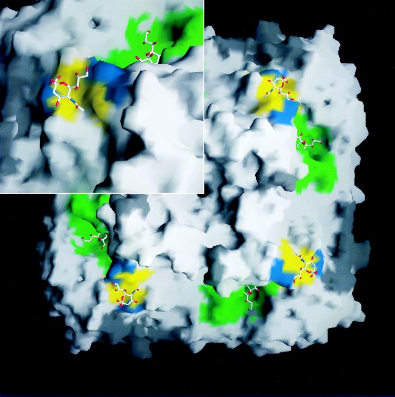
A molecular surface rendered image (42) of a tetrameric head of A/tern/Australia/G70C/75 N9 NA viewed from above the molecule. The active site residues interacting with the Neu5Ac moiety (twist-boat conformation) in the catalytic sites are show in green. The residues that interact with the Neu5Ac moiety (chair conformation) in the HB sites are colored yellow (conserved in all avian strains) and blue (conserved in N9 stains). (Inset) A magnified region (×1.6) in the vicinity of these two sites of one subunit, illustrating the deep pocket of the catalytic site and the flat surface of the HB site of the enzyme.

(A) A stereo drawing (43) of the refined x-ray atomic model of the Neu5Ac (orange) bound in the HA site of A/tern/Australia/G70C/75 NA with Neu5Ac soaked at 4°C, showing all the amino acids (green), and water molecules (red) making contact with the moiety. Atomic interactions are shown in broken lines. Oxygen, nitrogen, and carbon atoms are colored red, blue, and black, respectively. The protein backbone is represented by a yellow tube. (B) The two-Fo-Fc electron density map (blue caged-mesh contour at 1.6 σ level) of the Neu5Ac in the same orientation, using the refined phases of the complex, where Fo and Fc are the observed and calculated structure factors, respectively.
References
Publication types
MeSH terms
Substances
Associated data
- Actions
LinkOut - more resources
Full Text Sources
