. 1997 Oct 28;94(22):11839-44.
doi: 10.1073/pnas.94.22.11839.

The amino-terminal region of Tyk2 sustains the level of interferon alpha receptor 1, a component of the interferon alpha/beta receptor

Affiliations

The amino-terminal region of Tyk2 sustains the level of interferon alpha receptor 1, a component of the interferon alpha/beta receptor

M C Gauzzi et al. Proc Natl Acad Sci U S A. .

Abstract

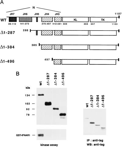
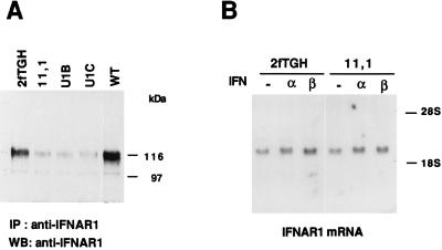
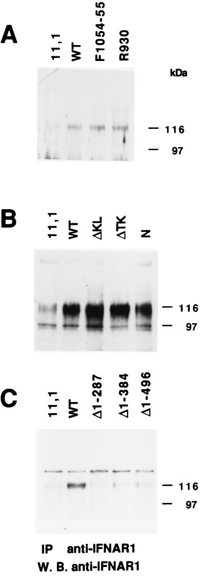
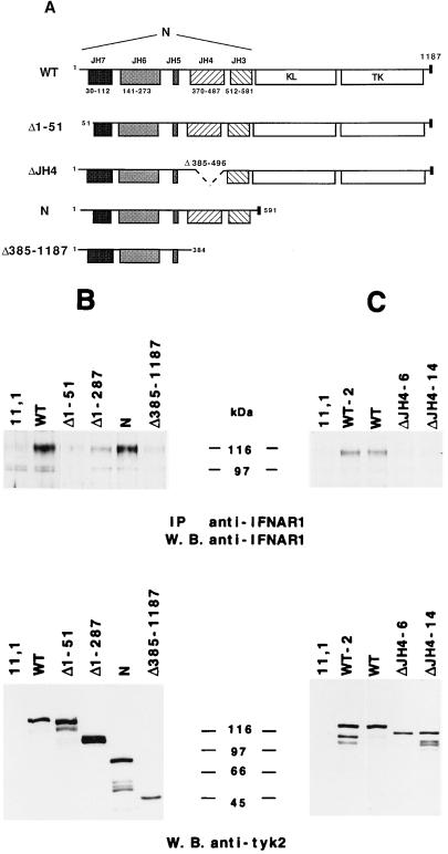
Tyk2 belongs to the Janus kinase (JAK) family of receptor associated tyrosine kinases, characterized by a large N-terminal region, a kinase-like domain and a tyrosine kinase domain. It was previously shown that Tyk2 contributes to interferon-alpha (IFN-alpha) signaling not only catalytically, but also as an essential intracellular component of the receptor complex, being required for high affinity binding of IFN-alpha. For this function the tyrosine kinase domain was found to be dispensable. Here, it is shown that mutant cells lacking Tyk2 have significantly reduced IFN-alpha receptor 1 (IFNAR1) protein level, whereas the mRNA level is unaltered. Expression of the N-terminal region of Tyk2 in these cells reconstituted wild-type IFNAR1 level, but did not restore the binding activity of the receptor. Studies of mutant Tyk2 forms deleted at the N terminus indicated that the integrity of the N-terminal region is required to sustain IFNAR1. These studies also showed that the N-terminal region does not directly modulate the basal autophosphorylation activity of Tyk2, but it is required for efficient in vitro IFNAR1 phosphorylation and for rendering the enzyme activatable by IFN-alpha. Overall, these results indicate that distinct Tyk2 domains provide different functions to the receptor complex: the N-terminal region sustains IFNAR1 level, whereas the kinase-like domain provides a function toward high affinity ligand binding.


Figures

Figure 1
Figure 1
(A) Schematic representation of wt and deleted Tyk2 proteins. The position and size of the JH domains and the first amino acid of each mutant protein are indicated. The small black box corresponds to the VSV-G epitope. (B) Basal in vitro kinase activity of wt and deleted Tyk2 forms. Whole cell extracts of the indicated transfectant were immunoprecipitated with anti-VSV-G antibodies. One-third of the immunoprecipitate was subjected to an in vitro kinase assay (Left) and the remaining material was directly resuspended in SDS/sample buffer. Samples were then fractionated on SDS/7% polyacrylamide gel, transferred to Hybond C-super membrane and subjected to autoradiography (Left) or probed with a monoclonal anti-VSV-G antibody, followed by goat anti-mouse 125I-conjugated, and subjected to autoradiography (Right). Phosphorylated and iodinated bands were quantified with a PhosphorImager.
Figure 2
Figure 2
IFNAR1 expression in 2fTGH cells and derivatives. (A) Whole cell extracts (1 mg) were immunoprecipitated with the anti-IFNAR1 monoclonal EA12 antibody, fractionated on SDS/7% polyacrylamide gel and blotted with the anti-IFNAR1 monoclonal GB8 antibody. (B) Northern transfers of 10 μg of total RNA extracted from subconfluent untreated (−), IFN-α, or IFN-β treated cells (4 hr) were hybridized to an IFNAR1 cDNA specific probe. Comparable signals in each lane were obtained with a β-actin probe (not shown).
Figure 3
Figure 3
IFNAR1 level in 11,1 expressing mutated forms of Tyk2. Anti-IFNAR1 immunoprecipitation and Western blot analysis were performed as in Fig. 2A. (A) IFNAR1 level in cells expressing the K930R point mutant, and the Y1054F/Y1055F mutant. (B) IFNAR1 level in cells expressing Tyk2 mutants deleted of either or both kinase domains. Five milligrams of lysates were processed. (C) IFNAR1 levels in cells expressing the N-terminal mutants, as indicated. The slower migrating band was seen only in this experiment.
Figure 4
Figure 4
(A) Schematic representation of four N-terminal deleted Tyk2 mutants: Δ1–51, ΔJH4, N, and Δ385–1187. (B and C) IFNAR1 level in 11,1 cells expressing mutated forms of Tyk2. (Upper) IFNAR1 protein level was analyzed in each cell line as described in Fig. 2. (Lower) The level of expression of each mutant protein was analyzed by direct anti-Tyk2 Western blot from 20 μg of crude extract.
Figure 5
Figure 5
Binding of IFN-α2 to 11,1 cells expressing deleted Tyk2 forms. Scatchard plot of direct binding of 125I-labeled IFN-α2c at 37°C.

References

    1. Ihle J N. Adv Immunol. 1995;60:1–35. - PubMed
    1. Leaman D W, Leung S, Li X, Stark G R. FASEB J. 1996;10:1578–1588. - PubMed
    1. Velazquez L, Fellous M, Stark G R, Pellegrini S. Cell. 1992;70:313–322. - PubMed
    1. Müller M, Briscoe J, Laxton C, Guschin D, Ziemiecki A, Silvennoinen O, Harpur A G, Barbieri G, Witthuhn B A, Schindler C, Pellegrini S, Wilks A F, Ihle J N, Stark G R, Kerr I M. Nature (London) 1993;366:129–166. - PubMed
    1. Uzé G, Luftalla G, Gresser I. Cell. 1990;60:225–234. - PubMed

Publication types

LinkOut - more resources