. 1997 Oct 28;94(22):12030-5.
doi: 10.1073/pnas.94.22.12030.

A gene spans the pseudoautosomal boundary in mice

Affiliations

A gene spans the pseudoautosomal boundary in mice

S Palmer et al. Proc Natl Acad Sci U S A. .

Abstract

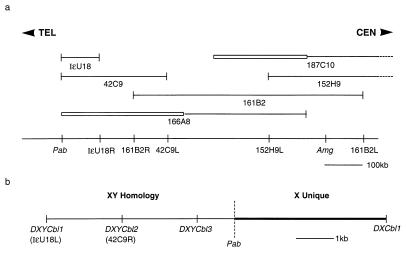
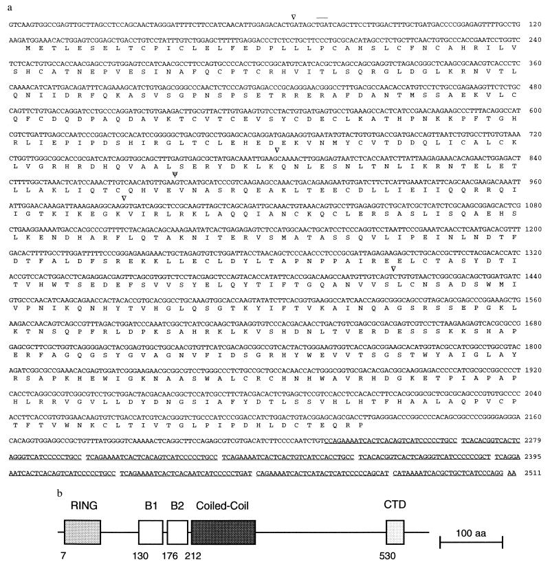
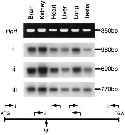
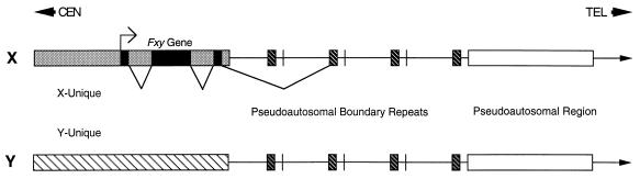
The X and Y chromosomes of the mouse, like those of other mammals, are heteromorphic over most of their length, but at the distal ends of the chromosomes is a region of sequence identity, the pseudoautosomal region (PAR), where the chromosomes pair and recombine during male meiosis. The point at which the PAR diverges into X- and Y-specific sequences is called the pseudoautosomal boundary. We have completed a genomic walk from the X-specific Amelogenin gene to the PAR. Analysis of this region revealed that the pseudoautosomal boundary of mice is located within an intron of a transcribed gene that encodes a novel RING finger protein. The first three of the exons of the gene are located on the X chromosome whereas the 3' exons of the gene are located on both X and Y chromosomes. This unusual arrangement may indicate that the gene is in a state of transition from pseudoautosomal to X-unique and provides evidence for a process of attrition of the pseudoautosomal region on the Y chromosome.


Figures

Figure 1
Figure 1
Physical map of the distal X chromosome and pseudoautosomal boundary region of the mouse. (a) Overlapping YAC clones covering the region between Amg and the pseudoautosomal boundary (Pab). Boxed regions of YACs indicate the presence of non-X chromosomal DNA. The STSs derived from these YACs and their relative distances are indicated below the line. The suffix L or R on the STS name indicates the left or right arm of the YAC from which the STS is derived. The dotted lines on the YACs 187C10 and 152H9 indicate that they extend beyond the map. TEL and CEN indicate telomeric and centromeric directions, respectively. (b) A high-resolution map of the pseudoautosomal boundary showing the positions of the flanking STSs.
Figure 2
Figure 2
Physical linkage of DXYCbl1 to a known pseudoautosomal locus. Southern analysis of three C57BL/6 male DNAs cut with PacI and hybridized with DXYCbl1, pMov15/1 and Amg. (a) A blot probed with DXYCbl1 then reprobed in b with pMov15/1 showing the same bands hybridizing. The smallest allele shown in lane two is 500 kb. The lower hybridizing bands in b are derived from other loci containing the macrosatellite repeat including a 400-kb band that is thought to be from chromosome 9. (c) A similar blot probed with Amg showing specific hybridization at 800 kb. The lower smear is nonspecific hybridization to the main bulk of DNA. (d) The same blot shown in c rehybridized with pMov15/1 demonstrating that different bands are identified by this probe. Size markers are Saccharomyces cerevisiae chromosomes (strain YPH148).
Figure 3
Figure 3
Southern analysis of the region surrounding the pseudoautosomal boundary. Male and female DNAs from mice of the FVB and C57BL/6 strains and from a female C57BL/10 mouse digested with BamHI and EcoRV were analyzed by Southern blotting and probed with DNA fragments derived from either side of the pseudoautosomal boundary and from an autosomal gene as a loading control. (Middle) A blot probed with DXCbl1 shows that this STS is X-unique and gives a 2:1 ratio of hybridization intensity in female and male lanes. (Top) The same blot stripped and reprobed with DXYCbl1 showing hybridization to the same X-linked band as DXCbl1 in the BamHI cut lanes and an additional, more intense, band at 6.5 kb derived from the pseudoautosomal repeats in the C57BL/10 and C57BL/6 lanes. The repeat band appears to be absent in the FVB female and of an altered allele size in the FVB male. The single 12-kb repeat band in the C57BL/10 female demonstrates that the repeat unit must be at least this size and is probably larger because there are no junction fragments. (Bottom) The same blot stripped and reprobed with a fragment of the autosomal gene Tcp-1 (31) to show equivalence of loading between male and female tracks.
Figure 4
Figure 4
Structure of Fxy. (a) DNA and encoded protein sequence of the Fxy gene. Introns are indicated by arrowheads and ψ indicates the intron that contains the pseudoautosomal boundary. Not all the positions of introns 3′ to the boundary have been defined. The DXYMov15 repeats in the 3′-untranslated region of the gene are underlined. An in-frame termination codon present upstream of the putative translation initiating methionine is overlined. (b) Schematic representation of the protein encoded by Fxy. Structural features associated with RING finger proteins (24) are indicated. (Bar = 100 amino acids.)
Figure 5
Figure 5
Expression of the Fxy gene. PCR primer sets (i, ii, and iii) derived from the sequence of Fxy, or as a control Hprt, were used to amplify cDNA made from the indicated tissues. For i, ii, and iii, the products were electrophoresed on an agarose gel, blotted to a membrane, and hybridized with Fxy cDNA. For Hprt a photograph of an ethidium bromide stained gel is shown. Ψ indicates the position of the pseudoautosomal boundary. Multiple introns are present between all the primer pairs and no product is amplified from genomic DNA. The sequences of the primers are described in Materials and Methods.
Figure 6
Figure 6
Schematic representation of the mouse pseudoautosomal boundary region. The three exons at the 5′ end of the gene are X-unique (solid area) but the 3′ region of the gene (dark hatched area) is present on the X and Y chromosomes and within a repeat region just distal to the boundary. Note that for simplicity the 3′ end of the gene is shown as a single block whereas it is actually interrupted by several introns and only four of the variable number of pseudoautosomal repeats (22, 23) are shown. TEL and CEN indicate telomeric and centromeric directions, respectively.

References

    1. Ohno S. Sex Chromosomes and Sex-Linked Genes. Berlin: Springer; 1967.
    1. Burgoyne P S. Hum Genet. 1982;61:85–90. - PubMed
    1. Ellis N A, Goodfellow P N. Trends Genet. 1989;5:406–410. - PubMed
    1. Rouyer F, Simmler M C, Johnsson C, Vergnaud G, Cooke H J, Weissenbach J. Nature (London) 1986;319:291–295. - PubMed
    1. Goodfellow P J, Pym B, Mohandas T, Shapiro L. Am J Hum Genet. 1984;36:777–782. - PMC - PubMed

Publication types

MeSH terms

Associated data