Comparative Study
. 1997 Oct 28;94(22):12075-80.
doi: 10.1073/pnas.94.22.12075.

Cell cycle-dependent colocalization of BARD1 and BRCA1 proteins in discrete nuclear domains

Affiliations
Comparative Study

Cell cycle-dependent colocalization of BARD1 and BRCA1 proteins in discrete nuclear domains

Y Jin et al. Proc Natl Acad Sci U S A. .

Abstract

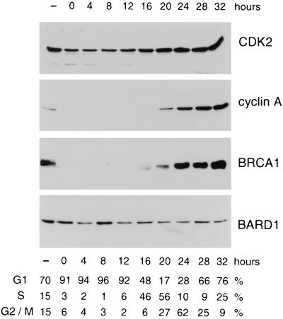
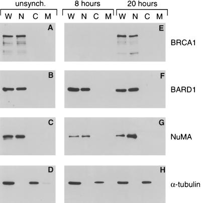
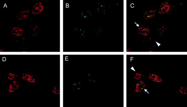
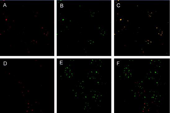
Germ-line mutations of the BRCA1 gene predispose women to early-onset breast and ovarian cancer by compromising the gene's presumptive function as a tumor suppressor. Although the biochemical properties of BRCA1 polypeptides are not understood, their expression pattern and subcellular localization suggest a role in cell-cycle regulation. When resting cells are induced to proliferate, the steady-state levels of BRCA1 increase in late G1 and reach a maximum during S phase. Moreover, in S phase cells, BRCA1 polypeptides are hyperphosphorylated and accumulate into discrete subnuclear foci termed "BRCA1 nuclear dots." BRCA1 associates in vivo with a structurally related protein termed BARD1. Here we show that the steady-state levels of BARD1, unlike those of BRCA1, remain relatively constant during cell cycle progression. However, immunostaining revealed that BARD1 resides within BRCA1 nuclear dots during S phase of the cell cycle, but not during the G1 phase. Nevertheless, BARD1 polypeptides are found exclusively in the nuclear fractions of both G1- and S-phase cells. Therefore, progression to S phase is accompanied by the aggregation of nuclear BARD1 polypeptides into BRCA1 nuclear dots. This cell cycle-dependent colocalization of BARD1 and BRCA1 indicates a role for BARD1 in BRCA1-mediated tumor suppression.


Figures

Figure 1
Figure 1
Expression of BARD1 polypeptides during cell cycle progression. Contact-inhibited T24 cells were released from growth arrest by replating at low density. Synchronized cell populations were then lysed at the indicated time points, and equivalent aliquots of each lysate were subjected to Western analyses with antibodies specific for CDK2, cyclin A, BRCA1, and BARD1. The cell cycle distribution of the synchronized cultures at each time point was estimated by FACS analysis, as tabulated beneath the immunoblots.
Figure 5
Figure 5
BARD1 polypeptides reside within the nuclear compartment of G1- and S-phase cells. Whole cell (W), nuclear (N), cytoplasmic (C), and membrane (M) fractions were prepared from unsynchronized T24 cells (A–D), as well as from contact-inhibited cells that had been released from growth arrest for either 8 hr or 20 hr (E–H). Equivalent aliquots of each fraction were then subjected to Western analyses with antibodies specific for BRCA1 (A and E), BARD1 (B and F), nuclear matrix protein NuMA (C and G), and α-tubulin (D and H).
Figure 3
Figure 3
Cell cycle specificity of BARD1 immunostaining. Synchronized populations of T24 cells were costained with propidium iodide and either the BRCA1-specific MS110 monoclonal antibody (A–D) or the BARD1-specific monoclonal antibody (E–H). The composite images of two-color staining are illustrated. The cells were harvested at either 8 hr (A and E) or 20 hr (B–D and F–H) after synchronous release from growth arrest. In some experiments the monoclonal antibodies were preabsorbed with an excess of the BRCA1 immunogen (GST-BR304; C), the BARD1 immunogen (GST-EE; G) or the parental GST polypeptide (D and H).
Figure 2
Figure 2
A punctate pattern of nuclear staining observed with BRCA1- and BARD1-specific antibodies. Asynchronous HBL-100 cells were stained with propidium iodide to highlight the nucleus (red, A and D) and with either the BRCA1-specific MS110 monoclonal antibody (green, B) or a BARD1-specific mouse polyclonal antiserum (green, E). C is a composite image of A and B, and F is a composite of D and E; in the composite images yellow is produced where the red and green signals overlap. Punctate staining of BRCA1 and BARD1 is observed in some (arrows) but not all (arrowheads) interphase nuclei (C and F).
Figure 4
Figure 4
BARD1 is a component of the BRCA1 nuclear dots. Asynchronous HBL-100 cells were costained with an affinity-purified BARD1-specific rabbit antiserum (red, A and D) and either a BRCA1-specific (green, B) or PML-specific (green, E) monoclonal antibody. The composite images of two-color staining demonstrate that BARD1 colocalizes with BRCA1 (C) but not with PML (F).

References

    1. Hall J M, Lee M K, Newman B, Morrow J E, Anderson L A, Huey B, King M-C. Science. 1990;250:1684–1689. - PubMed
    1. Miki Y, Swensen J, Shattuck-Eidens D, Futreal P A, Harshman K, et al. Science. 1994;266:66–71. - PubMed
    1. Chen C-f, Li S, Chen Y, Chen P-L, Sharp Z D, Lee W-H. J Biol Chem. 1996;271:32863–32868. - PubMed
    1. Thakur S, Zhang H B, Peng Y, Le H, Carroll B, Ward T, Yao J, Farid L M, Couch F J, Wilson R B, Weber B L. Mol Cell Biol. 1997;17:444–452. - PMC - PubMed
    1. Koonin E V, Altschul S F, Bork P. Nat Genet. 1996;13:266–267. - PubMed

Publication types

LinkOut - more resources