. 1997 Oct 28;94(22):12094-9.
doi: 10.1073/pnas.94.22.12094.

Selective induction of p53 and chemosensitivity in RB-deficient cells by E1A mutants unable to bind the RB-related proteins

Affiliations

Selective induction of p53 and chemosensitivity in RB-deficient cells by E1A mutants unable to bind the RB-related proteins

A V Samuelson et al. Proc Natl Acad Sci U S A. .

Abstract

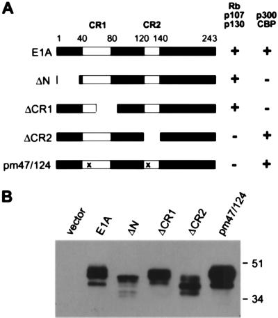
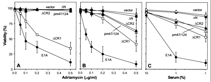
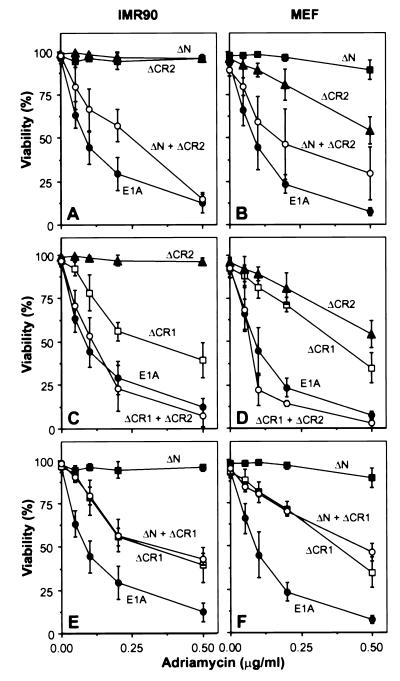
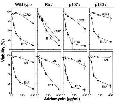
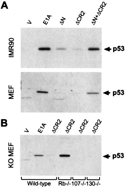
The adenovirus E1A oncoprotein renders primary cells sensitive to the induction of apoptosis by diverse stimuli, including many anticancer agents. E1A-expressing cells accumulate p53 protein, and p53 potentiates drug-induced apoptosis. To determine how E1A promotes chemosensitivity, a series of E1A mutants were introduced into primary human and mouse fibroblasts using high-titer recombinant retroviruses, allowing analysis of E1A in genetically normal cells outside the context of adenovirus infection. Mutations that disrupted apoptosis and chemosensitivity separated into two complementation groups, which correlated precisely with the ability of E1A to associate with either the p300/CBP or retinoblastoma protein families. Furthermore, E1A mutants incapable of binding RB, p107, and p130 conferred chemosensitivity to fibroblasts derived from RB-deficient mice, but not fibroblasts from mice lacking p107 or p130. Hence, inactivation of RB, but not p107 or p130, is required for chemosensitivity induced by E1A. Finally, the same E1A functions that promote drug-induced apoptosis also induce p53. Together, these data demonstrate that p53 accumulation and chemosensitivity are linked to E1A's oncogenic potential, and identify a strategy to selectively induce apoptosis in RB-deficient tumor cells.


Figures

Figure 1
Figure 1
Structure and expression of E1A mutants. (A) The E1A 243R contains two conserved regions (CR1 and CR2, white boxes). In the E1A mutants, deletions are indicated by gaps, and point mutations by an “x.” ΔN, ΔCR1, and ΔCR2 are deletions of amino acids 2–36, 68–85, and 120–140, respectively. pm47/124 mutant has a tyrosine to histidine and cysteine to glycine changes at amino acids 47 and 124, respectively. Cellular proteins able to interact with each E1A mutant in coimmunoprecipitations are indicated (31). (B) E1A was introduced into IMR90 cells using LPC-based retroviral vectors. After selection in puromycin, E1A levels were determined by Western blot analysis. E1A is highly phosphorylated and undergoes posttranslational modification, which accounts for variable migration in SDS gels (33). Note that the ΔN mutant was able to coimmunoprecipitate similar levels of RB as wild-type E1A (data not shown). Infection efficiencies were >50% before selection and >95% of the cells in the selected cells expressed E1A as determined by immunofluorescence (data not shown). Each E1A mutant localized to the nucleus (data not shown).
Figure 2
Figure 2
Multiple regions of E1A are required for chemosensitivity. Primary mouse (MEF) (A and C) or human (IMR90) (B) fibroblasts were infected with an empty vector (vector, ○), vectors expressing full-length E1A (•), or the following mutants: ΔN (▪), ΔCR1 (□), ΔCR2 (▴), and pm47/pm124 (▵). Infected populations were plated in multiwell dishes and treated with the indicated concentrations of adriamycin (A and B) or serum (C). Cell viability was determined 24 h following adriamycin treatment or 48 h after serum withdrawal. Previous studies demonstrated that cell death under these conditions results from apoptosis (1, 7), and this was confirmed by visualizing chromatin condensation using 4′,6-diamidino-2-phenylindole (data not shown). Each value represents the mean ± SD from at least three separate experiments.
Figure 3
Figure 3
Separate E1A functions cooperate to confer chemosensitivity. IMR90 or MEF cell populations expressing E1A (•), ΔN (▪), ΔCR1 (□), ΔCR2 (▴), ΔN and ΔCR2 (○ in A and B), ΔCR1 and ΔCR2 (○ in C and D), or ΔN and ΔCR1 (○ in E and F) were generated by retroviral infection. Multiple E1A mutants were introduced sequentially as described in Materials and Methods. Cell populations were treated with adriamycin and viability was determined 24 h later by trypan blue exclusion. Each value represents the mean ± SD of the data from at least three separate experiments.
Figure 4
Figure 4
Inactivation of RB by CR2 is required for chemosensitivity. Wild-type, RB−/−, p107−/−, and p130−/− MEFs expressing either E1A (•), ΔCR2 (○, Upper), or ΔN (○, Lower) were generated by retroviral infection. E1A and E1A mutants were expressed at similar levels (data not shown). Cell viability was determined 24 h after adriamycin treatment. Each point represents the mean ± SD from at least three separate experiments.
Figure 5
Figure 5
E1A and p53 accumulation. (A) p53 expression in populations of IMR90s or MEFs expressing the empty vector (V), full-length E1A, ΔN, ΔCR2, or coexpressing ΔN and ΔCR2 was examined by immunoblotting using polyclonal antibodies specific for human or mouse p53, respectively. (B) p53 expression in wild-type, RB−/−, p107−/−, or p130−/− MEF populations expressing the indicated E1A proteins. No increase in p53 was observed in RB−/−, p107−/−, or p130−/− MEFs without E1A, and E1A induced p53 in MEFs of all three genotypes (data not shown.)

References

    1. Lowe S W, Ruley H E, Jacks T, Housman D E. Cell. 1993;74:957–967. - PubMed
    1. Dive C, Hickman J A. Br J Cancer. 1991;64:192–196. - PMC - PubMed
    1. Evan G I, Wyllie A H, Gilbert C S, Littlewood T D, Land H, Brooks M, Waters C, Penn L Z, Hancock D C. Cell. 1992;69:119–128. - PubMed
    1. Ko L J, Prives C. Genes Dev. 1996;10:1054–1072. - PubMed
    1. Lowe S W. Curr Opin Oncol. 1995;7:547–553. - PubMed

Publication types