. 1997 Oct 28;94(22):12100-5.
doi: 10.1073/pnas.94.22.12100.

Restoration of beta-adrenergic signaling in failing cardiac ventricular myocytes via adenoviral-mediated gene transfer

Affiliations

Restoration of beta-adrenergic signaling in failing cardiac ventricular myocytes via adenoviral-mediated gene transfer

S A Akhter et al. Proc Natl Acad Sci U S A. .

Abstract

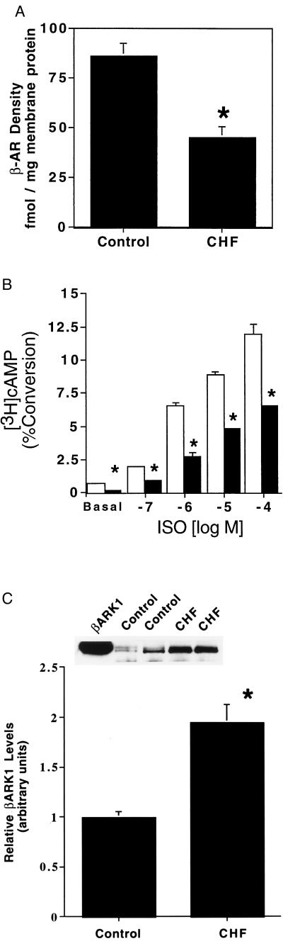
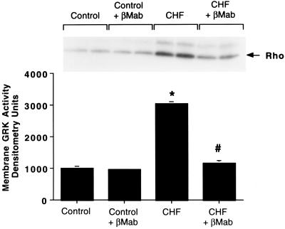
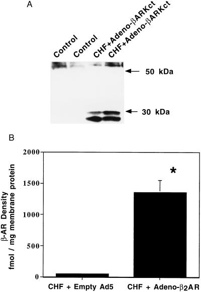
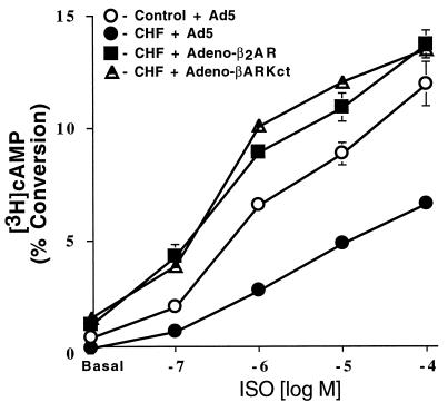
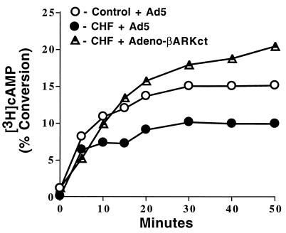
Cardiovascular gene therapy is a novel approach to the treatment of diseases such as congestive heart failure (CHF). Gene transfer to the heart would allow for the replacement of defective or missing cellular proteins that may improve cardiac performance. Our laboratory has been focusing on the feasibility of restoring beta-adrenergic signaling deficiencies that are a characteristic of chronic CHF. We have now studied isolated ventricular myocytes from rabbits that have been chronically paced to produce hemodynamic failure. We document molecular beta-adrenergic signaling defects including down-regulation of myocardial beta-adrenergic receptors (beta-ARs), functional beta-AR uncoupling, and an up-regulation of the beta-AR kinase (betaARK1). Adenoviral-mediated gene transfer of the human beta2-AR or an inhibitor of betaARK1 to these failing myocytes led to the restoration of beta-AR signaling. These results demonstrate that defects present in this critical myocardial signaling pathway can be corrected in vitro using genetic modification and raise the possibility of novel inotropic therapies for CHF including the inhibition of betaARK1 activity in the heart.


Figures

Figure 1
Figure 1
β-AR signaling defects in ventricular myocytes isolated from chronically paced rabbit hearts. (A) β-AR density was determined from myocardial sarcolemmal membranes prepared from ventricular myocytes isolated from control (sham-operated) or CHF (paced) rabbits (n = 5 in each group; ∗, P < 0.05 vs. control). (B) Basal and ISO-stimulated intracellular adenylyl cyclase activity was determined for control (open bars) and CHF ventricular myocytes (filled bars) (n = 5 myocyte preparations each; ∗ P < 0.05 vs. control). (C) Levels of expression of βARK1 in whole-cell extracts were assessed by protein immunoblotting. Shown is the average data for five myocyte preparations isolated from control (sham) and CHF (paced) rabbit hearts. βARK1 densitometry values from control immunoblots were arbitrarily set to 1 and all values were normalized to controls. The Inset shows a representative protein immunoblot from two control and two CHF preparations with purified βARK1 used as a marker (∗, P < 0.05 vs. control).
Figure 2
Figure 2
Myocyte membrane GRK activity. GRK activity was determined by an in vitro rhodopsin phosphorylation assay for control (sham) and CHF (paced) ventricular myocyte membrane extracts in the absence and presence of a βARK mAb (βMab) which selectively inhibits βARK activity. Shown is the mean of five separate myocyte preparations with the inset depicting a representative experiment done in duplicate. Phosphorylated rhodopsin is marked by the arrow (∗, P < 0.05 vs. control; #, P < 0.05 vs. CHF).
Figure 3
Figure 3
Expression of the adenoviral transgenes in failing ventricular myocytes. (A) Representative Western blot displaying expression of the βARKct peptide in only Adeno-βARKct infected myocytes. (B) Total β-AR density determined for CHF ventricular myocyte membranes infected with Empty Ad5 (n = 4) or Adeno-β2AR (n = 5) (∗, P < 0.005 vs. CHF + Ad5).
Figure 4
Figure 4
Intracellular cAMP production in failing ventricular myocytes following adenoviral-mediated gene transfer. After 36 hr of infection with the appropriate adenovirus, control (sham) or CHF myocytes (n = 4 preparations each) were stimulated with increasing concentrations of ISO. Accumulation of cAMP was expressed as percent incorporation of the total [3H] uptake into [3H]cAMP. Responses for CHF cells infected with Adeno-β2AR and Adeno-βARKct were significantly elevated compared with both CHF cells (P < 0.005, ANOVA) and control cells (P < 0.005, ANOVA) infected with Ad5.
Figure 5
Figure 5
Time course of cAMP production. Assessment of the effect of Adeno-βARKct infection on β-AR desensitization via a time course of cAMP production in response to 10 μM ISO. Myocytes were isolated from control (sham) rabbit hearts or CHF (paced) rabbit hearts and infected with Empty Ad5 or Adeno-βARKct (n = 4 each) and cAMP was measured over the course of 50 min.

References

    1. Williams R S. N Engl J Med. 1995;332:817–818. - PubMed
    1. Bristow M R, Ginsburg R, Minobe W, Cubicciotti R, Sageman W S, Lurie K, Billingham M E, Harrison D C, Stinson E B. N Engl J Med. 1982;307:205–211. - PubMed
    1. Brodde O E, Michel M C, Zerkowski H R. Cardiovasc Res. 1995;30:570–584. - PubMed
    1. Ungerer M, Bohm M, Elce J S, Erdmann E, Lohse M L. Circulation. 1993;87:454–463. - PubMed
    1. Inglese J, Freedman N J, Koch W J, Lefkowitz R J. J Biol Chem. 1993;268:23735–23738. - PubMed

Publication types

MeSH terms