. 1997 Oct;181(4):331-41.
doi: 10.1007/s003590050119.

GABAergic disinhibition changes the recovery cycle of bat inferior collicular neurons

Affiliations

GABAergic disinhibition changes the recovery cycle of bat inferior collicular neurons

Y Lu et al. J Comp Physiol A. 1997 Oct.

Abstract

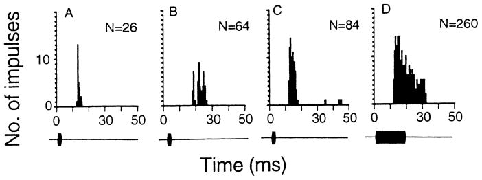
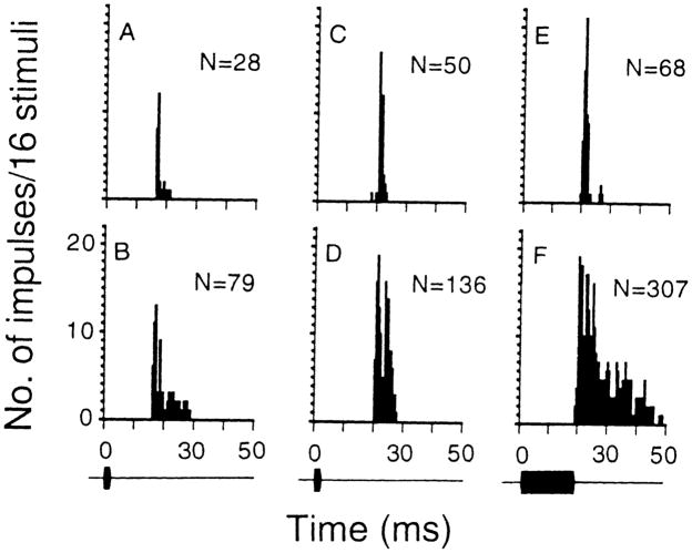
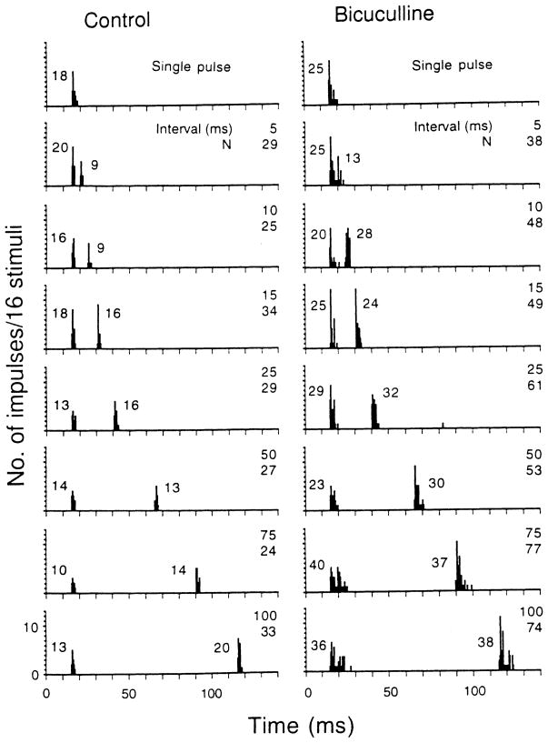
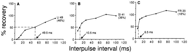
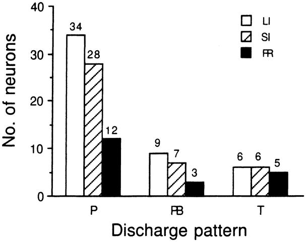
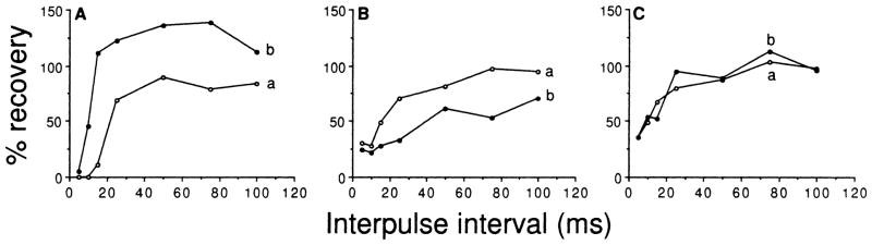
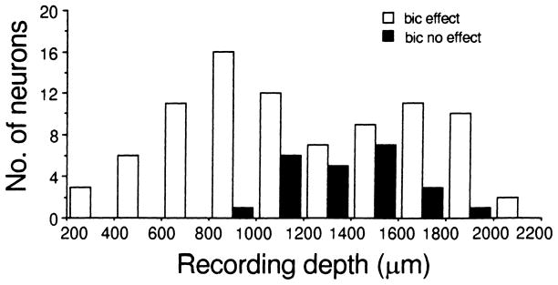
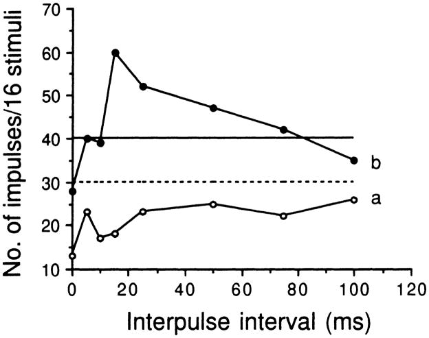
This study examines the contribution of GABAergic inhibition to the discharge pattern and recovery properties of 110 bat inferior collicular neurons by means of bicuculline application to their recording sites. When stimulated with single pulses, 74 (67%) neurons discharged one or two impulses (phasic responders), 19 (17%) discharged three to ten impulses (phasic bursters) and 17 (16%) discharged impulses throughout the entire stimulus duration (tonic responders). Bicuculline application changed phasic responders into phasic bursters or tonic responders, increased the number of impulses by 10-2000% and shortened the response latency of most neurons. When stimulated with pairs of sound pulses, the recovery cycles of these neurons can be described as: (1) long inhibition (n = 49, 45%); (2) short inhibition (n = 41, 37%); and (3) fast recovery (n = 20, 18%) based upon the 50% recovery time that was either longer than 20 ms, between 10 and 20 ms or shorter than 10 ms. Bicuculline application shortened the 50% recovery time of most neurons by 11-2350% allowing them to respond to pairs of sound pulses at very short interpulse intervals. These data demonstrate that GABAergic inhibition contributes significantly to auditory temporal processing.


Figures

Fig. 1
Fig. 1
AD The post-stimulus-time (PST) histograms showing the discharge patterns of three representative inferior collicular neurons: phasic responder (A), phasic burster (B) and tonic responder (C, D). When stimulated with acoustic stimuli of 3 or 19 (not shown) ms, the phasic responder discharged 1–2 impulses (A) and the phasic buster discharged 3–10 impulses (B). In contrast, the tonic responder discharged impulses throughout the entire duration of the acoustic stimulus (C, D). N: number of impulses discharged to 16 stimuli. The BF (kHz) and MT (dB SPL) of these three neurons were 59.9, 31 (A); 38.7, 33 (B) and 64.0, 52 (C, D). The stimulus intensity used to obtain these PST histograms was always 80 dB SPL. Bin width: 500 μs, sampling period: 300 ms. To highlight the discharge pattern, the sampling period beyond 50 ms is not shown
Fig. 2
Fig. 2
AF PST histograms showing the discharge patterns of two inferior collicular neurons obtained with 80-dB SPL sounds before and after bicuculline application to their recording sites. The discharge patterns of AD were obtained with 3-ms sounds and those in E, F were obtained with 19-ms sounds. The discharge pattern of one collicular neuron (BF: 43.5 kHz, MT: 22 dB SPL) changed from phasic responder (A) into phasic burster (B) after bicuculline application. The discharge pattern of another neuron (BF: 35.1 kHz, MT: 47 dB SPL) changed from phasic responder (C, E) into tonic responder (D, F) after bicuculline application such that this neuron discharged impulses with a duration longer than the presented pulses. N: total number of impulses in the histogram. Bin width: 500 μs, sampling period: 300 ms. To highlight the change in discharge pattern, the sampling period beyond 50 ms is not shown
Fig. 3
Fig. 3
PST histograms showing the discharge pattern of an inferior collicular neuron obtained with single pulses (top histogram of both columns) and with an identical pulse pair (BF pulses of 80–80 dB SPL, 3 ms duration with 0.5-ms rise-decay times) delivered at different interpulse intervals before (control, left column) and after (bicuculline, right column) iontophoretic application of bicuculline to the recording site. All stimuli were delivered at 30° contralateral to the neuron’s recording site. In the top histogram of both columns, the number of impulses discharged to 16 single pulses is shown. In the remaining histograms, the number of impulses elicited by the first and the second pulse in 16 presentations are shown. In the right upper corner of each histogram, the interpulse interval (ms) is shown by the upper number and the total number of impulses discharged to both presented pulses is shown by the lower number (N). When the neuron’s discharge to the first and second pulses was inseparable, the neuron’s discharge to the second pulse was calculated as the difference between the neuron’s discharge to both pulses (i.e., N) and its discharge to the first pulse alone (shown at the top of each column). The BF (kHz) and MT(dB SPL) of this neuron was 39.5, 25. Bin width: 500 μs, sampling period: 300 ms. To save space, the sampling period beyond 140 ms is not shown
Fig. 4
Fig. 4
AC Three types of recovery cycles obtained from the inferior collicular neurons of Eptesicus fuscus. Long inhibition recovery cycle (LI) has a 50% recovery time longer than 20 ms (A). Short inhibition recovery cycle (SI) has a 50% recovery time between 10 and 20 ms (B). Fast recovery cycle (FR) has a 50% recovery time shorter than 10 ms (C). The number and percentage of neurons of each type is shown at each curve. The 50% recovery time for each representative curve is shown by the arrow. Ordinates and abscissas represent percentage recovery and interpulse interval in ms
Fig. 5
Fig. 5
Bar histograms showing the distribution of different types of recovery cycles of inferior collicular neurons according to their discharge patterns before the application of bicuculline to their recording sites. The number of neurons of each type of recovery cycle is shown atop each bar. P: phasic responder, PB: phasic burster, T: tonic responder, LI (unfilled bars): long inhibition recovery cycle, SI (hatched bars): short inhibition recovery cycle, FR (filled bars): fast recovery cycle
Fig. 6
Fig. 6
AC Recovery cycles of three representative inferior collicular neurons (A, B, C) determined before (curve a) and after (curve b) iontophoretic application of bicuculline to their recording sites. Bicuculline application either shortened (Ab), lengthened (Bb) or did not affect (Cb) their recovery cycles
Fig. 7
Fig. 7
Bar histogram showing the distribution of two types of inferior collicular neurons according to their recording depths (abscissa). Unfilled bars: inferior collicular neurons whose recovery cycles were affected by bicuculline application (bic effect). Solid bars: inferior collicular neurons whose recovery cycles were not affected by bicuculline application (bic no effect)
Fig. 8
Fig. 8
Variation in the number of impulses of an inferior collicular neuron discharged to paired identical pulses (80–80 dB SPL) obtained at different interpulse intervals (shown by abscissa) before (a: unfilled circle curve) and after (b: filled circle curve) iontophoretic application of bicuculline to its recording site. The dashed and solid horizontal lines represent the summation of the number of impulses discharged in response to the pulse and echo alone before and after bicuculline application to its recording site. The BF and MT of this neuron were 32.0 kHz and 21 dB SPL

Similar articles

Cited by

References

    1. Adams JC. Ascending projections to the inferior colliculus. J Comp Neurol. 1979;183:519–538. - PubMed
    1. Adams JC, Mugnaini E. Dorsal nucleus of the lateral lemniscus: a nucleus of GABAergic projection neurons. Brain Res Bull. 1984;13:585–590. - PubMed
    1. Aitkin LM, Prain SM. Medial geniculate body: unit responses in awake cats. J Neurophysiol. 1974;37:512–521. - PubMed
    1. Bormann J. Electrophysiology of GABAA and GABAB receptor subtypes. Trend Neurosci. 1988;11:112–116. - PubMed
    1. Casseday JH, Covey E. Frequency tuning properties of neurons in the inferior colliculus of an FM bat. J Comp Neurol. 1992;319:34–50. - PubMed

Publication types

LinkOut - more resources