. 1997 Nov 3;139(3):579-87.
doi: 10.1083/jcb.139.3.579.

Mapping and use of a sequence that targets DNA ligase I to sites of DNA replication in vivo

Affiliations

Mapping and use of a sequence that targets DNA ligase I to sites of DNA replication in vivo

M C Cardoso et al. J Cell Biol. .

Abstract

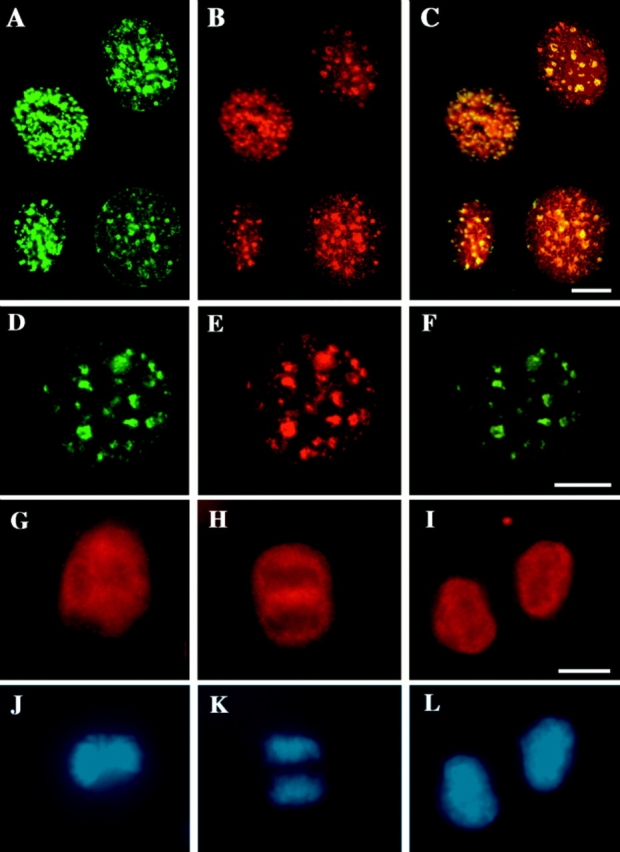
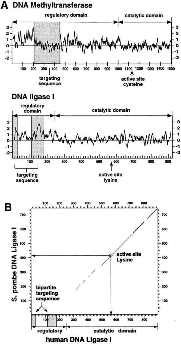
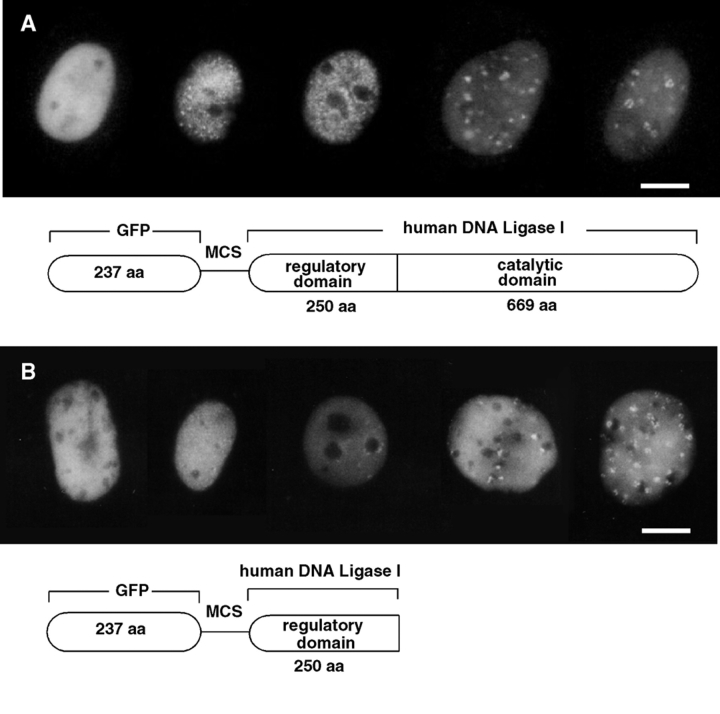
The mammalian nucleus is highly organized, and nuclear processes such as DNA replication occur in discrete nuclear foci, a phenomenon often termed "functional organization" of the nucleus. We describe the identification and characterization of a bipartite targeting sequence (amino acids 1-28 and 111-179) that is necessary and sufficient to direct DNA ligase I to nuclear replication foci during S phase. This targeting sequence is located within the regulatory, NH2-terminal domain of the protein and is dispensable for enzyme activity in vitro but is required in vivo. The targeting domain functions position independently at either the NH2 or the COOH termini of heterologous proteins. We used the targeting sequence of DNA ligase I to visualize replication foci in vivo. Chimeric proteins with DNA ligase I and the green fluorescent protein localized at replication foci in living mammalian cells and thus show that these subnuclear functional domains, previously observed in fixed cells, exist in vivo. The characteristic redistribution of these chimeric proteins makes them unique markers for cell cycle studies to directly monitor entry into S phase in living cells.


Figures

Figure 2
Figure 2
Localization of DNA ligase I throughout the cell cycle. Asynchronously growing mouse fibroblasts (C3H10T1/2 cells; A–C and G–L) and myoblasts (C2C12 cells; A–F) were pulse labeled with BrdU for 5 to 10 min (A–C) and formaldehyde (A–C, G, and J) or methanol fixed (D–F, H, I, K, and L). Cells were stained for DNA ligase I with the affinity-purified anti-DNA ligase I rabbit antibodies (see Fig. 1 red; B, E, and G–I), for sites of BrdU incorporation with anti-BrdU mouse monoclonal antibody (green; A), and for PCNA with anti-PCNA–specific mouse monoclonal antibody (green; D), and DNA was visualized by counterstaining with Hoechst 33258 dye (J–L). A–F show the distribution of DNA ligase I (B and E) in interphase cells relative to sites of ongoing DNA replication labeled with BrdU (A) and to PCNA (D), which has previously been shown to redistribute in S phase nuclei to replication centers (5, 7). A–C is a composite of cell nuclei at different stages of S phase. As can be better visualized in the overlay of the green and red images in C and F, DNA ligase I takes on a pattern of subnuclear foci that colocalize with sites of BrdU incorporation (C) and with PCNA (F). G–L show the distribution of DNA ligase I in mitotic cells. A cell in metaphase as evidenced by the absence of nuclear membrane and the alignment of the chromosomes in the metaphase plate in J, shows that DNA ligase I is excluded from the condensed chromosomes and upon nuclear envelope breakdown distributes in the cytoplasm (G). During chromatid separation and movement to the spindle poles at anaphase in K, DNA ligase I is still dispersed in the cytoplasm and excluded from the condensed chromosomes (H). At the end of telophase and cytokinesis, when the nuclear envelope reforms around the decondensing chromosomes (L), there is an immediate import of DNA ligase I into the nucleus as seen in I. Bars, 10 μm.
Figure 1
Figure 1
Purification and characterization of antibodies against DNA ligase I. (a) Immunoblot analysis of MEL whole cell extract (20 μg/lane) with anti-DNA ligase I antibodies. Lane 1, Preimmune serum; lane 2, affinity-purified antibodies eluted with 0.2 M glycine-HCl, pH 2.0; lane 3, affinity-purified antibodies eluted with 6 M guanidine hydrochloride. A band with an apparent molecular weight of 120 to 130 kD reacts specifically with the affinity-purified antibodies. (b) Specifity of the antibody (lane 1) was further tested by pre-incubation with specific (S, lane 2) and unspecific (U, lane 3) peptides at a 100-fold molar excess. The DNA ligase I signal is competed out only with the NH2-terminal DNA ligase I peptide and not with the same amount of an unrelated peptide, confirming the specificity of the antibody. (c) Species reactivity of anti-DNA ligase I antibodies: lane 1, C2C12 (mouse) cell extract; lane 2, L6E9 (rat) cell extract; lane 3, Cos 7 (monkey) cell extract; lane 4, HeLa (human) cell extract. A total of 50 μg of whole cell extracts was loaded in each lane. The anti-DNA ligase I antibodies specifically detect a protein band of similar size in rat and mouse cell extracts and slightly bigger in monkey and human cell extracts, which correspond most likely to the DNA ligase I protein from these species.
Figure 3
Figure 3
Mapping of the human DNA ligase I targeting sequence. (A) Sixteen different epitope-tagged deletion mutations of DNA ligase I were constructed. Their structure is schematically outlined, and their respective capability to associate with nuclear replication foci is indicated with + (targeting proficient) and − (targeting deficient). Numbers on the left refer to the amino acids of human DNA ligase I remaining in the deletion constructs. The structure of DNA ligase I is outlined on the top, showing the location of the regulatory NH2-terminal domain, which is dispensable for enzyme activity in vitro (43), and the position of the active site lysine residue 568 (20). Notice that the lower part of the graph is an enlargement of the first 263 amino acids to better display the results of the fine mapping. Shaded boxes highlight the bipartite targeting sequence that is necessary and sufficient for association with replication foci, as defined by these deletion constructs. As indicated, the first four constructs are full length or deletion mutants of DNA ligase I tagged with the Flutag epitope inserted at codons 307, 773, or between both. A representative example of one S phase pattern of these Flu-tagged proteins is shown in part (B, a–c). The following 12 DNA ligase I deletion mutants are fused at their COOH terminus to β-gal (amino acids 361–1,069) as described in 24. The β-gal part of the fusion proteins is not depicted again in the lower half of this graph. The black diamond represents a short NLS derived from SV40 large T antigen and was added at the NH2 terminus of some deletion constructs to compensate for the potential loss of their own NLS using a translational fusion vector. The expression of all listed fusion proteins was monitored by Western blot analysis (data not shown). (B) The subnuclear patterns of the Flu epitope-tagged and β-gal fusion proteins with human DNA ligase I were determined by transiently expressing the fusion construct into mouse C3H10T1/2 cells and double staining the formaldehyde-fixed cells for: DNA ligase I (red; a, d, and g) using an epitope-specific monoclonal antibody; DNA MTase (which redistributes to replication sites during S phase) using rabbit polyclonal antiserum (green; b, e, and h); and overlay of DNA ligase and DNA MTase staining (c, f, and i). a–c depict a nucleus of a cell transfected with full length DNA ligase I tagged at codon 773 with the Flu epitope (corresponding to the first construct in A) in late S phase, and the double exposure in c shows the colocalization of DNA ligase I and DNA MTase at nuclear replication foci. Both proteins show similar redistribution within the nucleus during the cell cycle, which also parallels the one of PCNA, as shown in Fig. 2. d–f show one example of a targeting-proficient fusion construct with β-gal (d; containing amino acids 1–28 and 111–263 of DNA ligase I) that colocalizes with DNA MTase (e) at replication foci as can be seen in the double exposure (f). g–i show a targeting- deficient fusion construct with β-gal (g; containing amino acids 62–212 of DNA ligase I) that takes on a dispersed distribution and does not redistribute during S phase to replication foci as visualized with DNA MTase antibodies (h) and also in the double exposure (i). Bars, 10 μm.
Figure 3
Figure 3
Mapping of the human DNA ligase I targeting sequence. (A) Sixteen different epitope-tagged deletion mutations of DNA ligase I were constructed. Their structure is schematically outlined, and their respective capability to associate with nuclear replication foci is indicated with + (targeting proficient) and − (targeting deficient). Numbers on the left refer to the amino acids of human DNA ligase I remaining in the deletion constructs. The structure of DNA ligase I is outlined on the top, showing the location of the regulatory NH2-terminal domain, which is dispensable for enzyme activity in vitro (43), and the position of the active site lysine residue 568 (20). Notice that the lower part of the graph is an enlargement of the first 263 amino acids to better display the results of the fine mapping. Shaded boxes highlight the bipartite targeting sequence that is necessary and sufficient for association with replication foci, as defined by these deletion constructs. As indicated, the first four constructs are full length or deletion mutants of DNA ligase I tagged with the Flutag epitope inserted at codons 307, 773, or between both. A representative example of one S phase pattern of these Flu-tagged proteins is shown in part (B, a–c). The following 12 DNA ligase I deletion mutants are fused at their COOH terminus to β-gal (amino acids 361–1,069) as described in 24. The β-gal part of the fusion proteins is not depicted again in the lower half of this graph. The black diamond represents a short NLS derived from SV40 large T antigen and was added at the NH2 terminus of some deletion constructs to compensate for the potential loss of their own NLS using a translational fusion vector. The expression of all listed fusion proteins was monitored by Western blot analysis (data not shown). (B) The subnuclear patterns of the Flu epitope-tagged and β-gal fusion proteins with human DNA ligase I were determined by transiently expressing the fusion construct into mouse C3H10T1/2 cells and double staining the formaldehyde-fixed cells for: DNA ligase I (red; a, d, and g) using an epitope-specific monoclonal antibody; DNA MTase (which redistributes to replication sites during S phase) using rabbit polyclonal antiserum (green; b, e, and h); and overlay of DNA ligase and DNA MTase staining (c, f, and i). a–c depict a nucleus of a cell transfected with full length DNA ligase I tagged at codon 773 with the Flu epitope (corresponding to the first construct in A) in late S phase, and the double exposure in c shows the colocalization of DNA ligase I and DNA MTase at nuclear replication foci. Both proteins show similar redistribution within the nucleus during the cell cycle, which also parallels the one of PCNA, as shown in Fig. 2. d–f show one example of a targeting-proficient fusion construct with β-gal (d; containing amino acids 1–28 and 111–263 of DNA ligase I) that colocalizes with DNA MTase (e) at replication foci as can be seen in the double exposure (f). g–i show a targeting- deficient fusion construct with β-gal (g; containing amino acids 62–212 of DNA ligase I) that takes on a dispersed distribution and does not redistribute during S phase to replication foci as visualized with DNA MTase antibodies (h) and also in the double exposure (i). Bars, 10 μm.
Figure 4
Figure 4
(A) Comparison between the targeting sequences of DNA ligase I and of DNA MTase. A hydrophilicity plot was prepared for both enzymes, and the respective targeting sequences were highlighted by shaded boxes. The overall structure of both enzymes was outlined by delineating the respective regulatory and catalytic domains and indicating the position of the active site residues. The targeting sequences do not share any sequence homology and are on the contrary very different; the bipartite targeting sequence of the DNA ligase I is extremely hydrophilic, while the DNA MTase sequence falls into a rather hydrophobic domain. In both cases the targeting sequence is located in the protease-sensitive, regulatory domain and is dispensable for enzyme activity in vitro. (B) Mammalian DNA ligase-targeting sequence is not conserved in lower eukaryotic homologues. The amino acid sequence of the human DNA ligase I was compared with the Schizosaccharomyces pombe homologue using the DNA Strider program version 1.2 (C. Marck) and the results are displayed in a dot plot format. The yeast and human enzymes show a high degree of homology throughout the catalytic domain, which is outlined in the graph; however, no homologous sequences for the NH2-terminal domain of the human enzyme (including the targeting sequence) could be detected in the fission yeast enzyme. Similar results are obtained comparing the human to the budding yeast protein.
Figure 5
Figure 5
Visualization of DNA ligase I subnuclear localization in living cells. Asynchronous populations of mouse fibroblast and myoblast cells were transfected with plasmid DNA containing the full length (A) and the NH2-terminal 250 amino acids (B) of human DNA ligase I fused at the COOH terminus of the GFP. One day after DNA addition, cells were split onto glass bottom petri dishes, and the following days media was changed for a Hepes-buffered media. Cells expressing GFP-ligase fusion were screened under the microscope using an FITC filter and photographed. Below the micrographs are the respective schematic representations of the GFP fusion proteins with the full length human DNA ligase I (A) and with the NH2-terminal 250 amino acids of human DNA ligase I (B) containing the targeting sequence responsible for association with replication foci during S phase. The regulatory and catalytic domains of DNA ligase I are depicted, and MCS stands for multiple cloning site, which provides appropriate restriction sites for translational fusions. Bars, 10 μm.
Figure 6
Figure 6
Model of a DNA replication and methylation factory. The DNA double strand (thick lines) is spooled through a multiprotein complex, often referred to as “replication factory,” which is attached to the nuclear matrix (17). The newly synthesized strands are represented by thin lines, and interruptions represent Okazaki fragments of the lagging strand. The numerous participating enzymes in these factories (only two are depicted) are organized in an assembly line-like fashion, which ensures that, upon passage through these factories, DNA is fully replicated, all Okazaki fragments are ligated, and all methyl groups (CH3) are added to the new strand at hemimethylated sites. This organization is in part achieved by the tethering of DNA ligase I and DNA MTase to the respective sites of these factories via the targeting sequences mapped in these enzymes (see Fig. 3 and reference 24). Targeting sequences are depicted as separate domains since, in both cases (DNA ligase I and DNA MTase), they are protease-sensitive domains, dispensable for enzyme activity in vitro and they are necessary and sufficient for localization at replication foci.

References

    1. Banks GR, Barker DG. DNA ligase-AMP adducts: identification of yeast DNA ligase polypeptides. Biochim Biophys Acta. 1985;826:180–185. - PubMed
    1. Barnes DE, Johnston LH, Kodama K, Tomkinson AE, Lasko DD, Lindahl T. Human DNA ligase I cDNA: cloning and functional expression in Saccharomyces cerevisiae. . Proc Natl Acad Sci USA. 1990;87:6679–6683. - PMC - PubMed
    1. Barnes DE, Tomkinson AE, Lehmann AR, Webster AD, Lindahl T. Mutations in the DNA ligase I gene of an individual with immunodeficiencies and cellular hypersensitivity to DNA-damaging agents. Cell. 1992;69:495–503. - PubMed
    1. Bentley D, Selfridge J, Millar JK, Samuel K, Hole N, Ansell JD, Melton DW. DNA ligase I is required for fetal liver erythropoiesis but is not essential for mammalian cell viability. Nat Genet. 1996;13:489–491. - PubMed
    1. Bravo R, MacDonald-Bravo H. Existence of two populations of cyclin/proliferating cell nuclear antigen during the cell cycle: association with DNA replication sites. J Cell Biol. 1987;105:1549–1554. - PMC - PubMed

Publication types