. 1997 Nov 3;139(3):717-28.
doi: 10.1083/jcb.139.3.717.

Mal3, the fission yeast homologue of the human APC-interacting protein EB-1 is required for microtubule integrity and the maintenance of cell form

Affiliations

Mal3, the fission yeast homologue of the human APC-interacting protein EB-1 is required for microtubule integrity and the maintenance of cell form

J D Beinhauer et al. J Cell Biol. .

Abstract

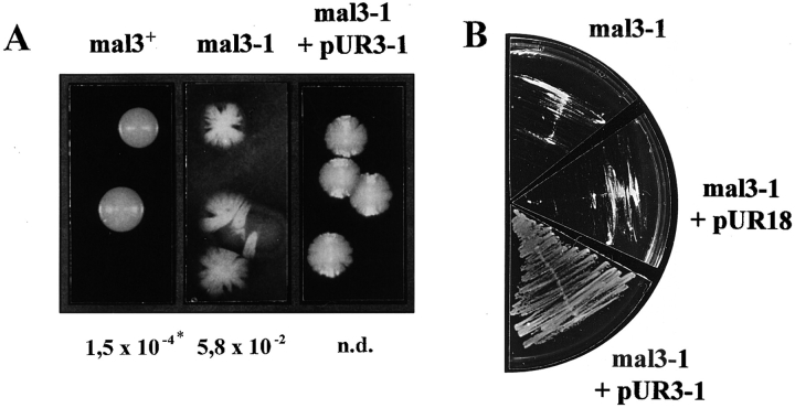
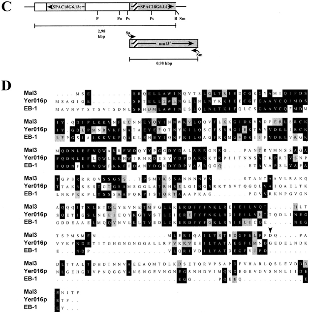
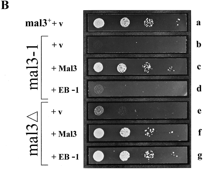
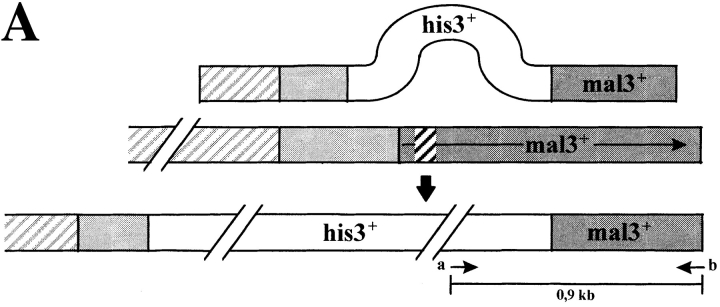
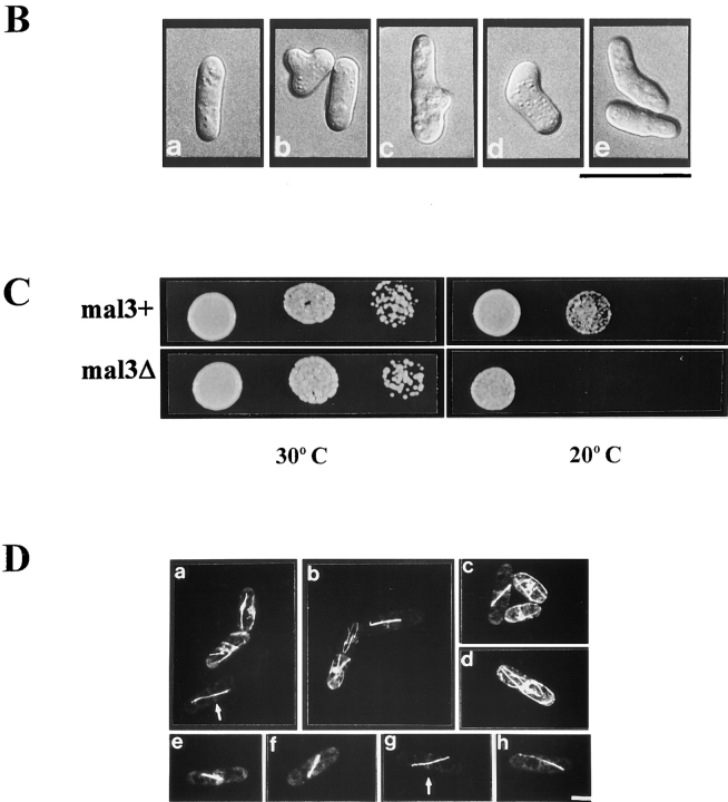
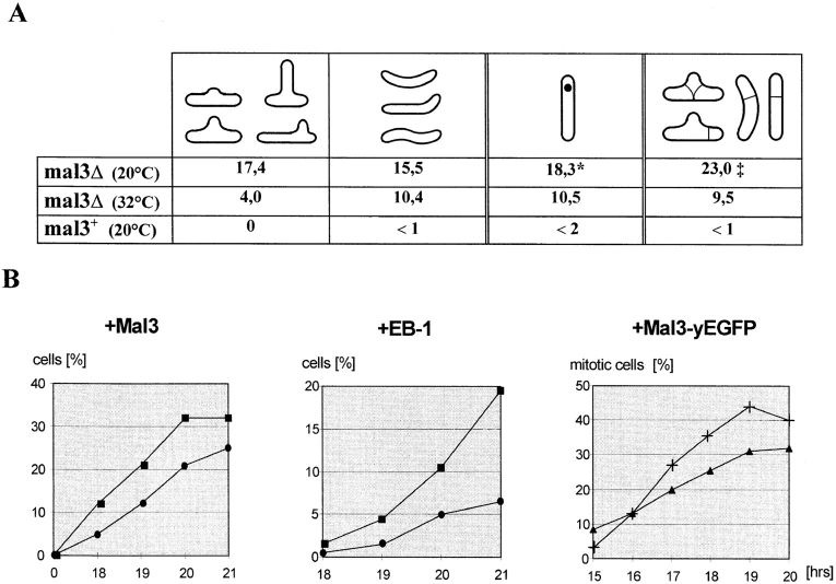
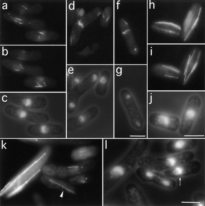
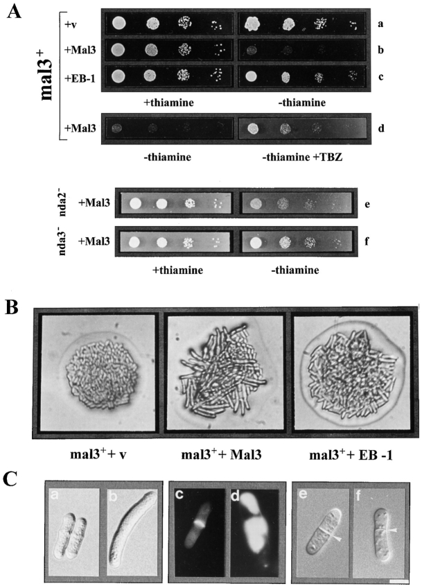
Through a screen designed to isolate novel fission yeast genes required for chromosome segregation, we have identified mal3+. The mal3-1 mutation decreased the transmission fidelity of a nonessential minichromosome and altered sensitivity to microtubule-destabilizing drugs. Sequence analysis revealed that the 35-kD Mal3 is a member of an evolutionary conserved protein family. Its human counterpart EB-1 was identified in an interaction screen with the tumour suppressor protein APC. EB-1 was able to substitute for the complete loss of the mal3+ gene product suggesting that the two proteins might have similar functions. Cells containing a mal3 null allele were viable but showed a variety of phenotypes, including impaired control of cell shape. A fusion protein of Mal3 with the Aequorea victoria green fluorescent protein led to in vivo visualization of both cytoplasmic and mitotic microtubule structures indicating association of Mal3 with microtubules. The absence of Mal3 protein led to abnormally short, often faint cytoplasmic microtubules as seen by indirect antitubulin immunofluorescence. While loss of the mal3+ gene product had no gross effect on mitotic spindle morphology, overexpression of mal3+ compromised spindle formation and function and led to severe growth inhibition and abnormal cell morphology. We propose that Mal3 plays a role in regulating the integrity of microtubules possibly by influencing their stability.


Figures

Figure 1
Figure 1
Phenotypic characterization of the mal3-1 strain, isolation of mal3 + gene and Mal3 amino acid sequence. (A) Sectoring phenotypes of mal3 +, mal3-1, and mal3-1 transformed with plasmid pUR3-1 strains grown at 24°C are shown. *Minichromosome loss rate; n. d., not determined. (B) While the mal3-1 strain or mal3-1 plus vector (pUR18) are unable to grow on TBZ containing EMM medium at 30°C, this defect is rescued by plasmid pUR3-1. (C) Diagrammatic representation of genomic DNA insert of pUR3-1 (top) and full length mal3 + gene obtained by PCR (bottom). PCR primers have been marked by arrows, ORF by grey boxes and a putative intron in SPAC18G6.14/mal3 + by a striped box. P, PvuI; Pa, PacI; Ps, PstI; B, BamHI; Sm, SmaI; Sa, SalI. (D) Amino acid sequence comparison of fission yeast Mal3, budding yeast Yer016p (GenBank accession number P40013) and human EB-1 (GenBank accession number U24166). Identical and similar amino acids are indicated by black and grey boxes, respectively. The arrow head indicates the end of the Mal3 truncated protein in pUR3-1. The mal3 + sequence data are available from EMBL/GenBank/DDBJ under accession number Y09518.
Figure 1
Figure 1
Phenotypic characterization of the mal3-1 strain, isolation of mal3 + gene and Mal3 amino acid sequence. (A) Sectoring phenotypes of mal3 +, mal3-1, and mal3-1 transformed with plasmid pUR3-1 strains grown at 24°C are shown. *Minichromosome loss rate; n. d., not determined. (B) While the mal3-1 strain or mal3-1 plus vector (pUR18) are unable to grow on TBZ containing EMM medium at 30°C, this defect is rescued by plasmid pUR3-1. (C) Diagrammatic representation of genomic DNA insert of pUR3-1 (top) and full length mal3 + gene obtained by PCR (bottom). PCR primers have been marked by arrows, ORF by grey boxes and a putative intron in SPAC18G6.14/mal3 + by a striped box. P, PvuI; Pa, PacI; Ps, PstI; B, BamHI; Sm, SmaI; Sa, SalI. (D) Amino acid sequence comparison of fission yeast Mal3, budding yeast Yer016p (GenBank accession number P40013) and human EB-1 (GenBank accession number U24166). Identical and similar amino acids are indicated by black and grey boxes, respectively. The arrow head indicates the end of the Mal3 truncated protein in pUR3-1. The mal3 + sequence data are available from EMBL/GenBank/DDBJ under accession number Y09518.
Figure 2
Figure 2
Suppression of the TBZ sensitivity of the mal3-1 and mal3Δ strains by mal3 + and EB-1. (A) Diagrammatic representation of cloning of EB-1 cDNA. The positions of the five oligonucleotides used to construct the 5′ end (lightly shaded box) of EB-1 not present in the original cDNA (dark shaded box) are indicated. X, XbaI; A, AvaI; P, PstI; S, StuI; N, NotI. (B) The mal3 + strain transformed with vector (a), the mal3-1 strain transformed with vector (b) or with plasmids expressing mal3 +(c) or EB-1 (d) and the mal3Δ strain transformed with vector (e), or with plasmids expressing mal3 + (f) or EB-1 (g) were all grown for 18 h at 30°C in thiamine-less EMM medium, diluted in water and serial dilutions (1:10) starting with 104 cells were spotted on selective EMM medium with 10 μg/ml TBZ and incubated for 3 d at 30°C.
Figure 2
Figure 2
Suppression of the TBZ sensitivity of the mal3-1 and mal3Δ strains by mal3 + and EB-1. (A) Diagrammatic representation of cloning of EB-1 cDNA. The positions of the five oligonucleotides used to construct the 5′ end (lightly shaded box) of EB-1 not present in the original cDNA (dark shaded box) are indicated. X, XbaI; A, AvaI; P, PstI; S, StuI; N, NotI. (B) The mal3 + strain transformed with vector (a), the mal3-1 strain transformed with vector (b) or with plasmids expressing mal3 +(c) or EB-1 (d) and the mal3Δ strain transformed with vector (e), or with plasmids expressing mal3 + (f) or EB-1 (g) were all grown for 18 h at 30°C in thiamine-less EMM medium, diluted in water and serial dilutions (1:10) starting with 104 cells were spotted on selective EMM medium with 10 μg/ml TBZ and incubated for 3 d at 30°C.
Figure 3
Figure 3
Phenotypic characterization of mal3Δ strain and in vivo localization of Mal3 protein. (A) Diagrammatic representation and PCR analysis of the mal3 + disruption. Diploid strain YP41 was transformed with a linear 3.2-kb DNA fragment where 800 bp of the mal3 + gene had been replaced by the 1.9-kb his3 + gene. Spores were grown and their DNA analysed by PCR for the presence of the mal3 + disruption. The positions of the PCR primers a and b are shown in the bottom diagram. The 900-bp DNA fragment generated by PCR can be obtained only if the linear mal3 + disruption fragment has integrated correctly, as the DNA sequence homologous to primer b is not present on the 3.2-kb DNA fragment used for replacement. a and b show the PCR product of his and his+ spore DNA, respectively. M, DNA molecular mass markers (0.94 and 0.83 kb). (B) Photomicrographs of mal3Δ cells. A wild-type strain (a) and the mal3Δ strain (b–e) were grown logarithmically at 24°C. (C) Serial dilution patch test for cold-sensitivity of mal3 +and mal3Δ strains. Dilutions shown were tenfold. Strains were incubated at 30°C and 20°C for 3 and 5 d, respectively. (D) In vivo localization of Mal3 protein. A wild-type S. pombe strain expressing Mal3-yEGFP under the control of the nmt1 + promoter was pregrown on solid EMM thiamine medium, resuspended in liquid EMM medium with 0.05 μM thiamine, grown for 16 h at 30°C and photographed directly. Bars: (B) 20 μm; (D) 5 μm.
Figure 3
Figure 3
Phenotypic characterization of mal3Δ strain and in vivo localization of Mal3 protein. (A) Diagrammatic representation and PCR analysis of the mal3 + disruption. Diploid strain YP41 was transformed with a linear 3.2-kb DNA fragment where 800 bp of the mal3 + gene had been replaced by the 1.9-kb his3 + gene. Spores were grown and their DNA analysed by PCR for the presence of the mal3 + disruption. The positions of the PCR primers a and b are shown in the bottom diagram. The 900-bp DNA fragment generated by PCR can be obtained only if the linear mal3 + disruption fragment has integrated correctly, as the DNA sequence homologous to primer b is not present on the 3.2-kb DNA fragment used for replacement. a and b show the PCR product of his and his+ spore DNA, respectively. M, DNA molecular mass markers (0.94 and 0.83 kb). (B) Photomicrographs of mal3Δ cells. A wild-type strain (a) and the mal3Δ strain (b–e) were grown logarithmically at 24°C. (C) Serial dilution patch test for cold-sensitivity of mal3 +and mal3Δ strains. Dilutions shown were tenfold. Strains were incubated at 30°C and 20°C for 3 and 5 d, respectively. (D) In vivo localization of Mal3 protein. A wild-type S. pombe strain expressing Mal3-yEGFP under the control of the nmt1 + promoter was pregrown on solid EMM thiamine medium, resuspended in liquid EMM medium with 0.05 μM thiamine, grown for 16 h at 30°C and photographed directly. Bars: (B) 20 μm; (D) 5 μm.
Figure 4
Figure 4
Phenotypes of the mal3Δ strain and Mal3 and EB-1 overproducing strains. (A) mal3Δ cells were grown at 20 or 32°C in liquid YE5S medium and the percentage of cells with abnormal cell morphology, a displaced nucleus and misplaced septum was determined. The mal3 + control strain was grown at 20°C. *Percentage of all interphase cells with a displaced nucleus; percentage of septated cells with a misplaced septum. (B) Wild-type strains overproducing Mal3, EB-1 or Mal3-yEGFP were grown for 0–21 h at 30°C in expression inducing medium and the percentage of cells with altered cell shape (•), a displaced nucleus (▪), a mitotic spindle (▴) and aberrant spindles out of the total number of spindles (+) was determined.
Figure 5
Figure 5
Antitubulin immunofluorescence images of mal3Δ cells cultured at 20°C. a, b, d, f, h, i, and k show antitubulin staining while c, e, g, j, and l show DAPI and phase contrast images. a and b represent two different focal planes of three mal3Δ cells showing several short microtubules around the location of the nucleus shown in c. (d) Strong post-anaphase-array staining in the upper, bent cell. (f) Displaced post-anaphase-array. h–i show antitubulin immunofluorescence images of two mal3 + cells grown at 20°C at different focal planes, while j shows DAPI staining. k and l show a mixed culture of mal3Δ and elongated cdc25-22 cells. k shows antitubulin while l shows DAPI staining. cdc25-22 cells were grown at 25°C in YE5S to early log phase, shifted to the restrictive temperature for 210 min, returned to 20°C in 30 s by immersion in an ice bath and mixed at a ratio of 1:4 with the mal3Δ culture grown at 20°C inYE5S. k shows four mal3Δ cells and an elongated cdc25-22 cell. The abnormally faint spindle in the mal3Δ cell is indicated by an arrow head in k. The small arrow in l indicates a mal3Δ cell with a misplaced nucleus. Bars, 5 μm.
Figure 6
Figure 6
Phenotypic characterization of overexpression of Mal3 and EB-1 proteins. (A) Serial dilution patch test for inhibition of colony formation by overproduction of Mal3 and EB-1. Dilutions shown were tenfold. A mal3 + strain was transformed with either the insert-less vector (a) or plasmids expressing mal3 + (b) or EB-1 (c) from the nmt1 + promoter. Strains were grown for 18 h at 30°C in thiamineless medium and then plated on EMM medium with (+) or without thiamine (−).The growth inhibition phenotype of Mal3 overexpression was suppressed by 10 μg/ml TBZ (d) or the presence of the α- and β-tubulin mutants, nda2-KM52 and nda3-KM311, respectively (e and f). Colonies were photographed after incubation for 3 d at 30 or 29°C (mutant tubulin strains). (B) Photomicrographs of colonies of wild-type cells transformed with vector control (v), or plasmids expressing mal3 + or EB-1. Strains were pregrown as in A before streaking cells on thiamineless plates and incubating them for 1.5 d at 30°C. (C) mal3 + was overexpressed in a wild-type strain for 0 (a, c, and e), 19 (f), 21 (b), or 24 (d) h at 30°C. c and d show staining of septa by calcofluor. Septa in e and f are indicated by arrowheads. Bar, 5 μm.
Figure 7
Figure 7
Overexpression of mal3 + results in defects in form and function of the mitotic spindle. Wild-type cells overexpressing mal3 + were grown at 32°C for 21 h in inducing conditions. Each panel shows three different images of the same cells: in the first, containing the lettering, the relative location of the chromatin and the cell outline is shown by combining DAPI fluorescence and phase contrast images; in the second the microtubules (red) and the SPBs (green) are shown; the third panel shows the position of the SPBs (red) relative to the chromatin (green). a shows three strongly staining spindles of roughly equal length. The chromosomes are highly condensed and randomly positioned along the spindle axis. In each case bars of tubulin staining emanate from the SPB away from the main body of the spindle. b, d, and g show examples of cells where the two half spindles appear to be losing their interdigitation presumably leading to the complete loss of interdigitation seen in g. In b the smaller chromosome III has wandered from the spindle axis, indicating that the microtubules are not contacting the kinetochores. e shows a bent interphase cell which has not separated from its partner during the previous cytokinesis. c and f show examples of defects during the early stages of spindle formation. Bar, 5 μm.

References

    1. Barbet N, Muriel WJ, Carr AM. Versatile shuttle vectors and genomic libraries for use with Schizosaccharomyces pombe. . Gene (Amst) 1992;114:59–66. - PubMed
    1. Boguski MS, Lowe TMJ, Tolstoshev CM. dbEST-database for “Expressed Sequence Tags.” . Nat Genet. 1993;4:332–333. - PubMed
    1. Belmont LD, Mitchison TJ. Identification of a protein that interacts with tubulin dimers and increases the catastrophe rate of microtubules. Cell. 1996;84:623–631. - PubMed
    1. Chang F, Wollard A, Nurse P. Isolation and characterization of fission yeast mutants defective in the assembly and placement of the contractile ring. J Cell Sci. 1996;109:132–142. - PubMed
    1. Cormack BP, Bertram G, Egerton M, Gow NA, Falkow S, Brown AJ. Yeast-enhanced green fluorescent protein (yEGFP) a reporter of gene expression in Candida albicans. Microbiology. 1997;143:303–311. - PubMed

Publication types

MeSH terms

Substances

Associated data