. 1997 Nov 3;139(3):749-58.
doi: 10.1083/jcb.139.3.749.

ERM (ezrin/radixin/moesin)-based molecular mechanism of microvillar breakdown at an early stage of apoptosis

Affiliations

ERM (ezrin/radixin/moesin)-based molecular mechanism of microvillar breakdown at an early stage of apoptosis

T Kondo et al. J Cell Biol. .

Abstract

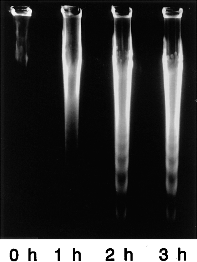
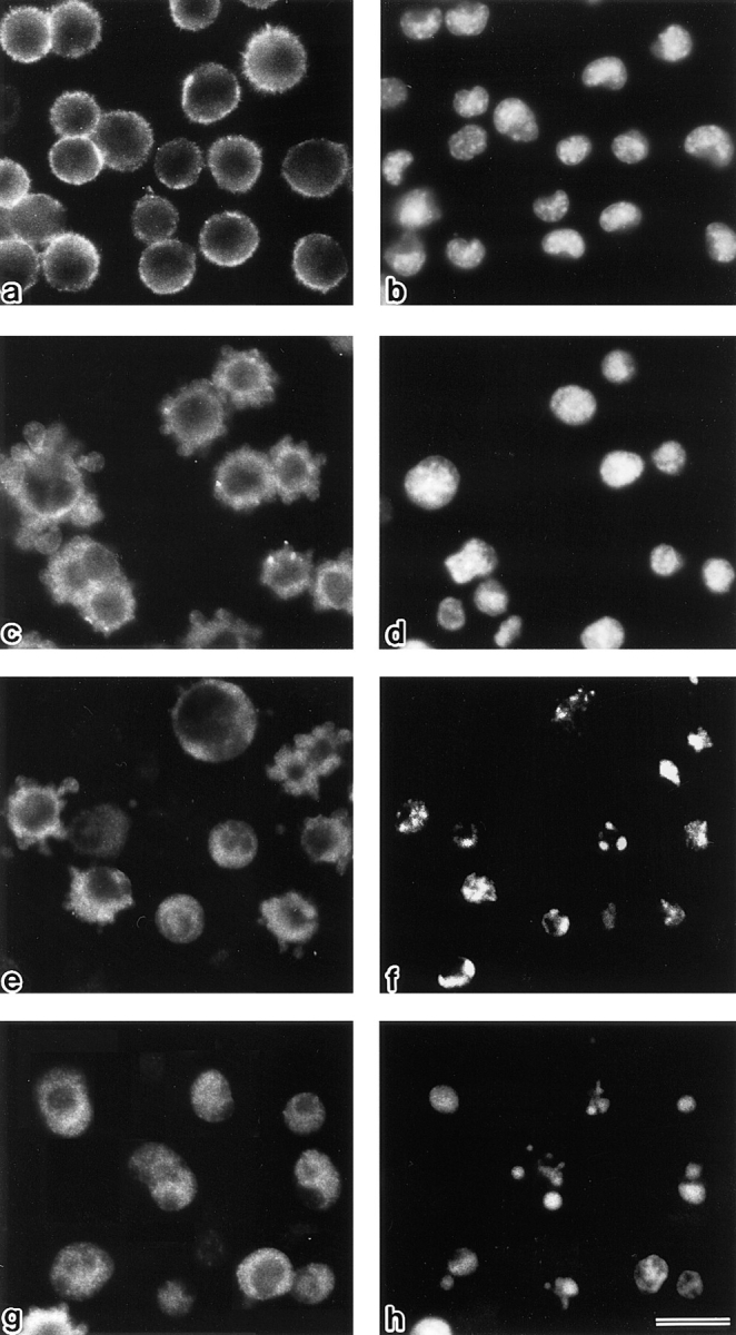
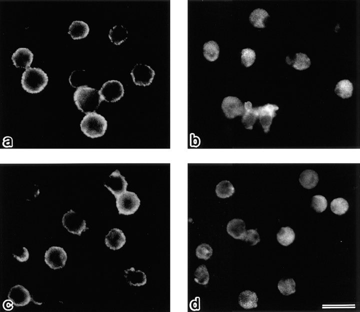
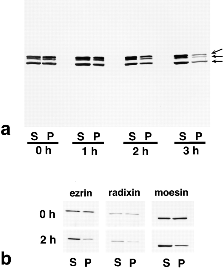
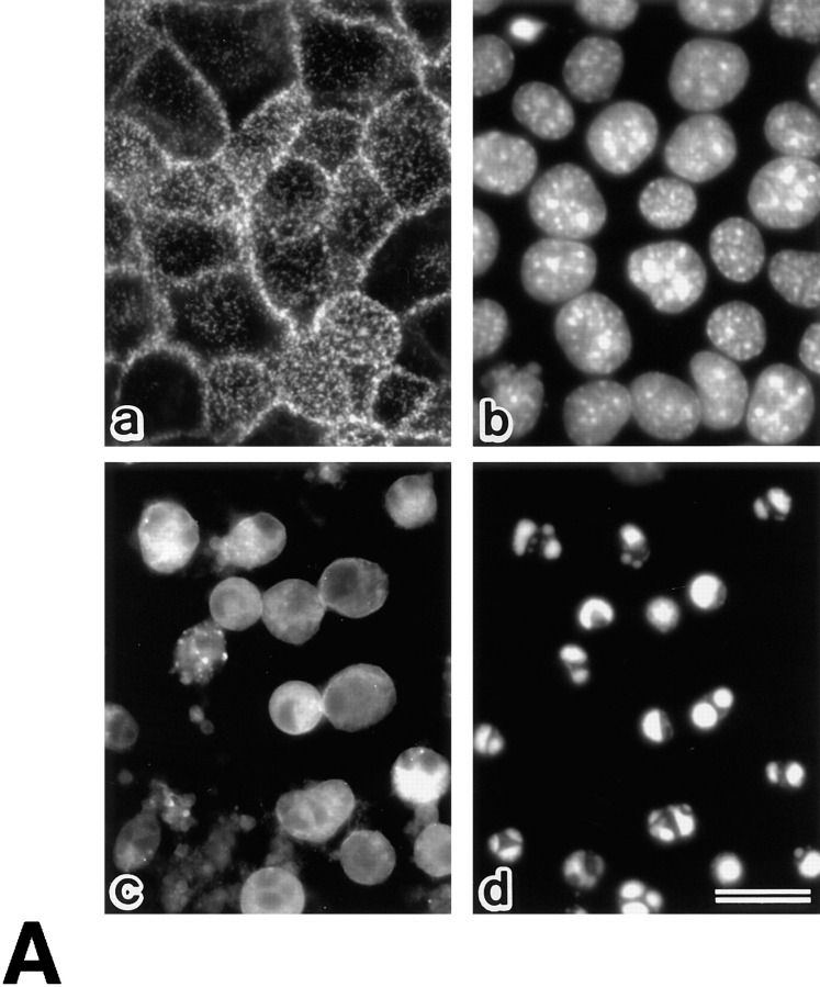
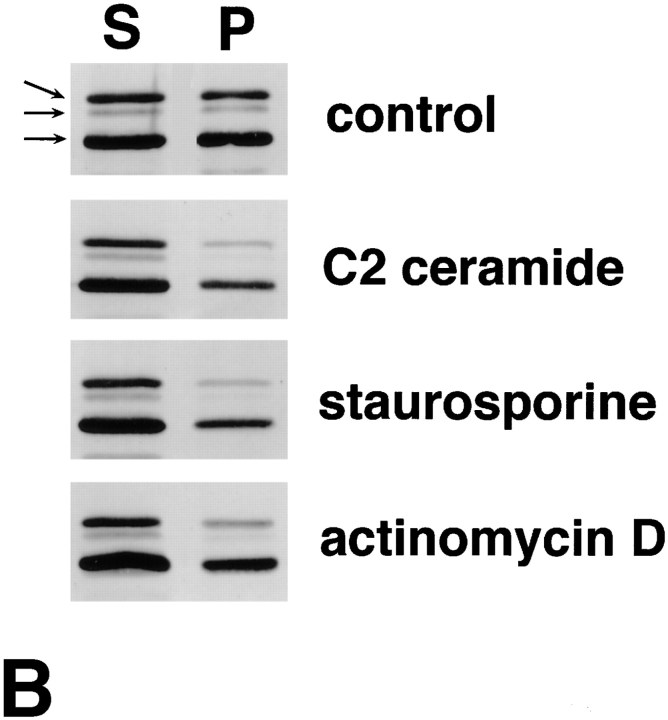
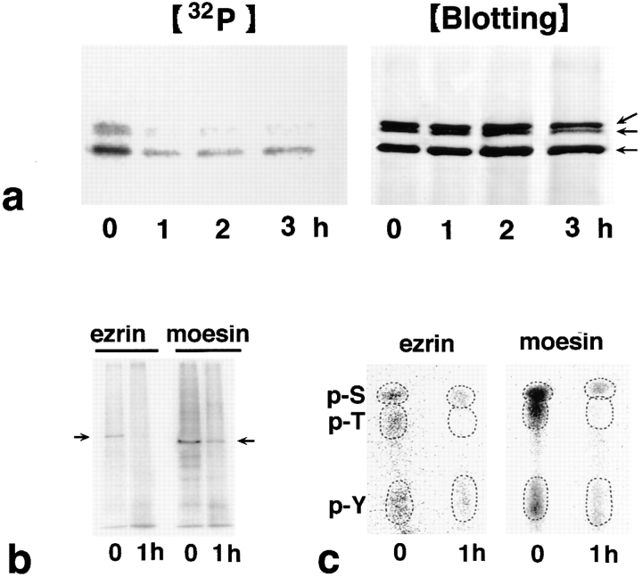
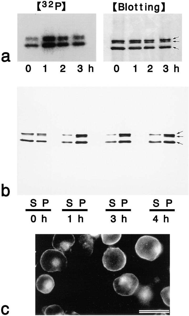
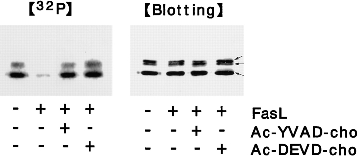
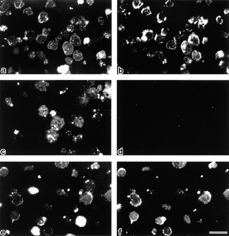
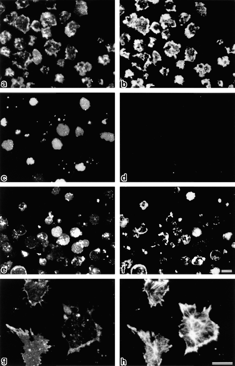
Breakdown of microvilli is a common early event in various types of apoptosis, but its molecular mechanism and implications remain unclear. ERM (ezrin/radixin/moesin) proteins are ubiquitously expressed microvillar proteins that are activated in the cytoplasm, translocate to the plasma membrane, and function as general actin filament/plasma membrane cross-linkers to form microvilli. Immunofluorescence microscopic and biochemical analyses revealed that, at the early phase of Fas ligand (FasL)-induced apoptosis in L cells expressing Fas (LHF), ERM proteins translocate from the plasma membranes of microvilli to the cytoplasm concomitant with dephosphorylation. When the FasL-induced dephosphorylation of ERM proteins was suppressed by calyculin A, a serine/threonine protein phosphatase inhibitor, the cytoplasmic translocation of ERM proteins was blocked. The interleukin-1beta-converting enzyme (ICE) protease inhibitors suppressed the dephosphorylation as well as the cytoplasmic translocation of ERM proteins. These findings indicate that during FasL-induced apoptosis, the ICE protease cascade was first activated, and then ERM proteins were dephosphorylated followed by their cytoplasmic translocation, i.e., microvillar breakdown. Next, to examine the subsequent events in microvillar breakdown, we prepared DiO-labeled single-layered plasma membranes with the cytoplasmic surface freely exposed from FasL-treated or nontreated LHF cells. On single-layered plasma membranes from nontreated cells, ERM proteins and actin filaments were densely detected, whereas those from FasL-treated cells were free from ERM proteins or actin filaments. We thus concluded that the cytoplasmic translocation of ERM proteins is responsible for the microvillar breakdown at an early phase of apoptosis and that the depletion of ERM proteins from plasma membranes results in the gross dissociation of actin-based cytoskeleton from plasma membranes. The physiological relevance of this ERM protein-based microvillar breakdown in apoptosis will be discussed.


Figures

Figure 1
Figure 1
FasL-induced apoptosis in LHF cells. After incubation of LHF cells with FasL for 0, 1, 2, and 3 h, the chromosomal DNA was extracted and analyzed by gel electrophoresis. Judging from the DNA fragmentation ladder formation, apoptosis was induced within 2–3 h.
Figure 2
Figure 2
Behavior of radixin in LHF cells at 0 h (a and b), 1 h (c and d), 2 h (e and f), and 3 h (g and h) after FasL stimulation. LHF cells were doubly stained with antiradixin mAb, R21 (a, c, e, and g) and DAPI (b, d, f, and h). After 1 h of incubation, radixin translocated from the plasma membrane to the cytoplasm, and the nuclear change, such as chromatin condensation, began to be observed after 2 h of incubation. Bar, 20 μm.
Figure 3
Figure 3
Behavior of ezrin and moesin in LHF cells at 0 h (a and c) and 2 h (b and d) after FasL stimulation. LHF cells were stained with ezrin-specific pAb TK90 (a and b) or moesin-specific mAb M22 (c and d). Behavior of ezrin and moesin during apoptosis was the same as that of radixin (see Fig. 2). Bar, 20 μm.
Figure 4
Figure 4
Solubility of ERM proteins in LHF cells at 0, 1, 2, and 3 h after addition of FasL. LHF cells were homogenized in physiological saline and centrifuged to separate the soluble and insoluble fractions into the supernatant (S) and pellet (P), respectively. Equivalent amounts of supernatant and pellet were applied to SDS-PAGE and subsequently subjected to immunoblotting. To detect all members of ERM proteins, pAb TK89 was used (a). To detect ezrin, radixin, and moesin separately, mAb M11, mAb R21, and mAb M22 were used, respectively (b). ERM proteins translocated from the insoluble (P) to the soluble fraction (S) as apoptosis proceeded. Arrows in a indicate ezrin, radixin, and moesin, respectively, from the top.
Figure 5
Figure 5
Behavior of ERM proteins during apoptosis in mouse epithelial cells (MTD-1A) and human promyelocytic leukemic cells (HL-60). (A) Immunofluorescence micrographs of MTD-1A cells at 0 h (a and b) and 16 h (c and d) incubation with staurosporine. MTD-1A cells were doubly stained with antiradixin mAb R21 (a and c) and DAPI (b and d). After 16 h of incubation, radixin translocated from apical microvilli and lateral cell–cell contact sites to the cytoplasm, and nuclear changes such as chromatin condensation were observed. Ezrin and moesin showed the same behavior as radixin (data not shown). (B) Solubility of ERM proteins in HL-60 cells at 0 (control) and 16 h incubation with C2 ceramide, staurosporine, or actinomycin D. HL-60 cells were homogenized in physiological saline and centrifuged to separate the soluble and insoluble fractions into the supernatant (S) and pellet (P), respectively. Equivalent amounts of supernatant and pellet were applied to SDS-PAGE and subsequently subjected to immunoblotting with pAb TK89 capable of recognizing all ERM proteins. ERM proteins translocated from the insoluble (P) to the soluble fraction (S) as apoptosis proceeded. Arrows indicate ezrin, radixin, and moesin, respectively, from the top. Bar, 20 μm.
Figure 5
Figure 5
Behavior of ERM proteins during apoptosis in mouse epithelial cells (MTD-1A) and human promyelocytic leukemic cells (HL-60). (A) Immunofluorescence micrographs of MTD-1A cells at 0 h (a and b) and 16 h (c and d) incubation with staurosporine. MTD-1A cells were doubly stained with antiradixin mAb R21 (a and c) and DAPI (b and d). After 16 h of incubation, radixin translocated from apical microvilli and lateral cell–cell contact sites to the cytoplasm, and nuclear changes such as chromatin condensation were observed. Ezrin and moesin showed the same behavior as radixin (data not shown). (B) Solubility of ERM proteins in HL-60 cells at 0 (control) and 16 h incubation with C2 ceramide, staurosporine, or actinomycin D. HL-60 cells were homogenized in physiological saline and centrifuged to separate the soluble and insoluble fractions into the supernatant (S) and pellet (P), respectively. Equivalent amounts of supernatant and pellet were applied to SDS-PAGE and subsequently subjected to immunoblotting with pAb TK89 capable of recognizing all ERM proteins. ERM proteins translocated from the insoluble (P) to the soluble fraction (S) as apoptosis proceeded. Arrows indicate ezrin, radixin, and moesin, respectively, from the top. Bar, 20 μm.
Figure 6
Figure 6
Dephosphorylation of ERM proteins in LHF cells during Fas-mediated apoptosis. (a) Time course of apoptosis-associated dephosphorylation of ERM proteins. LHF cells were metabolically labeled with [32P]orthophosphate. At 0, 1, 2, and 3 h after FasL stimulation, the cell lysate was immunoprecipitated with anti-ERM pAb, TK89, and analyzed by autoradiography ([32P]) and immunoblotting ([Blotting]). Equal amounts of protein were applied to each lane as revealed in accompanying immunoblots with anti-ERM pAb, TK89. The phosphorylation level of ERM proteins markedly decreased after a 1-h incubation. Arrows indicate ezrin, radixin, and moesin, respectively, from the top. (b) Autoradiography of antiezrin pAb (TK90) or antimoesin mAb (M22) immunoprecipitates from 32P-labeled LHF cells. Immunoprecipitated ezrin and moesin were phosphorylated and dephosphorylated before and 1 h after FasL stimulation, respectively (arrows). (c) Phosphoamino acid analysis. 32P-labeled phosphorylated ezrin or moesin bands (arrows in b) were excised from membranes and processed for phosphoamino acid analysis. The positions of phosphoserine (p-S), phosphothreonine (p-T), and phosphotyrosine (p-Y) were determined by autoradiography through comparison with the ninhydrin staining profiles on unlabeled phosphoamino acid standards. In both ezrin and moesin, phosphothreonine appeared to be preferentially dephosphorylated after 1 h of incubation with FasL, although the levels of phosphoserine and phosphotyrosine were also decreased.
Figure 7
Figure 7
Effects of calyculin A on FasL-induced changes of ERM proteins in LHF cells. (a) Suppression of FasL-induced dephosphorylation of ERM proteins by calyculin A. LHF cells were metabolically labeled with [32P]orthophosphate. At 0, 1, 2, and 3 h after FasL stimulation in the presence of calyculin A, the cell lysate was immunoprecipitated with anti-ERM pAb, TK89, and then analyzed by autoradiography ([32P]) and immunoblotting ([Blotting]). Equal amounts of protein were applied to each lane as revealed in accompanying immunoblots with anti-ERM pAb, TK89. The FasL-induced dephosphorylation of ERM proteins (see Fig. 6 a) was suppressed, while conversely the phosphorylation level was elevated. Arrows indicate ezrin, radixin, and moesin, respectively, from the top. (b) Suppression of the FasL-induced cytoplasmic translocation of ERM proteins by calyculin A. Calyculin A was added to LHF cells together with FasL, and, after incubations for 1, 2, 3, and 4 h, ERM proteins were partitioned into soluble (S) and insoluble (P) fractions as described in Fig. 4 a. Immunoblots with anti-ERM pAb, TK89, revealed that FasL-induced cytoplasmic translocation of ERM proteins was suppressed and that conversely calyculin A increased the amounts of insoluble ERM proteins. Arrows indicate ezrin, radixin, moesin, respectively, from the top. (c) Antiradixin mAb immunofluorescence micrograph of LHF cells after 2 h of incubation of FasL and calyculin A. Radixin still remained on plasma membranes. Ezrin and moesin showed the same changes as radixin (data not shown). Bar, 20 μm.
Figure 8
Figure 8
Suppression of FasL-induced dephosphorylation of ERM proteins by ICE protease inhibitors. LHF cells were metabolically labeled with [32P]orthophosphate for 3 h, and then ICE protease inhibitors (Ac-YVAD-cho or Ac-DEVD-cho) were added at 300 μM. After 1 h incubation, FasL was added. At 3 h of incubation, the cell lysate was immunoprecipitated with anti-ERM pAb, TK89, and analyzed by autoradiography ([32P]) and immunoblotting ([Blotting]). Equal amounts of protein were applied to each lane as revealed in accompanying immunoblots with anti-ERM pAb, TK89. The FasL-induced dephosphorylation of ERM proteins was suppressed by both inhibitors. Arrows indicate ezrin, radixin, and moesin, respectively, from the top.
Figure 9
Figure 9
Detection of ERM proteins on single-layered plasma membranes isolated on cover glasses. After vital labeling of plasma membranes proper with DiO, single-layered plasma membranes with the cytoplasmic surface freely exposed were prepared from nontreated (a and b), FasL–1 h–treated (c and d), and FasL/calyculin A–1 h–treated (e and f) LHF cells as described in Materials and Methods. These preparations were immunofluorescently labeled with anti-ERM pAb, TK89. Each single-layered plasma membrane was detected by DiO signal (a, c, and e). ERM signal was intense from plasma membranes of nontreated cells (b), whereas it was undetectable from those of FasL-treated cells (d). Calyculin A suppressed the FasL-induced depletion of ERM proteins from plasma membranes (f). Bar, 20 μm.
Figure 10
Figure 10
Detection of actin filaments on the single-layered plasma membranes isolated on cover glasses. After vital labeling of plasma membranes proper with DiO, single-layered plasma membranes with the cytoplasmic surface freely exposed were prepared from nontreated (a, b, g, and h), FasL–1 h–treated (c and d), and FasL/calyculin A–1 h–treated (e and f) LHF cells as described in Materials and Methods. These preparations were labeled with rhodamine phalloidin to visualize actin filaments (b, d, f, and h). Each single-layered plasma membrane was detected by DiO signal (a, c, e, and g). Actin filaments were densely associated with plasma membranes of nontreated cells (b and h), whereas they were undetectable from those of FasL-treated cells (d). Calyculin A suppressed the FasL-induced dissociation of actin filaments from plasma membranes (f). Bars: (a–f) 10 μm; (g and h) 15 μm.

References

    1. Alnemri ES, Livingston DJ, Nicholoson DW, Salvensen G, Thornberry NA, Wong WW, Yuan J. Human ICE-CED-3 protease nomenclature. Cell. 1996;87:171. - PubMed
    1. Arpin M, Algrain M, Louvard D. Membrane-actin microfilament connections: an incresing diversity of players related to band 4.1. Curr Opin Cell Biol. 1994;6:136–141. - PubMed
    1. Berryman M, Gary R, Bretscher A. Ezrin oligomers are major cytoskeletal components of placental microvilli: a proposal for their involvement in cortical morphogenesis. J Cell Biol. 1995;131:1231–1242. - PMC - PubMed
    1. Boyle WJ, van der Geer P, Hunter T. Phosphopeptide mapping and phosphoamino acid analysis by two-dimensional separation on thin-layer cellulose plates. Methods Enzymol. 1991;201:110–148. - PubMed
    1. Brancolini C, Benedetti M, Schneider C. Microfilament reorganization during apoptosis: the role of Gas2, a possible substrate for ICE-like proteases. EMBO (Eur Mol Biol Organ) J. 1995;14:5179–5190. - PMC - PubMed

Publication types

MeSH terms