. 1997 Nov 3;139(3):759-71.
doi: 10.1083/jcb.139.3.759.

Dismantling cell-cell contacts during apoptosis is coupled to a caspase-dependent proteolytic cleavage of beta-catenin

Affiliations

Dismantling cell-cell contacts during apoptosis is coupled to a caspase-dependent proteolytic cleavage of beta-catenin

C Brancolini et al. J Cell Biol. .

Abstract

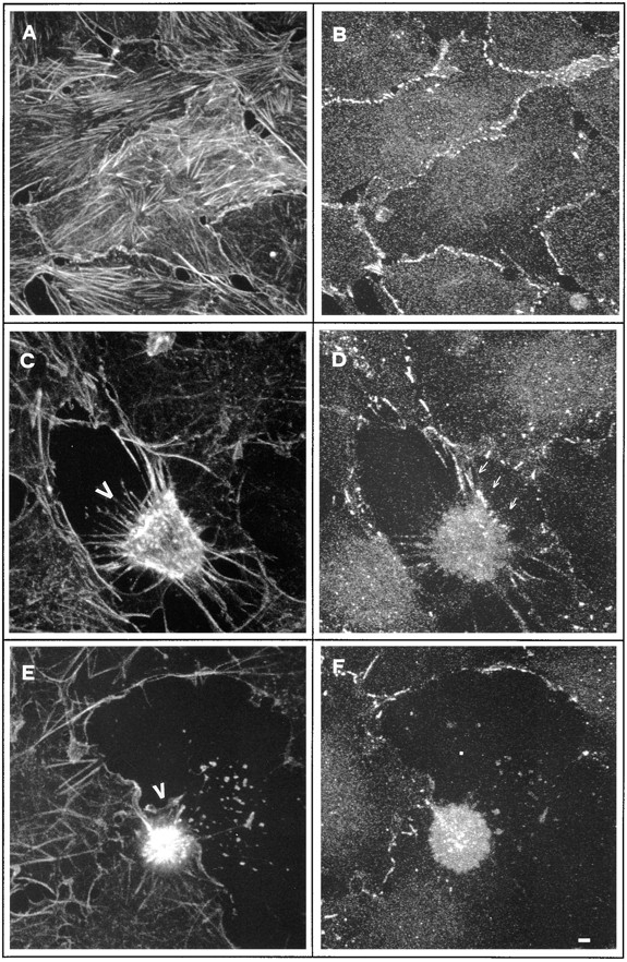
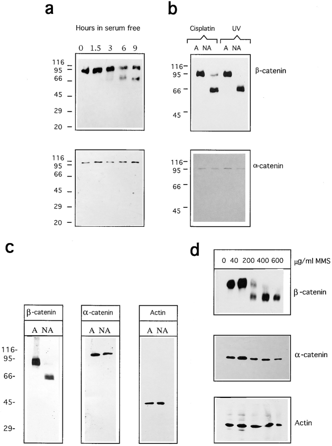
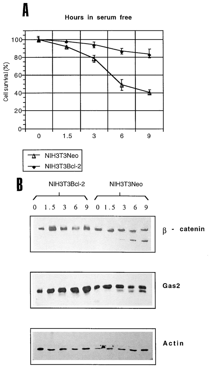
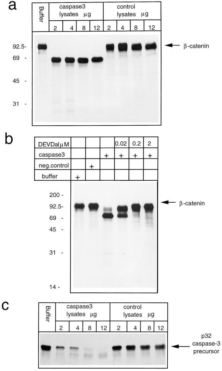
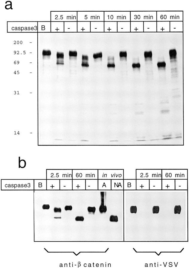
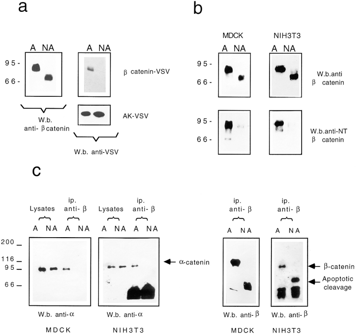
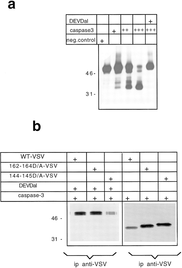
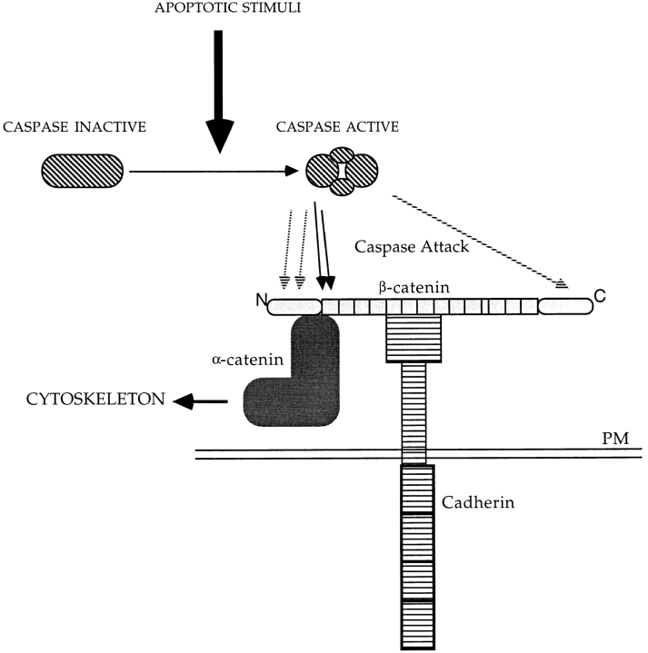
Cell death by apoptosis is a tightly regulated process that requires coordinated modification in cellular architecture. The caspase protease family has been shown to play a key role in apoptosis. Here we report that specific and ordered changes in the actin cytoskeleton take place during apoptosis. In this context, we have dissected one of the first hallmarks in cell death, represented by the severing of contacts among neighboring cells. More specifically, we provide demonstration for the mechanism that could contribute to the disassembly of cytoskeletal organization at cell-cell adhesion. In fact, beta-catenin, a known regulator of cell-cell adhesion, is proteolytically processed in different cell types after induction of apoptosis. Caspase-3 (cpp32/apopain/yama) cleaves in vitro translated beta-catenin into a form which is similar in size to that observed in cells undergoing apoptosis. beta-Catenin cleavage, during apoptosis in vivo and after caspase-3 treatment in vitro, removes the amino- and carboxy-terminal regions of the protein. The resulting beta-catenin product is unable to bind alpha-catenin that is responsible for actin filament binding and organization. This evidence indicates that connection with actin filaments organized at cell-cell contacts could be dismantled during apoptosis. Our observations suggest that caspases orchestrate the specific and sequential changes in the actin cytoskeleton occurring during cell death via cleavage of different regulators of the microfilament system.


Figures

Figure 1
Figure 1
Confocal analysis of the organization of actin in NIH3T3 cells induced to enter apoptosis by deprivation of survival factors. Growth-arrested NIH3T3 cells were cultured for 4 h in serum-free medium, fixed, and stained for actin filaments using phalloidin-FITC (a), or double stained with propidium iodide, to visualize nuclei (A, C, E, and G) and phalloidin-FITC (B, D, F, and H). Bars: (a) 5 μm; (A–H) 1 μm.
Figure 2
Figure 2
(a) NIH3T3 cells grown for 48 h in 0.5% FCS were incubated in serum-free medium for the indicated times (relative numbers and percentages of viable cells were determined by trypan blue dye exclusion). Data represent arithmetic means ± SD for three independent experiments. (b) Growth-arrested NIH3T3 cells were incubated in serum-free medium for the indicated times. Lysates from both adherent viable cells and nonadherent apoptotic cells were combined and Western blot analysis was performed. (c) Two-dimensional gel electrophoresis analysis showing actin patterns in adherent and nonadherent apoptotic cells.
Figure 2
Figure 2
(a) NIH3T3 cells grown for 48 h in 0.5% FCS were incubated in serum-free medium for the indicated times (relative numbers and percentages of viable cells were determined by trypan blue dye exclusion). Data represent arithmetic means ± SD for three independent experiments. (b) Growth-arrested NIH3T3 cells were incubated in serum-free medium for the indicated times. Lysates from both adherent viable cells and nonadherent apoptotic cells were combined and Western blot analysis was performed. (c) Two-dimensional gel electrophoresis analysis showing actin patterns in adherent and nonadherent apoptotic cells.
Figure 3
Figure 3
Confocal analysis of cell–cell contacts changes during apoptosis in NIH3T3 cells. Apoptosis was induced by culturing growth-arrested confluent NIH3T3 cells for 4 h in serum-free medium. Confluent growth-arrested NIH3T3 cells (A and B) and apoptotic NIH3T3 cells (C–F) were fixed and double stained for actin filaments using phalloidin-FITC (A, C, and E) and for β-catenin distribution (B, D, and E), using anti–mouse TRITC as secondary antibody. Bar, 1.5 μm.
Figure 4
Figure 4
Analysis of β-catenin cleavage during apoptosis in NIH3T3 and MDCK cells. (a) Growth-arrested NIH3T3 cells were incubated in serum-free medium for the indicated times. Lysates from both adherent viable cells and nonadherent apoptotic cells were combined and Western blot analysis was performed. (b) Density-arrested NIH3T3 cells were treated with 20 μg/ml of cisplatin or UV irradiated as described in Materials and Methods. After 24 h adherent (A) and nonadherent (NA) cells were processed separately and Western analysis was performed. (c) MDCK cells grown for 4 d in 10% FCS were incubated in serum-free medium for 12 h. Adherent (A) and nonadherent (NA) cells were processed separately and Western analysis was performed. (d) MDCK cells grown for 4 d in 10% FCS were treated for 4 h with the indicated amount of MMS. After 24 h, lysates from both adherent viable cells and nonadherent apoptotic cells were combined and Western blot analysis was performed.
Figure 5
Figure 5
(A) NIH3T3-NEO or NIH3T3 cell lines transfected with bcl-2 were grown for 48 h in 0.5% FCS and incubated in serum-free medium (relative numbers and percentages of viable cells were determined by trypan blue dye exclusion). Data represent arithmetic means ± SD for three independent experiments. (B) Growth-arrested NIH3T3-NEO and NIH3T3–bcl-2 cells were incubated in serum-free medium for the indicated times. Lysates from both adherent viable cells and nonadherent apoptotic cells were combined and Western blot analysis was performed.
Figure 6
Figure 6
In vitro protease assays. (a) 35S labeled in vitro translated β-catenin was incubated for 1 h at 37°C with caspase-3 buffer alone or with the indicated amounts of caspase-3–expressing bacteria lysates or control bacteria lysates. (b) 35S labeled in vitro translated β-catenin was incubated for 1 h at 37°C with caspase-3 buffer alone, with control bacteria lysates, with caspase-3–expressing bacteria lysates, or in the presence of the indicated amount of caspase-3–specific inhibitor Ac-DEVD-CHO. (c) 35S labeled in vitro translated caspase-3 precursor was incubated for 1 h at 37°C with caspase-3 buffer alone, or with the indicated amount of caspase-3–expressing bacterial lysates or control bacterial lysates.
Figure 7
Figure 7
(a) 35S labeled in vitro translated β-catenin was incubated for 1 h at 37°C with caspase-3 buffer (lane B), or with 2 μg of caspase-3–expressing bacterial lysates or control bacterial lysates for the indicated times. (b) Western immunoblots were performed on in vitro translated β-catenin and cellular lysates from (A) adherent nonapoptotic, and (NA) nonadherent apoptotic MDCK cells using antibodies against β-catenin or against VSV-tag. In vitro translated β-catenin was incubated for 1 h at 37°C with caspase-3 buffer (lane B) or with 2 μg of caspase-3–expressing bacterial lysates or control bacterial lysates.
Figure 8
Figure 8
(a) NIH3T3 cells transfected with β-catenin–VSV or with adenylate kinase were incubated in serum-free medium for 12 h. Adherent (A) and nonadherent (NA) cells were processed separately and Western analysis was performed using anti–β-catenin (aa 571–781) or anti–VSV-tag antibodies. (b) Growth-arrested NIH3T3 cells were incubated in serum-free medium for 12 h. Adherent (A) and nonadherent (NA) cells were processed separately and Western analysis was performed using anti–β-catenin (aa 571–781), or anti–amino-terminal β-catenin (aa 6–138) (McCrea et al., 1993). (c) NIH3T3 and MDCK cells were incubated for 12 h in serum-free medium. Adherent (A) and nonadherent (NA) cells were processed separately. Immunoprecipitation using anti–β-catenin was performed as described in Materials and Methods. The immunocomplexes were resolved in SDS-PAGE and processed for Western blot analysis with anti–α-catenin or anti–β-catenin as indicated.
Figure 9
Figure 9
(a) 35S labeled in vitro translated β-cateninΔCT was incubated for 1 h at 37°C with control bacterial lysates or with increasing amount of caspase-3–expressing bacterial lysates. (b) 35S labeled in vitro translated β-cateninΔCTwtVSV, β-cateninΔCT162–164D/AVSV, and the β-cateninΔCT144–145D/AVSV were incubated with caspase-3–expressing bacterial lysates or with caspase-3–expressing bacterial lysates in the presence of 200 nM Ac-DEVD-CHO. Immunoprecipitations were performed using anti-VSV antibody.
Figure 10
Figure 10
Schematic representation of β-catenin cleavage during apoptosis. Apoptotic stimuli lead to activation of the caspase proteases, removing both a segment of β-catenin from the carboxy-terminal domain and from the amino-terminal domain including the first armadillo repeat (black arrows). Cleaved β-catenin is released from α-catenin, thereby losing the connections to the cytoskeleton. Unmapped caspase cleavage sites are indicated by hatched arrows.

References

    1. Aberle H, Butz S, Stappert J, Weissig H, Kemler R, Hoschuetzky H. Assembling of the cadherin catenin complex in vitro with recombinant proteins. J Cell Sci. 1994;107:3655–3663. - PubMed
    1. Alnemri ES, Livingston DJ, Nicholson DW, Salvesen G, Thornberry NA, Wong WW, Yuan J. Human ICE/CED3 protease nomenclature. Cell. 1996;87:171. - PubMed
    1. Bardelli A, Longati P, Albero D, Goruppi S, Schneider C, Ponzetto C, Comoglio PM. HGF receptor associates with the antiapoptotic protein BAG-1 and prevents cell death. EMBO (Eur Mol Biol Organ) J. 1996;15:6205–6212. - PMC - PubMed
    1. Behrens J, von Kries JP, Kuhl M, Bruhn L, Wedlich D, Grosschedl R, Birchmeier W. Functional interaction of β-catenin with the transcription factor LEF-1 . Nature (Lond) 1996;382:225–230. - PubMed
    1. Beidler D, Tewari M, Friesen P, Poirier GG, Dixit V. The baculovirus p35 protein inhibits Fas- and tumor necrosis factor-induced apoptosis. J Biol Chem. 1995;270:16526–16528. - PubMed

Publication types

MeSH terms