. 1997 Nov 3;139(3):809-15.
doi: 10.1083/jcb.139.3.809.

Activated phosphatidylinositol 3-kinase and Akt kinase promote survival of superior cervical neurons

Affiliations

Activated phosphatidylinositol 3-kinase and Akt kinase promote survival of superior cervical neurons

K L Philpott et al. J Cell Biol. .

Abstract

The signaling pathways that mediate the ability of NGF to support survival of dependent neurons are not yet completely clear. However previous work has shown that the c-Jun pathway is activated after NGF withdrawal, and blocking this pathway blocks neuronal cell death. In this paper we show that over-expression in sympathetic neurons of phosphatidylinositol (PI) 3-kinase or its downstream effector Akt kinase blocks cell death after NGF withdrawal, in spite of the fact that the c-Jun pathway is activated. Yet, neither the PI 3-kinase inhibitor LY294002 nor a dominant negative PI 3-kinase cause sympathetic neurons to die if they are maintained in NGF. Thus, although NGF may regulate multiple pathways involved in neuronal survival, stimulation of the PI 3-kinase pathway is sufficient to allow cells to survive in the absence of this factor.


Figures
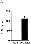
Figure 1
Figure 1
LY294002 does not kill SCG neurons maintained in NGF. Cells were treated with or without 50 μM LY294002 in the presence of NGF for 3 d. (A) Results are the mean ± SEM from three experiments. (B) Cells were maintained in NGF (a and b), NGF deprived (c and d), or maintained in NGF in the presence of 50 μM LY294002 (e and f) for 3 d. Phase micrographs are shown in a, c, and e, and Hoechst-stained cells are shown in b, d, and f. Bar, 30 μm.
Figure 1
Figure 1
LY294002 does not kill SCG neurons maintained in NGF. Cells were treated with or without 50 μM LY294002 in the presence of NGF for 3 d. (A) Results are the mean ± SEM from three experiments. (B) Cells were maintained in NGF (a and b), NGF deprived (c and d), or maintained in NGF in the presence of 50 μM LY294002 (e and f) for 3 d. Phase micrographs are shown in a, c, and e, and Hoechst-stained cells are shown in b, d, and f. Bar, 30 μm.
Figure 2
Figure 2
A dominant negative PI 3-kinase construct, Δp85, does not induce SCG neuron death. (A) Cells were microinjected and stained 24 h later for p85 (a), for guinea pig IgG (b), and Hoechst (c). (B) 200 cells/coverslip were injected with 100 ng/ml of either Δp85 or pCG. Cells were scored after 3 d for survival in the presence of NGF. Results are the mean of three independent experiments ± SEM. Bar, 20 μm.
Figure 2
Figure 2
A dominant negative PI 3-kinase construct, Δp85, does not induce SCG neuron death. (A) Cells were microinjected and stained 24 h later for p85 (a), for guinea pig IgG (b), and Hoechst (c). (B) 200 cells/coverslip were injected with 100 ng/ml of either Δp85 or pCG. Cells were scored after 3 d for survival in the presence of NGF. Results are the mean of three independent experiments ± SEM. Bar, 20 μm.
Figure 3
Figure 3
Activated PI 3-kinase protects SCG neurons from NGF withdrawal-induced death. (A) Cells were injected with 100 ng/ml of activated PI 3-kinase vector and stained 24 h later for the myc-tag epitope present on the PI 3-kinase construct (a), guinea pig IgG (b), and Hoechst (c). (B) 200 cells were injected per coverslip with 100 ng/ml of plasmid DNA and scored for survival 3 d later. Results are the mean of six experiments (five for Bcl-2) ± SEM. Bar, 20 μm.
Figure 3
Figure 3
Activated PI 3-kinase protects SCG neurons from NGF withdrawal-induced death. (A) Cells were injected with 100 ng/ml of activated PI 3-kinase vector and stained 24 h later for the myc-tag epitope present on the PI 3-kinase construct (a), guinea pig IgG (b), and Hoechst (c). (B) 200 cells were injected per coverslip with 100 ng/ml of plasmid DNA and scored for survival 3 d later. Results are the mean of six experiments (five for Bcl-2) ± SEM. Bar, 20 μm.
Figure 4
Figure 4
Akt Kinase protects SCG neurons from NGF withdrawal-induced death. 200 cells/coverslip were microinjected with plasmid DNA at 100 ng/ml, withdrawn from NGF 16–24 h later, and treated with no compound or with 10 μM LY294002 (LY) or 2 nM rapamycin (Rap) as indicated. Results are the mean of three experiments ± SEM.
Figure 5
Figure 5
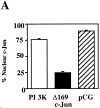
Activated PI 3-kinase does not affect the expression of c-Jun after NGF withdrawal. Cells were injected with activated PI 3-kinase, pCG, or Δ169 c-Jun. After 16–24 h they were withdrawn from NGF, fixed 24 h later, and stained for c-Jun (A) or phospho-c-Jun (B and C). The graphs in A and B represent data from three independent experiments each, in which 200 cells were injected per coverslip and scored positive for c-Jun or phospho-c-Jun if there was increased staining in the nucleus. (C) Injected cells were withdrawn from NGF (a–i) or maintained in NGF (j–l). Cells were injected with PI 3-kinase (a–c), pCG (d–f, and j–l), or Δ169 c-Jun (g–i). Cells were stained for phospho-c-Jun (a, d, g, and j), for guinea pig IgG (b, e, h, and k), or with Hoechst (c, f, i, and l). Bar, 20 μm.
Figure 5
Figure 5
Activated PI 3-kinase does not affect the expression of c-Jun after NGF withdrawal. Cells were injected with activated PI 3-kinase, pCG, or Δ169 c-Jun. After 16–24 h they were withdrawn from NGF, fixed 24 h later, and stained for c-Jun (A) or phospho-c-Jun (B and C). The graphs in A and B represent data from three independent experiments each, in which 200 cells were injected per coverslip and scored positive for c-Jun or phospho-c-Jun if there was increased staining in the nucleus. (C) Injected cells were withdrawn from NGF (a–i) or maintained in NGF (j–l). Cells were injected with PI 3-kinase (a–c), pCG (d–f, and j–l), or Δ169 c-Jun (g–i). Cells were stained for phospho-c-Jun (a, d, g, and j), for guinea pig IgG (b, e, h, and k), or with Hoechst (c, f, i, and l). Bar, 20 μm.

References

    1. Ahmed NN, Franke TF, Bellacosa A, Datta K, Gonzalez-Portal ME, Taguchi T, Testa JR, Tsichlis PN. The proteins encoded by c-akt and v-akt differ in post-translational modification, subcellular localization and oncogenic potential. Oncogene. 1993;8:1957–1963. - PubMed
    1. Bellacosa A, Testa JR, Staal SP, Tsichlis PN. A retroviral oncogene, akt, encoding a serine-threonine kinase containing an SH2-like region. Science (Wash DC) 1991;254:274–277. - PubMed
    1. Borasio GD, John J, Wittinghofer A, Barde YA, Sendtner M, Heumann R. ras p21 protein promotes survival and fiber outgrowth of cultured embryonic neurons. Neuron. 1989;2:1087–1096. - PubMed
    1. Borasio GD, Markus A, Wittinghofer A, Barde YA, Heumann R. Involvement of ras p21 in neurotrophin-induced response of sensory, but not sympathetic neurons. J Cell Biol. 1993;121:665–672. - PMC - PubMed
    1. Burgering BM, Coffer PJ. Protein kinase B (c-Akt) in phosphatidylinositol-3-OH kinase signal transduction. Nature (Lond) 1995;376:599–602. - PubMed

MeSH terms

Substances