. 1997 Nov 3;186(9):1487-94.
doi: 10.1084/jem.186.9.1487.

Requirements for both Rac1 and Cdc42 in membrane ruffling and phagocytosis in leukocytes

Affiliations

Requirements for both Rac1 and Cdc42 in membrane ruffling and phagocytosis in leukocytes

D Cox et al. J Exp Med. .

Abstract

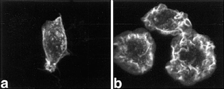
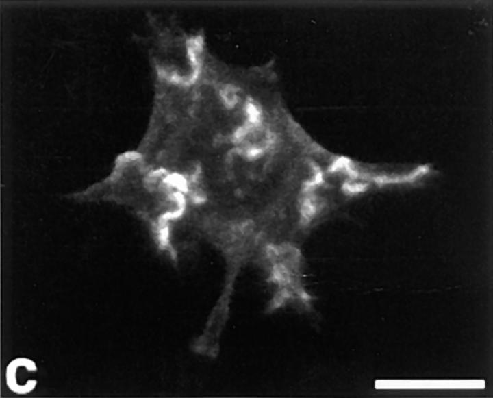
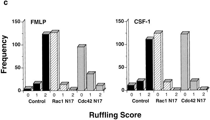
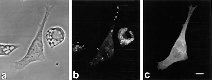
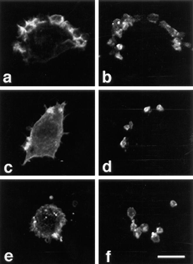
Specific pathways linking heterotrimeric G proteins and Fcgamma receptors to the actin-based cytoskeleton are poorly understood. To test a requirement for Rho family members in cytoskeletal events mediated by structurally diverse receptors in leukocytes, we transfected the full-length human chemotactic peptide receptor in RAW 264.7 cells and examined cytoskeletal alterations in response to the chemotactic peptide formyl-methionyl-leucyl-phenylalanine (FMLP), colony stimulating factor-1 (CSF-1), IgG-coated particles, and phorbol 12-myristate 13-acetate (PMA). Expression of Rac1 N17, Cdc42 N17, or the GAP domain of n-chimaerin inhibited cytoskeletal responses to FMLP and CSF-1, and blocked phagocytosis. Accumulation of F-actin- rich "phagocytic cups" was partially inhibited by expression of Rac1 N17 or Cdc42 N17. In contrast, PMA-induced ruffling was not inhibited by expression of Rac1 N17, but was blocked by expression of Cdc42 N17, indicating that cytoskeletal inhibition by these constructs was nonoverlapping. These results demonstrate differential requirements for Rho family GTPases in leukocyte motility, and indicate that both Rac1 and Cdc42 are required for Fcgamma receptor- mediated phagocytosis and for membrane ruffling mediated by structurally distinct receptors in macrophages.


Figures

Figure 1
Figure 1
FMLP or CSF-1 induces F-actin–rich ruffles in RAW LacR/ FMLPR.2 cells. Cells were fixed and stained with rhodamine-phalloidin as described in Materials and Methods. (a) Unstimulated. (b) Cells incubated with 100 nM FMLP for 1 min. (c) Cells incubated with 10 ng/ml CSF-1 for 5 min. Bar = 10 μm. (d) Inhibition of FMLP-induced ruffles by pertussis toxin (1 ng/ml for 24 h) in RAW LacR/FMLPR.2 cells. Solid bars, − pertussis toxin; striped bars, + pertussis toxin.
Figure 1
Figure 1
FMLP or CSF-1 induces F-actin–rich ruffles in RAW LacR/ FMLPR.2 cells. Cells were fixed and stained with rhodamine-phalloidin as described in Materials and Methods. (a) Unstimulated. (b) Cells incubated with 100 nM FMLP for 1 min. (c) Cells incubated with 10 ng/ml CSF-1 for 5 min. Bar = 10 μm. (d) Inhibition of FMLP-induced ruffles by pertussis toxin (1 ng/ml for 24 h) in RAW LacR/FMLPR.2 cells. Solid bars, − pertussis toxin; striped bars, + pertussis toxin.
Figure 2
Figure 2
Induction of expression of Myc-tagged fusion proteins in stable RAW cell transfectants. (a) 106 RAW cells (derived from a clone of RAW LacR/FMLPR.2 cells transfected with Myc-tagged Chimaerin-GAP) were incubated for 0, 5, or 15 h in the presence or absence of 10 mM IPTG, 50 μM zinc, and 2 mM butyrate. Cells were subjected to detergent lysis, SDS-PAGE, and immunoblotting with either anti-Myc or antiactin mAbs. Lanes 1 and 4, uninduced; lanes 2 and 3, induced with IPTG only; lanes 5 and 6, induced with IPTG, butyrate, and zinc. (b) Average levels of expression of Myc-tagged fusion proteins in individual cells. Induction of protein expression was performed as described in Materials and Methods and cells were fixed and stained with anti-Myc mAb and processed for microspectrofluorometry. Expression levels are shown in arbitrary fluorescence units. Nonspecific fluorescence (i.e., of uninduced cells or untransfected cells) was <10% of the total fluorescence signal. Data are expressed as the mean fluorescence (± SEM) of 150–200 Myc-positive cells and represent all Myc-positive cells observed in 5–10 high power fields from three separate experiments.
Figure 2
Figure 2
Induction of expression of Myc-tagged fusion proteins in stable RAW cell transfectants. (a) 106 RAW cells (derived from a clone of RAW LacR/FMLPR.2 cells transfected with Myc-tagged Chimaerin-GAP) were incubated for 0, 5, or 15 h in the presence or absence of 10 mM IPTG, 50 μM zinc, and 2 mM butyrate. Cells were subjected to detergent lysis, SDS-PAGE, and immunoblotting with either anti-Myc or antiactin mAbs. Lanes 1 and 4, uninduced; lanes 2 and 3, induced with IPTG only; lanes 5 and 6, induced with IPTG, butyrate, and zinc. (b) Average levels of expression of Myc-tagged fusion proteins in individual cells. Induction of protein expression was performed as described in Materials and Methods and cells were fixed and stained with anti-Myc mAb and processed for microspectrofluorometry. Expression levels are shown in arbitrary fluorescence units. Nonspecific fluorescence (i.e., of uninduced cells or untransfected cells) was <10% of the total fluorescence signal. Data are expressed as the mean fluorescence (± SEM) of 150–200 Myc-positive cells and represent all Myc-positive cells observed in 5–10 high power fields from three separate experiments.
Figure 3
Figure 3
Expression of Myc-tagged Rac1 N17, Cdc42 N17, or Chimaerin-GAP inhibits ruffling in response to FMLP. Cells were incubated with 100 nM FMLP for 1 min before fixation and staining with rhodamine-phalloidin to detect F-actin and mAb anti-Myc followed by FITC anti–mouse IgG to detect the indicated fusion proteins, as described in Materials and Methods. Rhodamine-phalloidin staining of a control cell is shown in a and of representative Myc-positive cells are shown in b–d. A field of cells, some of which express Chimaerin-GAP, is depicted in e (stained with anti-Myc) and f (stained with rhodamine-phalloidin). a, Control; b, Rac1 N17; c, Cdc42 N17; d–f, Chimaerin-GAP. Bar = 10 μm.
Figure 4
Figure 4
Ruffling indices and scores of FMLP- or CSF-1–stimulated RAW cells transfected with the indicated constructs. Narrow striped bars, Myc-expressing cells; solid bars, controls, derived from the same clones but which did not express the Myc epitope by indirect immunofluorescence. Ruffling indices of cells incubated in the absence of IPTG, zinc, and butyrate were indistinguishable from cells that were incubated with these agents but did not express Myc. a, FMLP; b, CSF-1. Data are expressed as the mean ± SEM, n = 3. The reduction of ruffling by Rac1 N17, Cdc42 N17, or Chimaerin-GAP was statistically significant (P <0.005). (c) Histograms of ruffling scores of individual cells stimulated with the indicated agonist. The extent of ruffling of each cell was scored using a scale of 0–2, where 0 indicates that no ruffles were present, 1 indicates that ruffling was confined to one area of the cell only (<25% of cell circumference), and 2 indicates that two or more discrete areas of the cell contained ruffles. 140 cells were counted for each construct.
Figure 4
Figure 4
Ruffling indices and scores of FMLP- or CSF-1–stimulated RAW cells transfected with the indicated constructs. Narrow striped bars, Myc-expressing cells; solid bars, controls, derived from the same clones but which did not express the Myc epitope by indirect immunofluorescence. Ruffling indices of cells incubated in the absence of IPTG, zinc, and butyrate were indistinguishable from cells that were incubated with these agents but did not express Myc. a, FMLP; b, CSF-1. Data are expressed as the mean ± SEM, n = 3. The reduction of ruffling by Rac1 N17, Cdc42 N17, or Chimaerin-GAP was statistically significant (P <0.005). (c) Histograms of ruffling scores of individual cells stimulated with the indicated agonist. The extent of ruffling of each cell was scored using a scale of 0–2, where 0 indicates that no ruffles were present, 1 indicates that ruffling was confined to one area of the cell only (<25% of cell circumference), and 2 indicates that two or more discrete areas of the cell contained ruffles. 140 cells were counted for each construct.
Figure 5
Figure 5
Inhibition of FcγR-mediated phagocytosis by Cdc42 N17. Phagocytosis assay was performed as described in Materials and Methods. (a) Phase-contrast micrograph. (b) Fluorescence micrograph of rhodamine anti–rabbit IgG-stained cells to indicate presence of IgG-RBCs. After fixation and permeabilization, uningested erythrocytes appear crenated. (c) Fluorescence micrograph of anti-Myc–stained cell to indicate expression of Myc-tagged Cdc42 N17. Note ingestion of IgG-RBCs in neighboring cells not expressing the Myc epitope.
Figure 6
Figure 6
Expression of Myc-tagged Rac1 N17, Cdc42 N17, or Chimaerin-GAP inhibits FcγR-mediated phagocytosis. Striped bars, Myc-expressing cells; solid bars, controls, which are derived from the same clones but did not express the Myc epitope by indirect immunofluorescence. Phagocytosis indices of cells incubated in the absence of IPTG, zinc, and butyrate were indistinguishable from cells that were incubated with these agents but did not express Myc. Data are expressed as the mean ± SEM, n = 3. The reduction of phagocytosis by Rac1 N17, Cdc42 N17, or Chimaerin-GAP was statistically significant (P <0.005).
Figure 7
Figure 7
Phagocytic cup formation is inhibited by expression of Rac1 N17 or Cdc42 N17. Cells were incubated with IgG-RBCs for 5 min at 37°C before fixation. Immunofluorescence using rhodamine-phalloidin and anti–rabbit IgG was performed as described in Materials and Methods. a, c, and e, rhodamine-phalloidin; b, d, and f, anti–rabbit IgG. a and b: control; c and d, Cdc42 N17; e and f, Rac1 N17. Bar = 10 μm.
Figure 8
Figure 8
PMA-induced ruffling is a Rac1-independent event in RAW cell transfectants. Cells were incubated with vehicle (a) or with 100 nM PMA (b–d), and stained with anti-Myc mAb and rhodamine-phalloidin as described in Materials and Methods. Rhodamine-phalloidin fluorescence is pictured. a, control; b, control incubated with PMA; c, Rac1 N17 incubated with PMA; d, Cdc42 N17 incubated with PMA. Bar = 10 μm.
Figure 9
Figure 9
Ruffling indices of PMA-stimulated RAW cells transfected with the indicated constructs. Striped bars, Myc-expressing cells; solid bars, controls, which are derived from the same clones but did not express the Myc epitope by indirect immunofluorescence. Ruffling indices of cells incubated in the absence of IPTG, zinc, and butyrate were indistinguishable from cells that were incubated with these agents but did not express Myc. Data are expressed as the mean ± SEM, n = 3. The reduction of ruffling by Cdc42 N17 or Chimaerin-GAP was statistically significant (P <0.005).

Similar articles

Cited by

References

    1. Tapon N, Hall A. Rho, Rac, and Cdc42 GTPases regulate the organization of the actin cytoskeleton. Curr Opin Cell Biol. 1997;9:86–92. - PubMed
    1. Lim L, Hall C, Monfries C. Regulation of actin cytoskeleton by Rho-family GTPases and their associated proteins. Sem Cell Dev Biol. 1996;7:699–706.
    1. Ridley AJ, Paterson HF, Johnston CL, Diekmann D, Hall A. The small GTP-binding protein rac regulates growth factor–induced membrane ruffling. Cell. 1992;70:401–410. - PubMed
    1. Allen WE, Jones GE, Pollard JW, Ridley AJ. Rho, Rac and Cdc42 regulate actin organization and cell adhesion in macrophages. J Cell Sci. 1997;110:707–720. - PubMed
    1. Kozma R, Ahmed S, Best A, Lim L. The Ras-related protein Cdc42Hs and bradykinin promote formation of peripheral actin microspikes and filopodia in Swiss 3T3 fibroblasts. Mol Cell Biol. 1995;15:1942–1952. - PMC - PubMed

Publication types

MeSH terms