. 1997 Nov 3;186(9):1503-12.
doi: 10.1084/jem.186.9.1503.

Specific activation of the cysteine protease CPP32 during the negative selection of T cells in the thymus

Affiliations

Specific activation of the cysteine protease CPP32 during the negative selection of T cells in the thymus

A Alam et al. J Exp Med. .

Abstract

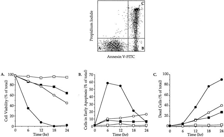
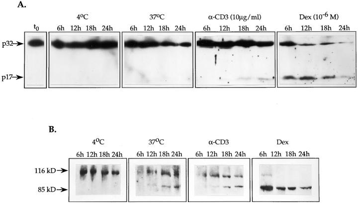
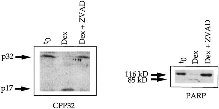
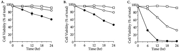
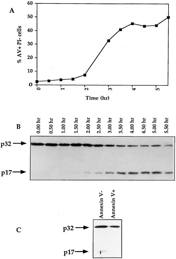
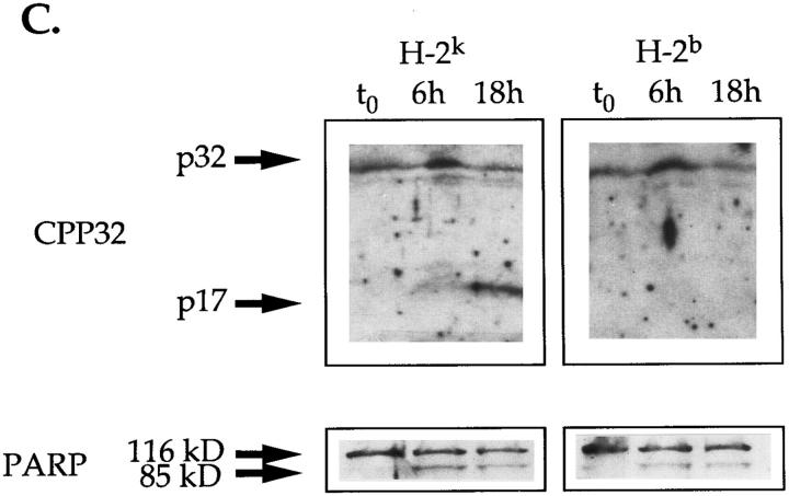
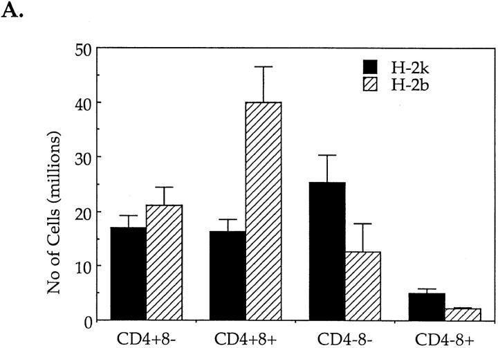
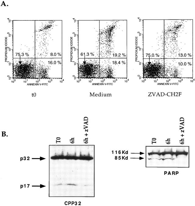
Cysteine proteases of the CED-3 and ICE family have been recently proposed as the ultimate executioners in several mammalian cell death pathways. Among them, the cysteine protease CPP32 has been shown to participate in programmed cell death (PCD), or apoptosis, affecting lymphoid cells in vitro. In the thymus, negative selection is a mechanism through which developing thymocytes expressing a TcR with high affinity for self peptide-MHC complexes are eliminated by PCD. In order to investigate the role of CPP32 in thymic apoptosis, isolated thymocytes were submitted to cell surface CD3 crosslinking by immobilized anti-CD3 mAb or to dexamethasone treatment. Although apoptosis occurred in the absence or after crosslinking with anti-CD3 mAb, specific activation of CPP32, as assessed by the extent of proteolytic cleavage of the p32 zymogen, was only detected in thymocytes cultured in the presence of the immobilized antibody or dexamethasone. This activation was a very early event during apoptosis as it occurred before the exposure of phosphatidyl serine to the upper side of the cell membrane. This was observed both in anti-CD3- and dexamethasone-induced apoptosis. Moreover, using mice transgenic for pigeon cytochrome C (PCC)-specific TcR, we were able to show that, after injection of PCC, the activation of CPP32 and cleavage of its substrate occurred in thymocytes obtained from mice expressing a permissive MHC haplotype for PCC presentation (H-2k). Moreover, PCC induced apoptosis was blocked by the caspase inhibitor zVAD. While spontaneous apoptosis was not accompanied by detectable levels of CPP32 processing, it was characterized by the proteolysis of poly(ADP-ribose) polymerase (PARP) and was blocked by the cysteine protease inhibitor, zVAD-CH2F. Taken together, these results support the concept that CPP32 is among the earliest effectors of the pathway leading to negative selection of autoreactive thymocytes. Our results also suggest the involvement of a distinct CPP32-like cysteine protease in spontaneous apoptosis of thymocytes.


Figures

Figure 1
Figure 1
Time course of apoptosis in isolated thymocytes cultured alone or in the presence of immobilized anti-CD3 mAbs or dexamethasone. The upper panel shows the flow cytometry measurement of annexin V binding (x axis) vs. propidium iodide uptake (y axis) on thymocytes cultured for 12 h in the presence of immobilized anti-CD3. Viable cells and cells in early apoptosis are annexin V− PI− (A) and annexin V+ PI− (B), respectively. Dead cells are annexin V+ PI+ (C). The lower panels display the evolution of cell viability (A), the percentage of cells in early apoptosis (B) and the mortality (C) in thymocyte populations incubated at 4°C (open square) or cultured at 37°C alone (closed squares), in the presence of immobilized anti-CD3 mAb (open circles) or with dexamethasone (closed circles). This experiment produced identical results in a total of four independent occasions.
Figure 2
Figure 2
In isolated thymocytes, CPP32 is activated during apoptosis induced by crosslinking of CD3 or exposure to dexamethasone. Time course of CPP32 activation (A) and cleavage of PARP (B) in thymocytes cultured at 4°C, or at 37°C alone, with immobilized anti-CD3 mAb or with dexamethasone for the indicated time. Cell lysates were electrophoresed, and substrate cleavage was visualized by immunoblotting as described in Materials and Methods. The results presented in this figure are representative of two independent experiments.
Figure 3
Figure 3
zVAD-CH2F inhibits the processing of CPP32 and the cleavage of PARP. CPP32 activation and cleavage of PARP in thymocytes cultured with dexamethasone for 6 h in the presence or absence of 100 μM zVAD-CH2F. Cell lysates were electrophoresed, and substrate cleavage was visualized by immunoblotting as described in Materials and Methods.
Figure 4
Figure 4
Inhibition of thymocyte apoptosis by zVAD-CH2F. In vitro, apoptosis in thymocytes cultured for 18 h at 37°C alone (A), with immobilized anti-CD3 mAb (B) or with dexamethasone (C) is inhibited by zVAD-CH2F. Cell viability in the presence (open circles) or absence (closed circles) of zVAD-CH2F was determined as in Fig. 1. Cells incubated at 4°C were used as a negative control (open squares). These experiments were repeated on two separate occasions with identical results.
Figure 5
Figure 5
The activation of CPP32 is a very early event of thymocyte apoptosis. (A and B) Thymocytes from C56BL/6 mice were isolated and incubated with dexamethasone. Samples were taken at 30-min intervals and analyzed for levels of apoptosis with annexin V-FITC and PI (A). Cell lysates were submitted to Western blotting analysis for the processing of CPP32 (B). (C) Thymocytes were cultured for 20 h in the presence of immobilized anti-CD3 mAb. Cells were then labeled with annexin V-FITC, and annexin V− and annexin V+ cells were sorted at 4°C with a FACStar® Plus. Sorted cells were lysed and extract contents were analyzed for processing of CPP32 by Western blotting.
Figure 6
Figure 6
Injection of PCC induce negative selection and activation of CPP32 in thymocytes of PCC-specific TcR transgenic mice of the H-2k, but not H-2b, haplotype. (A) Absolute number of cells in the different cell populations of thymuses isolated from H-2k and H-2b TcR transgenic mice 24 h after PCC injection. (B, top) Percentages of expression of CD4 and CD8 molecules by viable thymocytes (annexin V−) isolated from PCC-injected H-2k and H-2b TcR transgenic mice after 18 h of culture at 37°C. Thymuses from noninjected H-2k TcR transgenic mice contain 34.3 ± 2.5% of CD4+8 cells, 48.4 ± 5.7% of CD4+8+ cells and 15.0 ± 5.6% of CD48 cells. Thymuses from noninjected H-2b TcR transgenic mice contain 37.0 ± 1.8% of CD4+8 cells, 46.0 ± 4.1% of CD4+8+ cells and 14.8 ± 3.9% of CD48 cells. (B, bottom) Percentages of annexin V+ cells in CD4+8, CD4+8+ and CD48 thymocytes isolated from PCC-injected H-2k and H-2b TcR transgenic mice after 18 h of culture at 37°C. (C) Proteolytic cleavage of CPP32 and PARP in thymocytes isolated from PCC-injected H-2k and H-2b TcR transgenic mice. After 6 or 18 h of culture at 37°C, cells were lysed and the lysates were electrophoresed. The substrate cleavage was visualized by immunoblotting as described in Materials and Methods.
Figure 6
Figure 6
Injection of PCC induce negative selection and activation of CPP32 in thymocytes of PCC-specific TcR transgenic mice of the H-2k, but not H-2b, haplotype. (A) Absolute number of cells in the different cell populations of thymuses isolated from H-2k and H-2b TcR transgenic mice 24 h after PCC injection. (B, top) Percentages of expression of CD4 and CD8 molecules by viable thymocytes (annexin V−) isolated from PCC-injected H-2k and H-2b TcR transgenic mice after 18 h of culture at 37°C. Thymuses from noninjected H-2k TcR transgenic mice contain 34.3 ± 2.5% of CD4+8 cells, 48.4 ± 5.7% of CD4+8+ cells and 15.0 ± 5.6% of CD48 cells. Thymuses from noninjected H-2b TcR transgenic mice contain 37.0 ± 1.8% of CD4+8 cells, 46.0 ± 4.1% of CD4+8+ cells and 14.8 ± 3.9% of CD48 cells. (B, bottom) Percentages of annexin V+ cells in CD4+8, CD4+8+ and CD48 thymocytes isolated from PCC-injected H-2k and H-2b TcR transgenic mice after 18 h of culture at 37°C. (C) Proteolytic cleavage of CPP32 and PARP in thymocytes isolated from PCC-injected H-2k and H-2b TcR transgenic mice. After 6 or 18 h of culture at 37°C, cells were lysed and the lysates were electrophoresed. The substrate cleavage was visualized by immunoblotting as described in Materials and Methods.
Figure 7
Figure 7
zVAD-CH2F blocks early apoptosis and CPP32 activity in thymocytes undergoing negative selection. 3 wk-old H-2k transgenic mice were injected with PCC. 16 h later thymocytes were isolated and cultured with or without zVAD-CH2F. After 6 h, cells were compared for levels of apoptosis with annexin V-FITC and PI. Cell extracts were analyzed by Western blotting for processing of CPP32 and cleavage of PARP.

References

    1. Steller H. Mechanisms and genes of cellular suicide. Science (Wash DC) 1995;267:1445–1449. - PubMed
    1. Martin SJ, Green DR. Protease activation during apoptosis: death by a thousand cuts? . Cell. 1995;82:349–352. - PubMed
    1. Fraser A, Evan G. A license to kill. Cell. 1996;85:781–784. - PubMed
    1. Fernandes-Alnemri T, Litwack G, Alnemri ES. CPP32, a novel human apoptotic protein with homology to Caenorhabditis eleganscell death protein Ced-3 and mammalian interleukin-1 β-converting enzyme. J Biol Chem. 1994;269:30761–30764. - PubMed
    1. Tewari M, Quan LT, O'Rourke K, Desnoyers S, Zheng Z, Beidler DR, Poirier GG, Salvesen GS, Dixit VM. YAMA/CPP32β, a mammalian homolog of CED-3, is a Crma-inhibitable protease that cleaves the death substrate poly(ADP-ribose) polymerase. Cell. 1995;81:801–809. - PubMed

Publication types

MeSH terms