. 1997 Nov 3;186(9):1513-22.
doi: 10.1084/jem.186.9.1513.

Enforced Bcl-2 expression inhibits antigen-mediated clonal elimination of peripheral B cells in an antigen dose-dependent manner and promotes receptor editing in autoreactive, immature B cells

Affiliations

Enforced Bcl-2 expression inhibits antigen-mediated clonal elimination of peripheral B cells in an antigen dose-dependent manner and promotes receptor editing in autoreactive, immature B cells

J Lang et al. J Exp Med. .

Abstract

The mechanisms that establish immune tolerance in immature and mature B cells appear to be distinct. Membrane-bound autoantigen is thought to induce developmental arrest and receptor editing in immature B cells, whereas mature B cells have shortened lifespans when exposed to the same stimulus. In this study, we used Emu-bcl-2-22 transgenic (Tg) mice to test the prediction that enforced expression of the Bcl-2 apoptotic inhibitor in B cells would rescue mature, but not immature, B cells from tolerance induction. To monitor tolerance to the natural membrane autoantigen H-2Kb, we bred 3-83mudelta (anti-Kk,b) Ig Tg mice to H-2(b) mice or to mice expressing transgene-driven Kb in the periphery. In 3-83mudelta/bcl-2 Tg mice, deletion of autoreactive B cells induced by peripheral Kb antigen expression in the liver (MT-Kb Tg) or epithelia (KerIV-Kb Tg), was partly or completely inhibited, respectively. Furthermore, Bcl-2 protected peritoneal B-2 B cells from deletion mediated by acute antigen exposure, but this protection could be overcome by higher antigen dose. In contrast to its ability to block peripheral self-tolerance, Bcl-2 overexpression failed to inhibit central tolerance induced by bone marrow antigen expression, but instead, enhanced the receptor editing process. These studies indicate that apoptosis plays distinct roles in central and peripheral B cell tolerance.


Figures

Figure 1
Figure 1
Enforced Bcl-2 expression inhibits B cell deletion induced by acute challenge with membrane antigen in an antigen dose–dependent manner. NDTg and NDTg/bcl-2 mice were injected intraperitoneally with either PBS control, Kk-expressing tumor cells, or Kd tumor cells, and the peritoneal cells were analyzed 16 h later for the loss of 3–83 B cells using 54.1 anticlonotype and anti-IgM antibodies. The data from three experiments are presented as mean ± SEM. In some data points, error bars are not apparent because of their small range.
Figure 2
Figure 2
Enforced Bcl-2 expression blocks peripheral clonal deletion in 3–83μδ mice. (A and C) 3–83μδ Tg mice (top) or 3–83μδ/ bcl-2 Tg mice (bottom) bearing the indicated self-antigens were analyzed for Id+ B cells in lymph node (A) and spleen (C). (B and D) The compiled FACS® data as percentage of IgM+/Id+ cells in the lymphoid analysis gate is shown. Data are presented as mean ± SEM. The numbers below each bar represent the number of mice analyzed per group.
Figure 3
Figure 3
Effect of bcl-2 transgene on B cell turnover in peripheral antigen-expressing 3–83μδ Tg mice. Mice were fed BrdU-containing water for 7 d followed by staining of isolated lymph node and spleen cells with anti-BrdU and anti-B220 antibodies. Data are presented as percentage (mean ± SEM) of B220+ cells that were BrdU positive. Numbers of mice analyzed are indicated below bars. Mice lacking antigen were H-2d ND controls.
Figure 4
Figure 4
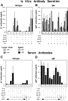
In vitro and in vivo analysis of antibody secretion. Lymph node and spleen cells from mice of the indicated genotypes were cultured in the presence or absence of 50 μg/ml LPS. After 3 d of culture, supernatants were analyzed by ELISA for Id+ (A) and IgM (B) antibodies. Antibody amounts were quantitated using 3–83 IgM transfectoma supernatant control. Similarly, Id+ (C) and IgM (D) serum antibodies were quantitated by ELISA. Data are presented as mean ± SEM with number of mice (n) analyzed shown. The table below the graph shows Tg genotypes (3–83, bcl-2, MT-Kb, and/or KerIV-Kb) and H-2 haplotypes (d, no antigen; b, antigen). In C and D the RAG-1 genotype of analyzed mice (+/+ or −/−) is indicated.
Figure 5
Figure 5
Enforced Bcl-2 expression fails to block central deletion induced by the ultralow affinity 3–83 ligand Dk. Lymph node and spleen cells from mice of the indicated genotypes were double stained for the presence of B cells bearing the 3–83 Tg BCR with 54.1 anti-Id and anti-IgM antibodies. The substantial population of IgM+ B cells present in the CDTg mice were clonotype and are presumably the result of receptor editing. The large percentage of B cells in the ND control in this experiment reflects the RAG deficiency of this particular mouse.
Figure 6
Figure 6
Appearance of immature B cells in spleen of CD (H-2b) bcl-2/RAG-1–deficient mice. Spleen cells from mice of the indicated genotypes were stained for presence of 3–83 B cells with anticlonotype (54.1) and anti-IgM antibodies (A), and anti-IgM and anti-B220 antibodies (B). The immature B cells have low levels of Id and lack detectable sIgM (arrows). The NDTg control used in this particular experiment was 3–83μδ homozygous, which consistently show reduced B cell populations. Data shown represent one of five similar experiments.
Figure 7
Figure 7
Increased percentage of IgDa, λ+ cells in CDTg mice with enforced Bcl-2 expression. 3–83μδ Tg mice were bred with bcl-2 Tg mice in the presence (CDTg) or absence (NDTg) of bone marrow antigen expression. To determine the extent of receptor editing, lymph node cells were double stained for Tg heavy chain (anti-IgDa) and endogenous light chain (anti-λ). (A) Each row illustrates FACS® analysis from independent experiments. (B) Summary data showing percentage of IgDa-positive, Id cells in both CDTg and CDTg mice with Bcl-2 overexpression. Data are presented as mean ± SEM.
Figure 8
Figure 8
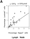
Bcl-2 overexpression increases λ/κ ratio of B cells in both 3–83 CDTg mice and non-Ig Tg mice. (A) Lymph node cells from CDTg and CDTg/bcl-2 mice were double stained with anti-IgDa and anti-λ antibodies. Percentage of lymph node cells staining positive for IgDa and negative for λ were scored as κ positive. (B) Increased percentage of λ-expressing B cells in (non-Ig Tg) bcl-2 Tg mice. The percentage of IgM+ cells that were also λ positive was determined. Data presented as mean ± SEM. Numbers above bars reflect number of mice examined.
Figure 8
Figure 8
Bcl-2 overexpression increases λ/κ ratio of B cells in both 3–83 CDTg mice and non-Ig Tg mice. (A) Lymph node cells from CDTg and CDTg/bcl-2 mice were double stained with anti-IgDa and anti-λ antibodies. Percentage of lymph node cells staining positive for IgDa and negative for λ were scored as κ positive. (B) Increased percentage of λ-expressing B cells in (non-Ig Tg) bcl-2 Tg mice. The percentage of IgM+ cells that were also λ positive was determined. Data presented as mean ± SEM. Numbers above bars reflect number of mice examined.

References

    1. Cory S. Regulation of lymphocyte survival by the bcl-2 gene family. Annu Rev Immunol. 1995;13:513–543. - PubMed
    1. Green DR, Scott DW. Activation-induced apoptosis in lymphocytes. Curr Opin Immunol. 1994;3:476–487. - PubMed
    1. Krammer PH, Behrmann I, Daniel P, Dhein J, Debatin KM. Regulation of apoptosis in the immune system. Curr Opin Immunol. 1994;2:279–289. - PubMed
    1. Thompson CB. Apoptosis in the pathogenesis and treatment of disease. Science (Wash DC) 1995;5203:1456–1462. - PubMed
    1. Nossal GJ. Negative selection of lymphocytes. Cell. 1994;2:229–239. - PubMed

Publication types

MeSH terms