. 1997 Nov 3;186(9):1615-20.
doi: 10.1084/jem.186.9.1615.

Wound healing is accelerated by agonists of adenosine A2 (G alpha s-linked) receptors

Affiliations

Wound healing is accelerated by agonists of adenosine A2 (G alpha s-linked) receptors

M C Montesinos et al. J Exp Med. .

Abstract

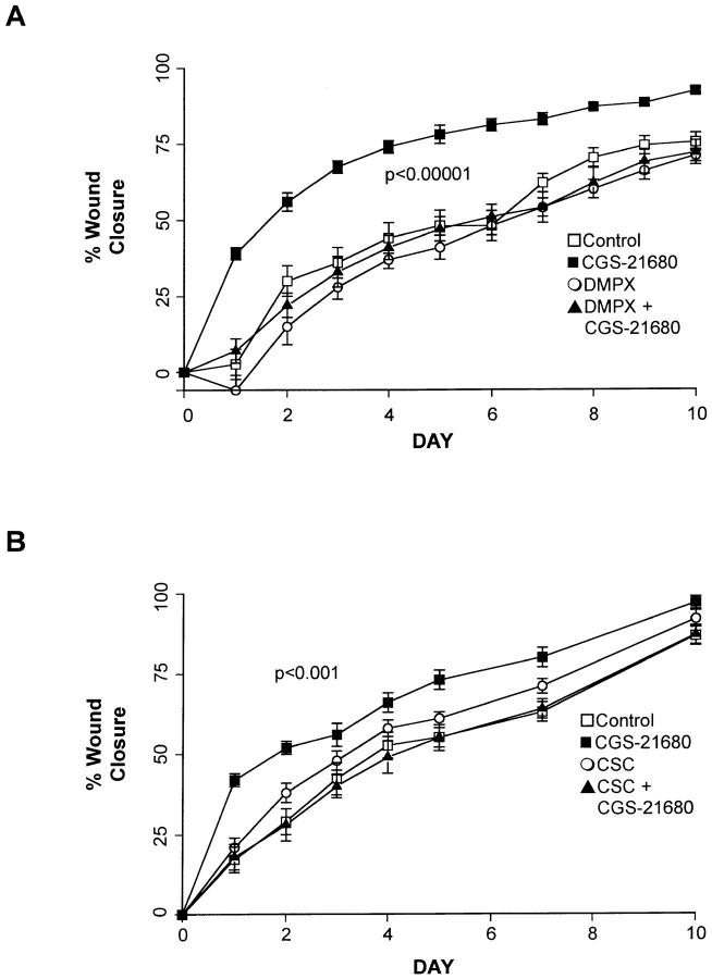
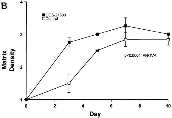
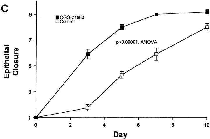
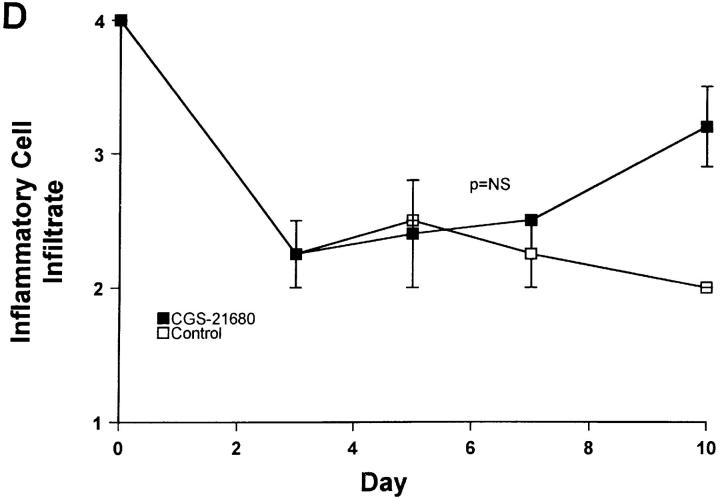
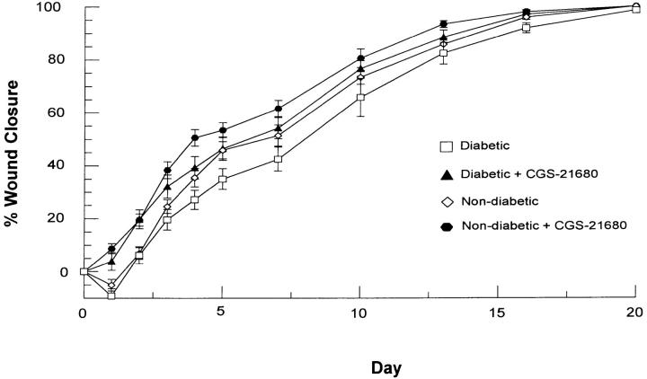
The complete healing of wounds is the final step in a highly regulated response to injury. Although many of the molecular mediators and cellular events of healing are known, their manipulation for the enhancement and acceleration of wound closure has not proven practical as yet. We and others have established that adenosine is a potent regulator of the inflammatory response, which is a component of wound healing. We now report that ligation of the G alpha s-linked adenosine receptors on the cells of an artificial wound dramatically alters the kinetics of wound closure. Excisional wound closure in normal, healthy mice was significantly accelerated by topical application of the specific A2A receptor agonist CGS-21680 (50% closure by day 2 in A2 receptor antagonists. In rats rendered diabetic (streptozotocin-induced diabetes mellitus) wound healing was impaired as compared to nondiabetic rats; CGS-21680 significantly increased the rate of wound healing in both nondiabetic and diabetic rats. Indeed, the rate of wound healing in the CGS-21680-treated diabetic rats was greater than or equal to that observed in untreated normal rats. These results appear to constitute the first evidence that a small molecule, such as an adenosine receptor agonist, accelerates wound healing in both normal animals and in animals with impaired wound healing.


Figures

Figure 1
Figure 1
Endothelial cells (HUVEC) and a fibroblast cell line (CCD-25sk) express message for adenosine receptor subtypes. RNA was isolated from confluent monolayers of either HUVEC or CCD-25sk cells, as described. cDNA was generated from the isolated mRNA by reverse transcriptase and the message for the adenosine receptors was amplified by antisense primers as described. Shown is one of two experiments yielding similar results.
Figure 2
Figure 2
The effect of the adenosine A2A agonist CGS-21680 (250 μg/ml) on wound closure. (A) Wounds were excised on the dorsum of mice and treated with carrier (1.5% methylcellulose), CGS-21680, the adenosine A2 antagonist DMPX (2.5 mg/ml), or their combination, as described. Wounds were traced daily and the area was determined after computer digitization of the wounds. (B) Wounds were excised on the dorsum of mice and treated with carrier (1.5% methylcellulose), CGS-21680, the adenosine A2 A antagonist CSC (250 μg/ml), or their combination, as described. Each point represents the mean (± SEM) of 10 wounds. Similar results were found in two other experiments.
Figure 3
Figure 3
Histologic analysis of wounds in control and CGS-21680–treated mice. Wounds were excised and mice were treated with topical application either of carrier or CGS-21680 in carrier. Analysis of fibroblast density (A), matrix density (B), epithelial closure (C), and inflammatory cell infiltrate (D) was carried out blindly, as described. Each point represents the mean (± SEM) of six wounds on three mice.
Figure 3
Figure 3
Histologic analysis of wounds in control and CGS-21680–treated mice. Wounds were excised and mice were treated with topical application either of carrier or CGS-21680 in carrier. Analysis of fibroblast density (A), matrix density (B), epithelial closure (C), and inflammatory cell infiltrate (D) was carried out blindly, as described. Each point represents the mean (± SEM) of six wounds on three mice.
Figure 4
Figure 4
The effect of the adenosine A2A agonist CGS-21680 on wound closure in normal and diabetic rats. Animals received a single injection of streptozotocin (60 mg/kg) followed 8 d later by excision of three wounds (2 cm in diameter) on the dorsum of each rat. Wounds were treated with carrier (1.5% methylcellulose/PBS, wt/vol) alone or CGS-21680 (250 μg/ml) in carrier. The wounds were traced at the indicated intervals and the area was determined after computer digitization of the wounds. Each point represents the mean (± SEM) of 9–21 wounds.

References

    1. Pierce GF, Mustoe TA. Pharmacologic enhancement of wound healing. Ann Rev Med. 1995;46:467–481. - PubMed
    1. Cronstein BN. Adenosine, an endogenous anti-inflammatory agent. J Appl Physiol. 1994;76:5–13. - PubMed
    1. Ethier MF, Chander V, Dobson JG., Jr Adenosine stimulates proliferation of human endothelial cells in culture. Am J Physiol. 1993;265:H131–H138. - PubMed
    1. Sexl V, Mancusi G, Baumgartner-Parzer S, Schutz W, Freissmuth M. Stimulation of human umbilical vein endothelial cell proliferation by A2-adenosine and beta 2-adrenoceptors. Br J Pharmacol. 1995;114:1577–1586. - PMC - PubMed
    1. Bull DA, Seftor EA, Hendrix MJ, Larson DF, Hunter GC, Putnam CW. Putative vascular endothelial cell chemotactic factors: comparison in a standardized migration assay. J Surg Res. 1993;55:473–479. - PubMed

Publication types

MeSH terms

Substances